. 2008 Aug;57(8):2149-57.
doi: 10.2337/db08-0176. Epub 2008 Apr 28.

Activation of peroxisome proliferator-activated receptor beta/delta inhibits lipopolysaccharide-induced cytokine production in adipocytes by lowering nuclear factor-kappaB activity via extracellular signal-related kinase 1/2

Affiliations

Activation of peroxisome proliferator-activated receptor beta/delta inhibits lipopolysaccharide-induced cytokine production in adipocytes by lowering nuclear factor-kappaB activity via extracellular signal-related kinase 1/2

Ricardo Rodríguez-Calvo et al. Diabetes. 2008 Aug.

Abstract

Objective: Chronic activation of the nuclear factor-kappaB (NF-kappaB) in white adipose tissue leads to increased production of pro-inflammatory cytokines, which are involved in the development of insulin resistance. It is presently unknown whether peroxisome proliferator-activated receptor (PPAR) beta/delta activation prevents inflammation in adipocytes.

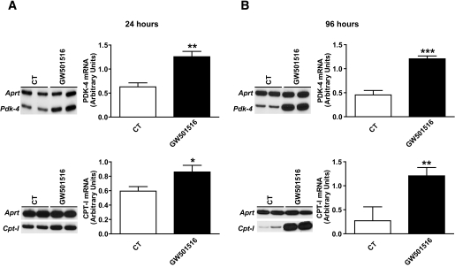
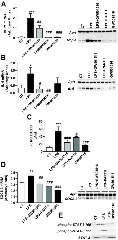
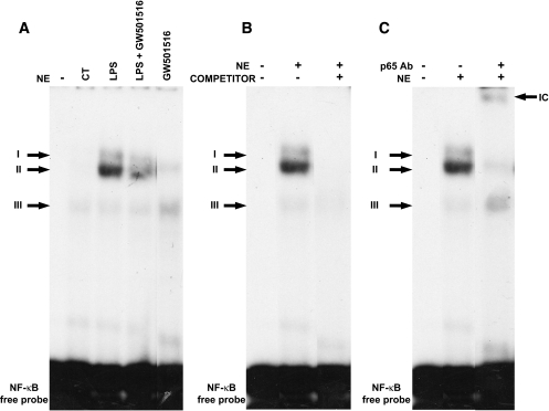
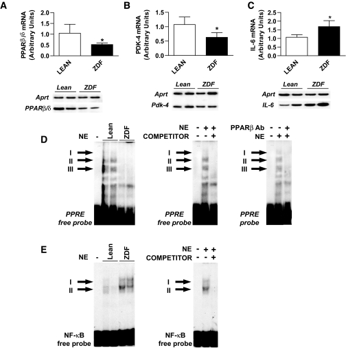
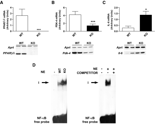
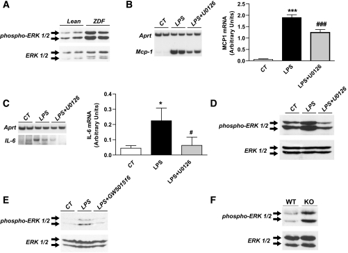
Research design and methods and results: First, we examined whether the PPARbeta/delta agonist GW501516 prevents lipopolysaccharide (LPS)-induced cytokine production in differentiated 3T3-L1 adipocytes. Treatment with GW501516 blocked LPS-induced IL-6 expression and secretion by adipocytes and the subsequent activation of the signal transducer and activator of transcription 3 (STAT3)-Suppressor of cytokine signaling 3 (SOCS3) pathway. This effect was associated with the capacity of GW501516 to impede LPS-induced NF-kappaB activation. Second, in in vivo studies, white adipose tissue from Zucker diabetic fatty (ZDF) rats, compared with that of lean rats, showed reduced PPARbeta/delta expression and PPAR DNA-binding activity, which was accompanied by enhanced IL-6 expression and NF-kappaB DNA-binding activity. Furthermore, IL-6 expression and NF-kappaB DNA-binding activity was higher in white adipose tissue from PPARbeta/delta-null mice than in wild-type mice. Because mitogen-activated protein kinase-extracellular signal-related kinase (ERK)1/2 (MEK1/2) is involved in LPS-induced NF-kappaB activation in adipocytes, we explored whether PPARbeta/delta prevented NF-kappaB activation by inhibiting this pathway. Interestingly, GW501516 prevented ERK1/2 phosphorylation by LPS. Furthermore, white adipose tissue from animal showing constitutively increased NF-kappaB activity, such as ZDF rats and PPARbeta/delta-null mice, also showed enhanced phospho-ERK1/2 levels.

Conclusions: These findings indicate that activation of PPARbeta/delta inhibits enhanced cytokine production in adipocytes by preventing NF-kappaB activation via ERK1/2, an effect that may help prevent insulin resistance.


Figures

FIG. 1.
FIG. 1.
The PPARβ/δ agonist GW501516 induces Pdk-4 and Cpt-I expression in 3T3-L1 adipocytes. Differentiated adipocytes were incubated in the presence or in the absence of 0.5 μmol/l GW501516 for either 24 h (A) or 96 h (B). Total RNA was isolated and analyzed by RT-PCR. A representative autoradiogram and the quantification normalized to the Aprt mRNA levels are shown. Data are means ± SD of five independent experiments. *P < 0.05, **P < 0.01, and ***P < 0.001 vs. control.
FIG. 2.
FIG. 2.
The PPARβ/δ agonist GW501516 prevents LPS-induced expression and secretion of pro-inflammatory cytokines. When indicated, differentiated adipocytes were incubated with 0.5 μmol/l GW501516 for 96 h and subsequently exposed to 100 ng/ml LPS for 24 h in the presence or in the absence of GW501516 or 10 μmol/l NF-κB inhibitor parthenolide (PARTH). Effects of GW501516 on the expression of Mcp-1 (A) and IL-6 (B). Total RNA was isolated and analyzed by RT-PCR. A representative autoradiogram and the quantification normalized to the Aprt mRNA levels are shown. C: Effect of GW501516 on the secretion of IL-6 to the culture media, as determined by ELISA. D: Effect of GW501516 on the expression of SOCS-3. E: Analysis of STAT3 and phospho-STAT3 by immunoblotting of nuclear protein extracts from 3T3-L1 adipocytes treated with 100 ng/ml LPS for 3 h in the presence or in the absence of 0.5 μmol/l GW501516. Data are means ± SD of five independent experiments. *P < 0.05, **P < 0.01, and ***P < 0.001 vs. control. #P < 0.05, ##P < 0.01, and ###P < 0.001 vs. LPS-exposed cells.
FIG. 3.
FIG. 3.
The PPARβ/δ agonist GW501516 prevents LPS-induced NF-κB activation in 3T3-L1 adipocytes. When indicated, differentiated adipocytes were incubated with 0.5 μmol/l GW501516 for 96 h and subsequently exposed to 100 ng/ml LPS for 1 h in the presence or in the absence of GW501516. A: Autoradiograph of EMSA performed with a 32P-labeled NF-κB nucleotide and crude nuclear protein extract (NE). Three specific complexes (I–III), based on competition with a molar excess of unlabeled probe (B), are shown. C: A supershift analysis performed by incubating NE with an antibody directed against the p65 subunit of NF-κB is also shown.
FIG. 4.
FIG. 4.
In white adipose tissue of ZDF rats, PPARβ/δ expression is reduced, whereas NF-κB activity is increased. Analysis of the mRNA levels of Pparβ/δ (A), Pdk-4 (B), and IL-6 (C) in white adipose tissue of lean and ZDF rats. Total RNA was isolated and analyzed by RT-PCR. A representative autoradiogram and the quantification normalized to the Aprt mRNA levels are shown. Data are means ± SD of five independent experiments. *P < 0.05. D: Autoradiograph of EMSA performed with a 32P-labeled PPRE nucleotide and crude nuclear protein extract (NE). Three specific complexes (I–III), based on competition with a molar excess of unlabeled probe (right), are shown. An analysis performed by incubating NE with an antibody directed against PPARβ/δ is also shown. This antibody does not shift the complex, but prevents its binding to the PPRE. E: Autoradiograph of EMSA performed with a 32P-labeled NF-κB nucleotide and NE. Two specific complexes, based on competition with a molar excess of unlabeled probe (right), are shown.
FIG. 5.
FIG. 5.
The PPARβ/δ-null mouse shows increased NF-κB activity in white adipose tissue. Analysis of the mRNA levels of Pparβ/δ (A), Pdk-4 (B), and Il-6 (C) in white adipose tissue of wild-type (wt) or PPARβ/δ-null (ko) mouse white adipose tissue. Total RNA was isolated and analyzed by RT-PCR. A representative autoradiogram and the quantification normalized to the Aprt mRNA levels are shown. Data are means ± SD of five independent experiments. *P < 0.05. D: Autoradiograph of EMSA performed with a 32P-labeled NF-κB nucleotide and crude nuclear protein extract (NE). One specific complex, based on competition with a molar excess of unlabeled probe (right), is shown.
FIG. 6.
FIG. 6.
PPARβ/δ regulates ERK1/2 phosphorylation in adipocytes and white adipose tissue. A: Analysis of ERK and phospho-ERK by immunoblotting of total protein extracts from white adipose tissue of lean and ZDF rats. The MEK1/2 inhibitor U0126 blocks NF-κB activation by LPS. Analysis of the mRNA levels of Mcp-1 (B) and IL-6 (C) in 3T3-L1 adipocytes. Differentiated adipocytes were treated with 100 ng/ml LPS for 6 h in the presence or in the absence of 10 μmol/l of the MEK1/2 inhibitor U0126. Total RNA was isolated and analyzed by RT-PCR. A representative autoradiogram and the quantification normalized to the Aprt mRNA levels are shown. Data are expressed as means ± SD of five independent experiments. *P < 0.05 and ***P < 0.001 vs. control; #P < 0.05 and ##P < 0.001 vs. LPS-exposed cells. Analysis of ERK and phospho-ERK by immunoblotting of total protein extracts from 3T3-L1 adipocytes treated with 100 ng/ml LPS for 10 min in the presence or in the absence of 10 μmol/l of U0126 (D) or 0.5 μmol/l GW501516 (E). F: Wild-type (wt) or PPARb/d-null (ko) mouse white adipose tissue.

References

    1. Wellen KE, Hotamisligil GS: Inflammation, stress, and diabetes. J Clin Invest 115: 1111–1119, 2005 - PMC - PubMed
    1. Hotamisligil GS, Shargill NS, Spiegelman BM: Adipose expression of tumor necrosis factor-alpha: direct role in obesity-linked insulin resistance. Science 259: 87–91, 1993 - PubMed
    1. Weisberg SP, McCann D, Desai M, Rosenbaum M, Leibel RL, Ferrante AW Jr: Obesity is associated with macrophage accumulation in adipose tissue. J Clin Invest 112: 1796–1808, 2003 - PMC - PubMed
    1. Shoelson SE, Lee J, Goldfine AB: Inflammation and insulin resistance. J Clin Invest 116: 1793–1801, 2006 - PMC - PubMed
    1. Pickup JC, Mattock MB, Chusney GD, Burt D: NIDDM as a disease of the innate immune system: association of acute-phase reactants and interleukin-6 with metabolic syndrome X. Diabetologia 40: 1286–1292, 1997 - PubMed

Publication types

MeSH terms