

Tightly coupled brain activity and cerebral ATP metabolic rate
- PMID: 18443293
- PMCID: PMC2359810
- DOI: 10.1073/pnas.0710766105
Tightly coupled brain activity and cerebral ATP metabolic rate
Abstract
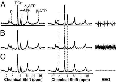
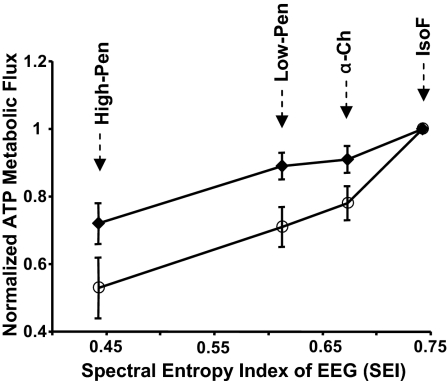
A majority of ATP in the brain is formed in the mitochondria through oxidative phosphorylation of ADP with the F(1)F(0)-ATP (ATPase) enzyme. This ATP production rate plays central roles in brain bioenergetics, function and neurodegeneration. In vivo (31)P magnetic resonance spectroscopy combined with magnetization transfer (MT) is the sole approach able to noninvasively determine this ATP metabolic rate via measuring the forward ATPase reaction flux (F(f,ATPase)). However, previous studies indicate lack of quantitative agreement between F(f,ATPase) and oxidative metabolic rate in heart and liver. In contrast, recent work has shown that F(f,ATPase) might reflect oxidative phosphorylation rate in resting human brains. We have conducted an animal study, using rats under varied brain activity levels from light anesthesia to isoelectric state, to examine whether the in vivo (31)P MT approach is suitable for measuring the oxidative phosphorylation rate and its change associated with varied brain activity. Our results conclude that the measured F(f,ATPase) reflects the oxidative phosphorylation rate in resting rat brains, that this flux is tightly correlated to the change of energy demand under varied brain activity levels, and that a significant amount of ATP energy is required for "housekeeping" under the isoelectric state. These findings reveal distinguishable characteristics of ATP metabolism between the brain and heart, and they highlight the importance of in vivo (31)P MT approach to potentially provide a unique and powerful neuroimaging modality for noninvasively studying the cerebral ATP metabolic network and its central role in bioenergetics associated with brain function, activation, and diseases.
Conflict of interest statement
The authors declare no conflict of interest.
Figures
References
-
- Boyer PD. What makes ATP synthase spin? Nature. 1999;402:247–249. - PubMed
-
- Erecinska M, Silver IA. ATP and brain function. J Cereb Blood Flow Metab. 1989;9:2–19. - PubMed
-
- Clarke DD, Sokoloff L. In: Basic Neruochemistry: Molecular, Cellular and Medical Aspects. Siegel GJ, et al., editors. Philadephia: Lippincott-Raven; 1999. pp. 633–669.
-
- Attwell D, Laughlin SB. An energy budget for signaling in the grey matter of the brain. J Cereb Blood Flow Metab. 2001;21:1133–1145. - PubMed
-
- Raichle ME, Mintun MA. Brain work and brain imaging. Annu Rev Neurosci. 2006;29:449–476. - PubMed
Publication types
MeSH terms
Substances
Grants and funding
LinkOut - more resources
Full Text Sources
Other Literature Sources
Medical
