. 2008 May 6;105(18):6620-5.
doi: 10.1073/pnas.0710516105. Epub 2008 Apr 28.

Bone marrow-derived circulating endothelial precursors do not contribute to vascular endothelium and are not needed for tumor growth

Affiliations

Bone marrow-derived circulating endothelial precursors do not contribute to vascular endothelium and are not needed for tumor growth

Susanna Purhonen et al. Proc Natl Acad Sci U S A. .

Abstract

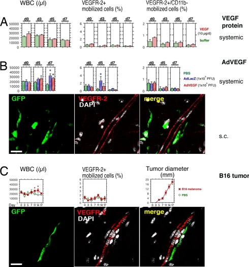
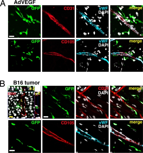
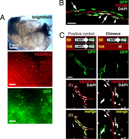
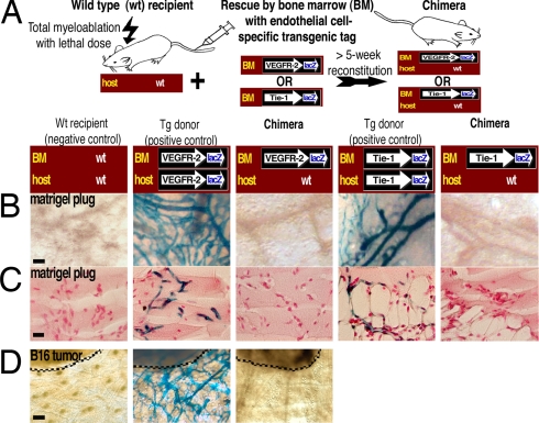
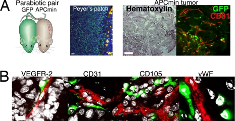
The mechanisms by which bone marrow (BM)-derived stem cells might contribute to angiogenesis and the origin of neovascular endothelial cells (ECs) are controversial. Neovascular ECs have been proposed to originate from VEGF receptor 2-expressing (VEGFR-2+) stem cells mobilized from the BM by VEGF or tumors, and it is thought that angiogenesis and tumor growth may depend on such endothelial precursors or progenitors. We studied the mobilization of BM cells to circulation by inoculating mice with VEGF polypeptides, adenoviral vectors expressing VEGF, or tumors. We induced angiogenesis by syngeneic melanomas, APCmin adenomas, adenoviral VEGF delivery, or matrigel plugs in four different genetically tagged universal or endothelial cell-specific chimeric mouse models, and subsequently analyzed the contribution of BM-derived cells to endothelium in a wide range of time points. To study the existence of circulating ECs in a nonmyeloablative setting, pairs of genetically marked parabiotic mice with a shared anastomosed circulatory system were created. We did not observe specific mobilization of VEGFR-2+ cells to circulation by VEGF or tumors. During angiogenesis, abundant BM-derived perivascular cells were recruited close to blood vessel wall ECs but did not form part of the endothelium. No circulation-derived vascular ECs were observed in the parabiosis experiments. Our results show that no BM-derived VEGFR-2+ or other EC precursors contribute to vascular endothelium and that cancer growth does not require BM-derived endothelial progenitors. Endothelial differentiation is not a typical in vivo function of normal BM-derived stem cells in adults, and it has to be an extremely rare event if it occurs at all.


Conflict of interest statement

The authors declare no conflict of interest.

Figures

Fig. 1.
Fig. 1.
Systemic VEGF or tumors do not promote the mobilization of VEGFR-2+ BM cells to circulation, and BM-derived VEGFR-2+ cells do not form part of the growing endothelium. Peripheral blood cells were isolated on indicated days, counted, and analyzed by FACS. Day 0 (d0) shows baseline levels before inoculation. The results are given as mean ± SE. The asterisks indicate statistical significance (P < 0.05). The s.c. angiogenesis was analyzed in chimeric mice with transgenic GFP-tagged BM. VEGFR-2+ cells were detected (antibody clone AVAS 12α1) and analyzed by multichannel confocal scanning. The nuclei are stained with DAPI (white) to recognize individual cells. (A) No mobilization of BM cell populations was observed in response to systemic treatment with VEGF polypeptides. (B) Systemic adenoviral administration caused nonspecific mobilization of cells from the BM, as can be seen in the WBC counts. The results on WBC mobilization were identical by using AdLacZ or AdVEGF. Statistically significant mobilization of VEGFR-2+ cells was observed on day 5 in the control mice treated with AdLacZ. Numerous GFP-tagged BM-derived perivascular cells (green) were recruited to the site of angiogenesis after s.c. AdVEGF. No VEGFR-2+ (red) ECs originating from the BM were discovered in the blood vessel endothelium. (Scale bar: 20 μm.) (C) The s.c. B16 tumors caused mobilization of hematopoietic cells from the BM on days 11–14, simultaneously to the highest growth rate of the tumors. Large numbers of BM-derived perivascular cells (green) were infiltrating the angiogenic tumor stroma. No VEGFR-2+ (red) vascular ECs originating from the BM were discovered. (Scale bar: 20 μm.)
Fig. 2.
Fig. 2.
All BM-derived cells recruited during AdVEGF- or tumor-induced angiogenesis are perivascular. ECs were detected using confocal scanning against CD31, CD105, or vWF. Transgenic GFP reporter is expressed in BM-derived cells. The nuclei are stained with DAPI (white) to recognize individual cells. No vascular ECs originating from the BM were detected. (Scale bars: 20 μm.) (A) Angiogenesis was induced in the chimeric hosts by s.c. AdVEGF. A time point of 14 days after administration is shown. (B) Angiogenesis was induced in the chimeric hosts by s.c. inoculation of B16 melanomas. A late time point of 21 days is shown. The 3D orthogonal projections (x–z and y–z axes) also are shown, and tumor blood vessel lumen (L) is indicated.
Fig. 3.
Fig. 3.
No BM-derived vascular ECs are present in matrigel plugs. All blood vessels observed within the plugs must be novel. (A) A high number of VEGFR-2+ (red; antibody clone AVAS 12α1) blood vessels and abundant BM-derived GFP+ cells can be seen. (Scale bars: 100 μm.) (B) At 1 week after the implantation of plug, the ECs (arrows) of the forming neovasculature (stained for CD105, red) could be seen in very close contact with the infiltrating BM-derived perivascular cells (arrowheads) when studied by multichannel confocal scanning. The nuclei are stained with DAPI (white). (Scale bar: 20 μm.) (C) Confocal scans of the whole mounts. The positive control is a transgenic C57BL/6-Tg(ACTB-EGFP)1Osb/J mouse, where all of the tissues including the blood vessel vessels are GFP+. The positive control confirms successful detection of GFP+ ECs. The ECs stain readily for VEGFR-2 (AVAS 12α1). In chimeric mice, where the WT C57BL/6 hosts are engrafted with transgenic GFP+ BM, no BM-derived VEGFR-2+ endothelium can be found. (Scale bar: 20 μm.)
Fig. 4.
Fig. 4.
Endothelial cell-specific genetic reporter systems identify no BM-derived VEGFR-2+ or Tie-1+ ECs in angiogenic neovasculature. (A) Strategy for the two EC-specific gene tag and BM-transplantation models. Transgenic β-gal expression is driven by the promoter for the endothelial cell-specific gene VEGFR-2 or Tie-1. WT mice serve as negative controls. Transgenic donor mice serve as positive controls and confirm successful detection of β-gal+ cells. (B) No BM-derived β-gal+ vascular ECs can be found in the chimeric mice, demonstrating that BM-derived cells do not differentiate to VEGFR-2+ or Tie-1+ vascular ECs. (Scale bar: 50 μm.) (C) Sections of the plugs were stained with Nuclear fast red to visualize the nuclei, thus also confirming equal cellularity within the matrigel plugs. (Scale bar: 20 μm.) (D) The mice were implanted with B16 melanomas (dashed lines). No BM-derived β-gal+ endothelium can be found in the chimeric mice, demonstrating that tumor angiogenesis or growth do not involve or require contribution from VEGFR-2+ circulating EC precursors. (Scale bar: 100 μm.)
Fig. 5.
Fig. 5.
No circulation-derived vascular ECs were observed in the parabiosed APCmin tumors. (A) Pairs of parabiotic mice that have a shared anastomosed circulatory system were created by cojoining GFP-expressing mice with young APCmin mice. Chimerism of lymphoid cells in the Peyer's patch of the APCmin mouse demonstrates the 50% blood cell chimerism in the system. Upon killing at 22–24 weeks of age, the parabiosed APCmin mice presented 20–55 adenomas per mouse. Overview pictures of the APCmin tumor are shown in brightfield and fluorescence. Note close association of the GFP+ circulating cells to the vascular endothelium (CD31 stain, red). (Scale bars: 50 μm.) (B) Confocal scans show that circulating cells did not contribute to host vascular endothelium. GFP-tagged cells in angiogenic APCmin tumor tissues are shown in green. The nuclei are stained with DAPI (white). (Scale bars: 20 μm.)

Comment in

References

    1. Carmeliet P. Angiogenesis in life, disease and medicine. Nature. 2005;438:932–936. - PubMed
    1. Asahara T, et al. Isolation of putative progenitor endothelial cells for angiogenesis. Science. 1997;275:964–967. - PubMed
    1. Asahara T, et al. VEGF contributes to postnatal neovascularization by mobilizing bone marrow-derived endothelial progenitor cells. EMBO J. 1999;18:3964–3972. - PMC - PubMed
    1. Takahashi T, et al. Ischemia- and cytokine-induced mobilization of bone marrow-derived endothelial progenitor cells for neovascularization. Nat Med. 1999;5:434–438. - PubMed
    1. Lin Y, Weisdorf DJ, Solovey A, Hebbel RP. Origins of circulating endothelial cells and endothelial outgrowth from blood. J Clin Invest. 2000;105:71–77. - PMC - PubMed

Publication types

MeSH terms