. 2008 May 6;105(18):6572-7.
doi: 10.1073/pnas.0800859105. Epub 2008 Apr 28.

Revealing the moonlighting role of NADP in the structure of a flavin-containing monooxygenase

Affiliations

Revealing the moonlighting role of NADP in the structure of a flavin-containing monooxygenase

Andrea Alfieri et al. Proc Natl Acad Sci U S A. .

Abstract

Flavin-containing monooxygenases (FMOs) are, after cytochromes P450, the most important monooxygenase system in humans and are involved in xenobiotics metabolism and variability in drug response. The x-ray structure of a soluble prokaryotic FMO from Methylophaga sp. strain SK1 has been solved at 2.6-A resolution and is now the protein of known structure with the highest sequence similarity to human FMOs. The structure possesses a two-domain architecture, with both FAD and NADP(+) well defined by the electron density maps. Biochemical analysis shows that the prokaryotic enzyme shares many functional properties with mammalian FMOs, including substrate specificity and the ability to stabilize the hydroperoxyflavin intermediate that is crucial in substrate oxygenation. On the basis of their location in the structure, the nicotinamide ring and the adjacent ribose of NADP(+) turn out to be an integral part of the catalytic site being actively engaged in the stabilization of the oxygenating intermediate. This feature suggests that NADP(H) has a moonlighting role, in that it adopts two binding modes that allow it to function in both flavin reduction and oxygen reactivity modulation, respectively. We hypothesize that a relative domain rotation is needed to bring NADP(H) to these distinct positions inside the active site. Localization of mutations in human FMO3 that are known to cause trimethylaminuria (fish-odor syndrome) in the elucidated FMO structure provides a structural explanation for their biological effects.


Conflict of interest statement

The authors declare no conflict of interest.

Figures

Fig. 1.
Fig. 1.
Biochemical properties of FMOs. (A) Scheme of the catalytic cycle. NADP+ that forms upon reduction of the flavin cofactor stays bound throughout the reaction and is essential for stabilization of C4a-hydroperoxyFAD (Bottom Right) (–6). Enzyme-bound NADP+ prevents FMO from working as an NADPH oxidase, which would produce H2O2 (dashed line). S and S-O indicate substrate and monooxygenated product, respectively. (B) Alignment of the sequences of Methylophaga FMO (MeFMO), Homo sapiens FMO3 isoform 1 (hFMO3), and S. pombe FMO (SpFMO). Sequence identities to hFMO3 are 31% for mFMO and 27% for S. pombe FMO. The red bars indicate the first Rossmann fold for FAD binding, the fingerprint sequence for FMOs (20), and the second Rossmann fold for NADP binding, respectively. The green asterisks mark residues that are part of the active site, whereas the red asterisks indicate residues that are mutated in patients affected by TMAU. The alignment was performed with ClustalW2 (22) and ESPript (23).
Fig. 2.
Fig. 2.
Absorbance spectra of mFMO. Trace A represents the fully oxidized form in the absence of NADP+. Trace B was obtained as described in Materials and Methods by the addition of an equimolar amount of NADPH under aerobic conditions and is consistent with formation of the C4a-hydroperoxyflavin intermediate. The presence of a small amount of oxidized enzyme because of the intrinsic NADPH-oxidase activity accounts for the shoulder observed at ≈450 nm. Traces C to G were sequentially recorded after 25 sec, 90 sec, 3 min, 10 min, and 18 min and show the slow decay of the C4a-hydroperoxyflavin intermediate, which is completed after 30 min.
Fig. 3.
Fig. 3.
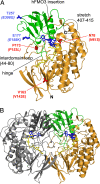
Overall crystal structure of mFMO. (A) Ribbon diagram of the monomer. FAD-binding domain (residues 8–169 and 281–450) is orange and NADP-binding domain (residues 170–280) is green. FAD is shown as yellow sticks and NADP+ as blue sticks. The positions of the long interdomain loop (residues 44–80), the hinge connecting the two domains, and the polypeptide stretch corresponding to residues 407–415 are outlined. mFMO residues corresponding to TMAU-causing mutations (17) and polymorphisms in hFMO3 (in parentheses) are in red and blue sticks, respectively. The position of a long insert in hFMO3 (residues 238–299; Fig. 1B) is also indicated. It is expected to occupy a surface-exposed position away from the active site. (B) Ribbon representation of the mFMO dimer. One monomer is shown in the same orientation and color as Fig. 3A; the other one is colored gray, with the NADP-binding domain in darker gray.
Fig. 4.
Fig. 4.
The active site of mFMO. (A) Surface representation of the enzyme with the catalytic site viewed from outside using the coloring scheme and approximately the same orientation of Fig. 3B. (B) Binding of the NADP+ nicotinamide ring. Only crucial residues are displayed. Oxygen is red, nitrogen is blue, carbon is differently colored depending on its belonging to FAD (yellow), NADP+ (cyan), FAD-binding domain (orange), NADP-binding domain (green). For the sake of clarity, only the backbone atoms of Trp-76 are shown. The reactive C4a and N5 atoms of the flavin and C4 of NADP+ nicotinamide are labeled. (C) Stereoscopic view of the active site of mFMO in approximately the same orientation as Fig. 4B and with the same color code. The red-colored electron density (2FoFc, contoured at 1.5 σ) was found in the dataset resulting from a soaking experiment (Table 2) and tentatively assigned to a dioxygen bound inside the cavity located in front of the flavin (here shown as a light blue transparent surface).
Fig. 5.
Fig. 5.
The role of NADP+ in the stabilization of C4a-hydroperoxyflavin intermediate. (A) Modeling experiment in which the hypothetical structure of C4a-hydroperoxyflavin was superimposed to the flavin in mFMO structure. The color code is the same as in Fig. 4B. Hypothetical hydrogen bonds involving the hydroperoxyflavin atoms are shown as blue dashed lines. The accommodation of the additional oxygen atoms of the C4a-adduct would require a shift of ≈1.5 Å of Asn-78 side chain (whose conformation in the native structure is shown as thin black stick). (B) Comparison of the NADP+-binding mode in S. pombe (Protein Data Bank ID code 2gv8) and Methylophaga FMOs. The picture was obtained by superimposing the Cα atoms of the two proteins and shows the FAD (yellow) and NADP+ (blue) molecules of mFMO together with FAD and NADP+ of the S. pombe enzyme (red). The N5 and C4a atoms of the flavin and C4 and C2 atoms of NADP+ are labeled.

Similar articles

Cited by

References

    1. Cashman JR. Some distinctions between flavin-containing and cytochrome P450 monooxygenases. Biochem Biophys Res Commun. 2005;338:599–604. - PubMed
    1. van Berkel WJ, Kamerbeek NM, Fraaije MW. Flavoprotein monooxygenases, a diverse class of oxidative biocatalysts. J Biotechnol. 2006;124:670–689. - PubMed
    1. Ziegler DM. An overview of the mechanism, substrate specificities, and structure of FMOs. Drug Metab Rev. 2002;34:503–511. - PubMed
    1. Poulsen LL, Ziegler DM. The liver microsomal FAD-containing monooxygenase. J Biol Chem. 1979;254:6449–6455. - PubMed
    1. Beaty NB, Ballou DP. The reductive half-reaction of liver microsomal FAD-containing monooxygenase. J Biol Chem. 1981;256:4611–4618. - PubMed

Publication types