

The placental syncytium and the pathophysiology of preeclampsia and intrauterine growth restriction: a novel assay to assess syncytial protein expression
- PMID: 18443340
- PMCID: PMC3671376
- DOI: 10.1196/annals.1434.015
The placental syncytium and the pathophysiology of preeclampsia and intrauterine growth restriction: a novel assay to assess syncytial protein expression
Abstract
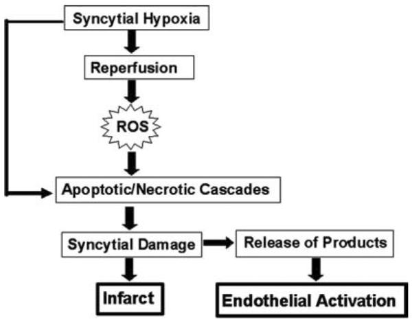
Preeclampsia is associated with an increased release of factors from the placental syncytium into maternal blood, including the antiangiogenic factors soluble fms-like tyrosine kinase-1 and soluble endoglin, the antifibrinolytic factor plasminogen activator inhibitor-1, prostanoids, lipoperoxides, cytokines, and microparticles. These factors are suggested to promote maternal endothelium dysfunction and are associated with placental damage in pregnancies also complicated with intrauterine growth restriction (IUGR). In this report, we briefly describe the interaction of syncytial factors with hypoxia, reactive oxygen species, and apoptosis in the pathophysiology of preeclampsia and IUGR. Given the critical role of the syncytium in these complications of pregnancy, we also present a novel methodology in which laser capture microdissection followed by Western blotting is used to assess levels of syncytial Fas ligand, a key protein in the apoptotic cascade.
Figures

Similar articles
-
Role of the syncytium in placenta-mediated complications of preeclampsia.Thromb Res. 2009 Sep;124(4):389-92. doi: 10.1016/j.thromres.2009.05.016. Epub 2009 Jun 16. Thromb Res. 2009. PMID: 19535132 Free PMC article. Review.
-
Placental mitochondrial content and function in intrauterine growth restriction and preeclampsia.Am J Physiol Endocrinol Metab. 2014 Feb 15;306(4):E404-13. doi: 10.1152/ajpendo.00426.2013. Epub 2013 Dec 17. Am J Physiol Endocrinol Metab. 2014. PMID: 24347055
-
Fas and FasL expression in placentas complicated with intrauterine growth retardation with and without preeclampsia.J Matern Fetal Neonatal Med. 2016;29(7):1154-9. doi: 10.3109/14767058.2015.1038702. Epub 2015 Apr 24. J Matern Fetal Neonatal Med. 2016. PMID: 25909501
-
The Role of Long Non-Coding RNAs in Trophoblast Regulation in Preeclampsia and Intrauterine Growth Restriction.Genes (Basel). 2021 Jun 25;12(7):970. doi: 10.3390/genes12070970. Genes (Basel). 2021. PMID: 34201957 Free PMC article. Review.
-
Divergent trophoblast invasion and apoptosis in placental bed spiral arteries from pregnancies complicated by maternal anemia and early-onset preeclampsia/intrauterine growth restriction.Am J Obstet Gynecol. 2006 Feb;194(2):557-63. doi: 10.1016/j.ajog.2005.07.035. Am J Obstet Gynecol. 2006. PMID: 16458661
Cited by
-
Angiogenic Imbalance and Inflammatory Biomarkers in the Prediction of Hypertension as Well as Obstetric and Perinatal Complications in Women with Gestational Diabetes Mellitus.J Clin Med. 2022 Mar 10;11(6):1514. doi: 10.3390/jcm11061514. J Clin Med. 2022. PMID: 35329840 Free PMC article.
-
Toxicity assessments of selected trichloroethylene and perchloroethylene metabolites in three in vitro human placental models.Reprod Toxicol. 2022 Apr;109:109-120. doi: 10.1016/j.reprotox.2022.03.003. Epub 2022 Mar 16. Reprod Toxicol. 2022. PMID: 35304307 Free PMC article.
-
Tissue factor activity in women with preeclampsia or SGA: a potential explanation for the excessive thrombin generation in these syndromes.J Matern Fetal Neonatal Med. 2018 Jun;31(12):1568-1577. doi: 10.1080/14767058.2017.1320543. Epub 2017 May 19. J Matern Fetal Neonatal Med. 2018. PMID: 28521572 Free PMC article.
-
Association between Placental Lesions, Cytokines and Angiogenic Factors in Pregnant Women with Preeclampsia.PLoS One. 2016 Jun 17;11(6):e0157584. doi: 10.1371/journal.pone.0157584. eCollection 2016. PLoS One. 2016. PMID: 27315098 Free PMC article.
-
Lipid rafts and cytoskeletal proteins in placental microvilli membranes from preeclamptic and IUGR pregnancies.J Membr Biol. 2011 Jun;241(3):127-40. doi: 10.1007/s00232-011-9369-3. Epub 2011 May 15. J Membr Biol. 2011. PMID: 21573936
References
-
- Redman CW, Sargent IL. Latest advances in understanding preeclampsia. Science. 2005;308:1592–1594. - PubMed
-
- Kaufmann P, et al. Endovascular trophoblast invasion: implications for the pathogenesis of intrauterine growth retardation and preeclampsia. Biol. Reprod. 2003;69:1–7. - PubMed
-
- Stepan H, et al. New insights into the biology of preeclampsia. Biol. Reprod. 2006;74:772–776. - PubMed
-
- Benirschke K. Remarkable placenta. Clin. Anat. 1998;11:194–205. - PubMed
-
- Benyo DF, et al. Expression of inflammatory cytokines in placentas from women with preeclampsia. J. Clin. Endocrinol. Metab. 2001;86:2505–2512. - PubMed
Publication types
MeSH terms
Substances
Grants and funding
LinkOut - more resources
Full Text Sources
Research Materials
Miscellaneous
