. 2008 May;19(5):877-83.
doi: 10.1681/ASN.2007050629.

HIV-1 upregulates VEGF in podocytes

Affiliations

HIV-1 upregulates VEGF in podocytes

Sonal Navin Korgaonkar et al. J Am Soc Nephrol. 2008 May.

Abstract

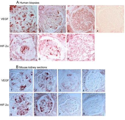
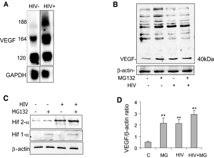
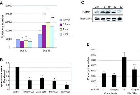
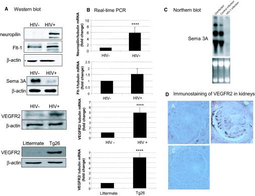
HIV-associated nephropathy (HIVAN) is characterized by collapsing FSGS. Because transgenic mice with podocyte-specific overexpression of the vascular endothelial growth factor 164 (VEGF164) isoform also develop collapsing FSGS, we sought to determine whether VEGF plays a role in HIVAN. Compared with controls, immunohistochemistry revealed that kidneys from HIV-1-transgenic mice (Tg26) and from patients with HIVAN had greater expression of both VEGF and its transcriptional regulator, hypoxia-inducible factor 2alpha (HIF-2alpha). Similarly, mRNA and protein levels of VEGF and HIF-2alpha were increased in HIV-infected podocytes in vitro, and this transcriptional upregulation was found to be stimulated by the HIV viral protein Nef in a Src kinase-and Stat3-dependent manner. HIV-1 also upregulated VEGFR2 and its co-receptor neuropilin-1 and suppressed the expression of semaphorin 3a in the podocyte. Exogenous VEGF stimulated proliferation and de-differentiation of podocytes, which are features of collapsing FSGS, and VEGFR2 neutralizing antibodies reversed these features in podocytes infected with HIV-1 or isolated from Tg26 mice. In conclusion, HIV-1 induces VEGF and VEGFR2 expression in podocytes, and this may be a critical step in the pathogenesis of HIVAN.


Figures

Figure 1.
Figure 1.
(A) Immunohistochemical staining for VEGF-A and HIF-2α in human kidney biopsies. (1 through 3) VEGF staining: Representative pictures from biopsy specimens from HIV-positive patients with HIVAN show VEGF-A staining localized mostly to podocytes (1 and 2), and HIV-negative patients with minimal-change disease show only background staining (3). (4) Control: IgG staining shows diffuse background staining. (5 through 7) HIF-2α staining: Representative pictures from biopsy specimens from HIV-positive patients with HIVAN show strong HIF-2α expression localized mostly to the podocyte (5 and 6), and HIV-negative patients with minimal-change disease show little podocyte staining for HIF-2α (7). (B) Immunostaining for VEGF and HIF-2α in kidneys from Tg26 mice: (1 through 4) VEGF staining in kidney sections from Tg26 mouse (1 and 2), from littermate (3), or stained with control IgG (4). (5 through 8) HIF-2α staining in kidneys sections from Tg26 mouse (5 and 6), from littermate (7), or stained with control IgG (8). Bright light microscopy (Olympus BX60). Magnification, ×20.
Figure 2.
Figure 2.
(A) HIV-1 upregulates expression of the VEGF isoforms 164 and 188. mRNA levels for VEGF-A and glyceraldehyde-3-phosphate dehydrogenase (GAPDH) were analyzed in control vector-infected podocytes and HIV-infected podocytes by Northern blotting. (B) HIV upregulates expression of VEGF165 at the protein level, and inhibition of proteasomal degradation with MG132 treatment upregulates VEGF165. This is a representative blot of three independent experiments. (C) HIV upregulates expression of HIF-2α at the protein level, and inhibition of proteasomal degradation upregulates HIF-2α and HIF-1α levels in control cells. This is a representative blot of three independent experiments. (D) Densitometric analysis of blots in B are summarized, n = 3, **P < 0.01 versus control.
Figure 3.
Figure 3.
(A) Effect of VEGF on podocyte proliferation. Podocytes were exposed to no VEGF or VEGF at indicated concentration. Cell number was determined at days 2 and 5. Bars represent mean cell number ± SE of three samples. ***P < 0.005 versus cells without VEGF treatment. (B) VEGF causes reduction of synaptopodin expression. Podocytes were incubated with or without VEGF for 2 d, and RNA was isolated. The synaptopodin/tubulin ratio was determined by real-time PCR. The fold of increase as compared with control podocytes is expressed. Means ± SEM of three independent experiments are shown; *P < 0.05 versus control. (C) VEGF stimulates MAPK phosphorylation. Podocytes were incubated with 1 nm of VEGF or no VEGF for 5, 10, 30, and 60 min. Total and phosphorylated MAPK was determined by Western blot using anti-MAPK and anti–p-MAPK. The representative blot of three independent experiments is shown. (D) Effect of recombinant sVEGFR1 on HIV-induced podocyte proliferation. Control and HIV-infected podocytes were treated with recombinant sVEGFR1 or control vehicle daily for 3 d, and then cell number was determined. **P < 0.01; n = 4 versus vehicle-treated cells.
Figure 4.
Figure 4.
(A) Effects of HIV-1 infection on protein expression of neuropilin-1, Flt1, VEGFR2, and Sema 3A in podocytes. Neuropilin-1, Flt1, VEGFR2, and Sema 3A expression was determined by Western blot in HIV-infected and control-infected podocytes. VEGFR2 expression was also examined in podocytes isolated from Tg26 mice and their littermates. These are representative blots of three independent experiments. (B) The neuropilin-1/tubulin, Flt1/tubulin, and VEGFR2/tubulin ratios were determined by real-time PCR. The fold of increase as compared with control podocytes is expressed. Means ± SEM of three independent experiments are shown. ****P < 0.001 versus control. (C) Northern blot analysis of Sema 3A expression was performed using RNA from podocytes infected with HIV-1 versus the control construct and uninfected podocytes. A representative blot of three independent experiments is shown demonstrating dramatic reduction in the expression of Sema 3A in response to the HIV-1 transgene as compared with the control infection (top). The ethidium bromide–stained RNA gel before transfer is shown to demonstrate that equivalent amounts of RNA were used for the analysis (bottom). (D) Immunostaining of VEGFR2 in kidney biopsies from patients with HIVAN (B) compared with patients with minimal-change disease (A) and IgG control (C).

Similar articles

Cited by

References

    1. Levine RJ, Maynard SE, Qian C, Lim KH, England LJ, Yu KF, Schisterman EF, Thadhani R, Sachs BP, Epstein FH, Sibai BM, Sukhatme VP, Karumanchi SA: Circulating angiogenic factors and the risk of preeclampsia. N Engl J Med 350: 672–683, 2004 - PubMed
    1. Takahashi A, Sasaki H, Kim SJ, Tobisu K, Kakizoe T, Tsukamoto T, Kumamoto Y, Sugimura T, Terada M: Markedly increased amounts of messenger RNAs for vascular endothelial growth factor and placenta growth factor in renal cell carcinoma associated with angiogenesis. Cancer Res 54: 4233–4237, 1994 - PubMed
    1. Khamaisi M, Schrijvers BF, De Vriese AS, Raz I, Flyvbjerg A: The emerging role of VEGF in diabetic kidney disease. Nephrol Dial Transplant 18: 1427–1430, 2003 - PubMed
    1. Houck KA, Leung DW, Rowland AM, Winer J, Ferrara N: Dual regulation of vascular endothelial growth factor bioavailability by genetic and proteolytic mechanisms. J Biol Chem 267: 26031–26037, 1992 - PubMed
    1. Ferrara N, Gerber HP, LeCouter J: The biology of VEGF and its receptors. Nat Med 9: 669–676, 2003 - PubMed

Publication types

MeSH terms

Substances

LinkOut - more resources