Expression of SDF-1 alpha and nuclear CXCR4 predicts lymph node metastasis in colorectal cancer
- PMID: 18443596
- PMCID: PMC2391124
- DOI: 10.1038/sj.bjc.6604363
Expression of SDF-1 alpha and nuclear CXCR4 predicts lymph node metastasis in colorectal cancer
Abstract
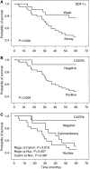
Although stromal cell-derived factor (SDF)-1 alpha and its receptor CXCR4 are experimentally suggested to be involved in tumorigenicity, the clinicopathological significance of their expression in human disease is not fully understood. We examined SDF-1 alpha and CXCR4 expression in colorectal cancers (CRCs) and their related lymph nodes (LNs), and investigated its relationship to clinicopathological features. Specimens of 60 primary CRCs and 27 related LNs were examined immunohistochemically for not only positivity but also immunostaining patterns for SDF-1 alpha and CXCR4. The relationships between clinicopathological features and SDF-1 alpha or CXCR4 expression were then analysed. Stromal cell-derived factor-1 alpha and CXCR4 expression were significantly associated with LN metastasis, tumour stage, and survival of CRC patients. Twenty-nine of 47 CXCR4-positive CRCs (61.7%) showed clear CXCR4 immunoreactivity in the nucleus and a weak signal in the cytoplasm (nuclear type), whereas others showed no nuclear immunoreactivity but a diffuse signal in the cytoplasm and at the plasma membrane (cytomembrane type). Colorectal cancer patients with nuclear CXCR4 expression showed significantly more frequent LN metastasis than did those with cytomembrane expression. Colorectal cancer patients with nuclear CXCR4 expression in the primary lesion frequently had cytomembrane CXCR4-positive tumours in their LNs. In conclusion, expression of SDF-1 alpha and nuclear CXCR4 predicts LN metastasis in CRCs.
Figures


Similar articles
-
Stromal cell derived factor-1 and CXCR4 expression in colorectal cancer promote liver metastasis.Cancer Biomark. 2015;15(6):869-79. doi: 10.3233/CBM-150531. Cancer Biomark. 2015. PMID: 26406413
-
[Significance of expression of stromal cell derived factor 1 and CXCR4 in invasive breast cancer].Zhonghua Bing Li Xue Za Zhi. 2008 Aug;37(8):529-35. Zhonghua Bing Li Xue Za Zhi. 2008. PMID: 19094464 Chinese.
-
CXCR4/SDF-1 axis is involved in lymph node metastasis of gastric carcinoma.World J Gastroenterol. 2011 May 21;17(19):2389-96. doi: 10.3748/wjg.v17.i19.2389. World J Gastroenterol. 2011. PMID: 21633638 Free PMC article.
-
[The expression and significance of trefoil factor 3 and SDF-1/CXCR4 biological axis in papillary thyroid carcinoma].Lin Chuang Er Bi Yan Hou Tou Jing Wai Ke Za Zhi. 2014 Jan;28(2):108-12. Lin Chuang Er Bi Yan Hou Tou Jing Wai Ke Za Zhi. 2014. PMID: 24738314 Chinese.
-
Clinical impact of minimal cancer cell detection in various colorectal cancer specimens.World J Gastroenterol. 2014 Sep 21;20(35):12458-61. doi: 10.3748/wjg.v20.i35.12458. World J Gastroenterol. 2014. PMID: 25253945 Free PMC article. Review.
Cited by
-
Expression of stromal cell-derived factor-1α is an independent risk factor for lymph node metastasis in early gastric cancer.Oncol Lett. 2011 Nov;2(6):1197-1202. doi: 10.3892/ol.2011.389. Epub 2011 Aug 19. Oncol Lett. 2011. PMID: 22848288 Free PMC article.
-
Lymphangiogenesis and cancer.Genes Cancer. 2011 Dec;2(12):1146-58. doi: 10.1177/1947601911423028. Genes Cancer. 2011. PMID: 22866206 Free PMC article.
-
A chemokine/chemokine receptor signature potentially predicts clinical outcome in colorectal cancer patients.Cancer Biomark. 2019;26(3):291-301. doi: 10.3233/CBM-190210. Cancer Biomark. 2019. PMID: 31524146 Free PMC article.
-
Characterization and Validation of Antibodies for Immunohistochemical Staining of the Chemokine CXCL12.J Histochem Cytochem. 2019 Apr;67(4):257-266. doi: 10.1369/0022155418818788. Epub 2018 Dec 18. J Histochem Cytochem. 2019. PMID: 30562126 Free PMC article.
-
Anti-HSP70 alleviates cell migration and proliferation in colorectal cancer cells (CRC) by targeting CXCR4 (in vitro study).Med Oncol. 2023 Jul 29;40(9):256. doi: 10.1007/s12032-023-02122-6. Med Oncol. 2023. PMID: 37516711
References
-
- Baggiolini M, Dewald B, Moser B (1997) Human chemokines: an update. Annu Rev Immunol 15: 675–705 - PubMed
-
- Brand S, Dambacher J, Beigel F, Olszak T, Diebold J, Otte JM, Goke B, Eichhorst ST (2005) CXCR4 and CXCL12 are inversely expressed in colorectal cancer cells and modulate cancer cell migration, invasion and MMP-9 activation. Exp Cell Res 310: 117–130 - PubMed
-
- Burger M, Glodek A, Hartmann T, Schmitt-Graff A, Silberstein LE, Fujii N, Kipps TJ, Burger JA (2003) Functional expression of CXCR4 (CD184) on small-cell lung cancer cells mediates migration, integrin activation, and adhesion to stromal cells. Oncogene 22: 8093–8101 - PubMed
-
- Cheng ZJ, Zhao J, Sun Y, Hu W, Wu YL, Cen B, Wu GX, Pei G (2000) beta-arrestin differentially regulates the chemokine receptor CXCR4-mediated signaling and receptor internalization, and this implicates multiple interaction sites between beta-arrestin and CXCR4. J Biol Chem 275: 2479–2485 - PubMed
-
- Fernandis AZ, Prasad A, Band H, Klosel R, Ganju RK (2004) Regulation of CXCR4-mediated chemotaxis and chemoinvasion of breast cancer cells. Oncogene 23: 157–167 - PubMed
MeSH terms
Substances
LinkOut - more resources
Full Text Sources
Other Literature Sources
Medical

