Clinical Trial
. 2008 May;19(5):496-510.
doi: 10.1089/hum.2007.0171.

Adoptive cell therapy for patients with melanoma, using tumor-infiltrating lymphocytes genetically engineered to secrete interleukin-2

Affiliations
Clinical Trial

Adoptive cell therapy for patients with melanoma, using tumor-infiltrating lymphocytes genetically engineered to secrete interleukin-2

Bianca Heemskerk et al. Hum Gene Ther. 2008 May.

Abstract

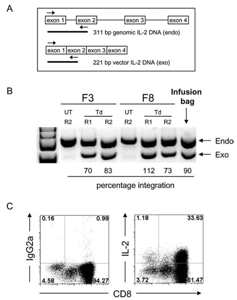
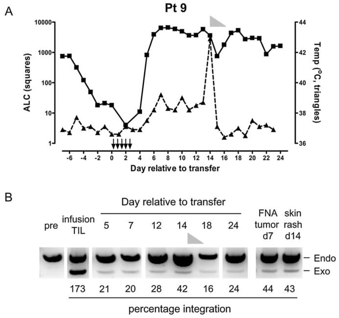
Adoptive cell transfer of tumor-infiltrating lymphocytes (TILs) after lymphodepletion mediates regression in 50% of patients with metastatic melanoma. In vivo persistence and telomere length of the transferred cells correlate with antitumor response. In an attempt to prolong the in vivo survival of the transferred cells, TILs were genetically engineered to produce interleukin (IL)-2. In vitro, these transduced TILs secreted IL-2 while retaining tumor specificity and exhibited prolonged survival after IL-2 withdrawal. In a phase I/II clinical trial, seven evaluable patients received transduced TILs and one patient experienced a partial response associated with in vivo persistence of IL-2-transduced TILs in circulating lymphocytes. An additional five patients received transduced TILs in conjunction with IL-2 administration. Persistence of IL-2-transduced TILs was observed in three patients, including one partial responder. The transgene DNA as well as vector-derived IL2 mRNA could be detected for 4 months in responding patients. The low response rate in this trial was possibly due to a reduction in telomere length in cells as a result of prolonged in vitro culture. In this study, insertion of the IL-2 gene into antitumor TILs increased their ability to survive after IL-2 withdrawal in vitro but did not increase their in vivo persistence or clinical effectiveness.


Figures

FIG. 1
FIG. 1
Transduction of patient TILs with a retroviral vector encoding human IL-2 cDNA. (A) The introduced IL-2 gene was confirmed by semiquantitative PCR, in which the primers flank exon 1 in the genomic DNA. The resulting PCR product from the endogenous gene is 311 bp, whereas the exogenous vector-derived IL-2 gene results in a smaller fragment of 221 bp. (B) Percentages of transduced TILs were assessed by quantifying the intensities of the endogenous (endo) IL-2 and the exogenous (exo) IL-2 signals and calculated as described in Materials and Methods. An example of two TIL fragments (F3 and F8) of patient 8 on day 9 of the first rapid expansion (R1) and day 14 of the second rapid expansion (R2) is shown as well as the infusion TIL sample of the combined fragments. (C) IL-2-transduced TILs secrete IL-2 in vitro as determined by intracellular cytokine staining. An infusion sample of transduced TILs from patient 9 is shown after a 6-hr culture in the absence of stimulation and in the presence of an exocytosis inhibitor. Left: Isotype control staining. Right: Percentage of IL-2-producing cells within the CD8+ and CD8 TIL population. UT, untransduced; Td, transduced.
FIG. 2
FIG. 2
IL-2-transduced TILs secrete large amounts of IL-2 and have prolonged survival after IL-2 withdrawal. (A) Patient (pt) TILs, either untransduced (UT) or transduced (Td) with the IL-2 gene from the day of infusion, were thawed and tested the next day in a 4-day assay for IL-2 production. Cells were cultured in medium alone and on anti-CD3 (OKT3 at 1 μg/ml)-coated plates, in the presence of anti-Tac to inhibit uptake of IL-2 by the TILs. IL-2 concentrations in the supernatants were determined by ELISA. Asterisks (*) denote that samples were not determined because of unavailability. (B) Untransduced (UT) and IL-2-transduced (Td) TILs were washed extensively on the day of infusion and placed in medium without IL-2. Viable cells were enumerated every 3–7 days by trypan blue exclusion and cell culture medium was refreshed weekly by replacing half of the spent medium with fresh medium.
FIG. 3
FIG. 3
IL-2-transduced TILs secrete more IL-2 and proliferate more extensively on autologous or HLA-matched tumors. (A) Tumor specificity was confirmed 7 days before infusion in an overnight coculture of untransduced (UT) or transduced (Td) patient TILs together with autologous tumor cell lines (when available), HLA-matched tumor cell lines, or HLA-mismatched tumor cell lines. Supernatants were tested for IFN-γ by ELISA. (B) On the day of infusion, untransduced or transduced patient TILs were cocultured with autologous tumor cell lines (when available) or with HLA-matched tumor cell lines. As a negative control, HLA-mismatched tumor cell lines were used. All wells received anti-Tac to block IL-2 uptake by the TILs. After 4 days, supernatants were assayed for IL-2 by ELISA. (C) The same TILs as in (B) were tested in a proliferation assay after stimulation with identical tumor targets as in (B). After 48 hr, wells were pulsed with 1 μCi of [3H]thymidine for 18 hr and incorporation was measured as counts per minute.
FIG. 4
FIG. 4
Persistence of IL-2-transduced TILs in vivo. DNA extracted from PBMCs was subjected to real-time quantitative TaqMan analysis to determine the percentage of transduced TILs in the circulation of patients at various time points after infusion. (A) Patient samples from cohort I. (B) Patient samples from cohort II. (C) Patient samples from cohort III. (D) Persistence of IL-2-transduced TILs in PBMC samples from the two responding patients, detected for at least 4 months. (E) IL-2 mRNA transcripts from vector-transduced TILs were determined by RT-PCR in PBMCs from patients with persistent TILs at various time points. β-Actin controls were determined for each sample and a representative β-actin is shown from the infusion TILs (1-month time point for patient 5). ND, not determined; PR, partial responder; NR, nonresponder.
FIG. 5
FIG. 5
Recovery of FoxP3-positive T cells after adoptive cell transfer with IL-2-transduced TILs. (A) Patient PBMCs were assessed by flow cytometry for the presence of FoxP3-positive T cells at various time points before transfer until 2 months post-transfer. Cells were stained with anti-CD3 and anti-CD4 surface markers, permeabilized, and stained for FoxP3 intracellularly. The frequency of FoxP3+ CD3+ T cells is shown. Patients who received transduced TILs without IL-2 are depicted as dashed gray lines with open symbols, whereas patients receiving concomitant IL-2 are represented as unbroken gray lines with gray symbols. The black line represents the mean of all patients. (B) The absolute number of FoxP3+ T cells was calculated for each patient on the basis of the absolute lymphocyte count (ALC) per microliter. Symbols are the same as in (A), and the black line represents the geometric mean.
FIG. 6
FIG. 6
IL-2-transduced TILs can traffic to tumor as well as skin. (A) Patient 9 experienced lymphocytosis between days 7 and 10 (absolute lymphocyte count [ALC], <4700/μl; squares). During the second week after transfer this patient developed a fever (temperature [Temp] in degrees Celsius; triangles) that peaked on day 14 concomitant with a skin rash. Steroid treatment (methylprednisone) is shown as a gray triangle from day 14 to day 18. Arrows depict IL-2 administration (n = 11). (B) PBMC samples as well as a fine needle aspirate (FNA) from a subcutaneous tumor (on day 7) and a sample of skin rash (on day 14) were assayed for the presence of IL-2-transduced TILs. Semiquantitative PCR amplified the endogenous (endo) IL-2 and the exogenous (exo) IL-2 genes within these samples and the percentage integration was calculated. Steroid treatment is indicated as a gray triangle.
FIG. 7
FIG. 7
Comparison of telomere lengths of patient TILs. Telomere length of the infusion TILs was assessed for patients within the IL-2-transduced cohort (SBIL2) and compared with responding and nonresponding patients from the historical cohort of patients receiving nonmyeloablative chemotherapy and TILs (NMA R and NMA NR, respectively). The telomere length of the responding patient from the IL-2-transduced TIL cohort (patient 9; patient 5 could not be assessed) is shown as a solid symbol. Statistical analysis was performed by Wilcoxon rank sum test.

Similar articles

Cited by

References

    1. AHMADZADEH M, ROSENBERG SA. IL-2 administration increases CD4+ CD25hi Foxp3+ regulatory T cells in cancer patients. Blood. 2006;107:2409–2414. - PMC - PubMed
    1. ANTONY PA, PICCIRILLO CA, AKPINARLI A, FINKELSTEIN SE, SPEISS PJ, SURMAN DR, PALMER DC, CHAN CC, KLEBANOFF CA, OVERWIJK WW, ROSENBERG SA, RESTIFO NP. CD8+ T cell immunity against a tumor/self-antigen is augmented by CD4+ T helper cells and hindered by naturally occurring T regulatory cells. J. Immunol. 2005;174:2591–2601. - PMC - PubMed
    1. CAVALIERI S, CAZZANIGA S, GEUNA M, MAGNANI Z, BORDIGNON C, NALDINI L, BONINI C. Human T lymphocytes transduced by lentiviral vectors in the absence of TCR activation maintain an intact immune competence. Blood. 2003;102:497–505. - PubMed
    1. DUDLEY ME, WUNDERLICH JR, ROBBINS PF, YANG JC, HWU P, SCHWARTZENTRUBER DJ, TOPALIAN SL, SHERRY R, RESTIFO NP, HUBICKI AM, ROBINSON MR, RAFFELD M, DURAY P, SEIPP CA, ROGERS-FREEZER L, MORTON KE, MAVROUKAKIS SA, WHITE DE, ROSENBERG SA. Cancer regression and autoimmunity in patients after clonal repopulation with antitumor lymphocytes. Science. 2002a;298:850–854. - PMC - PubMed
    1. DUDLEY ME, WUNDERLICH JR, YANG JC, HWU P, SCHWARTZENTRUBER DJ, TOPALIAN SL, SHERRY RM, MARINCOLA FM, LEITMAN SF, SEIPP CA, ROGERS-FREEZER L, MORTON KE, NAHVI A, MAVROUKAKIS SA, WHITE DE, ROSENBERG SA. A phase I study of nonmyeloablative chemotherapy and adoptive transfer of autologous tumor antigen-specific T lymphocytes in patients with metastatic melanoma. J. Immunother. 2002b;25:243–251. - PMC - PubMed

Publication types

MeSH terms