. 2008 May;27(9):2272-84.
doi: 10.1111/j.1460-9568.2008.06176.x.

Long-lasting alteration in mesocorticolimbic structures after repeated social defeat stress in rats: time course of mu-opioid receptor mRNA and FosB/DeltaFosB immunoreactivity

Affiliations

Long-lasting alteration in mesocorticolimbic structures after repeated social defeat stress in rats: time course of mu-opioid receptor mRNA and FosB/DeltaFosB immunoreactivity

Ella M Nikulina et al. Eur J Neurosci. 2008 May.

Abstract

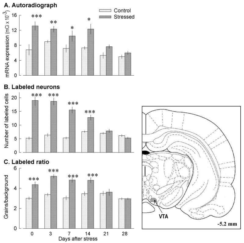
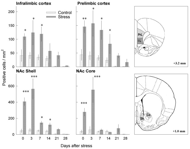
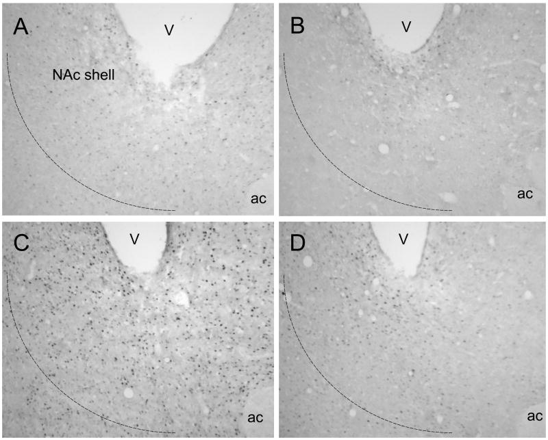
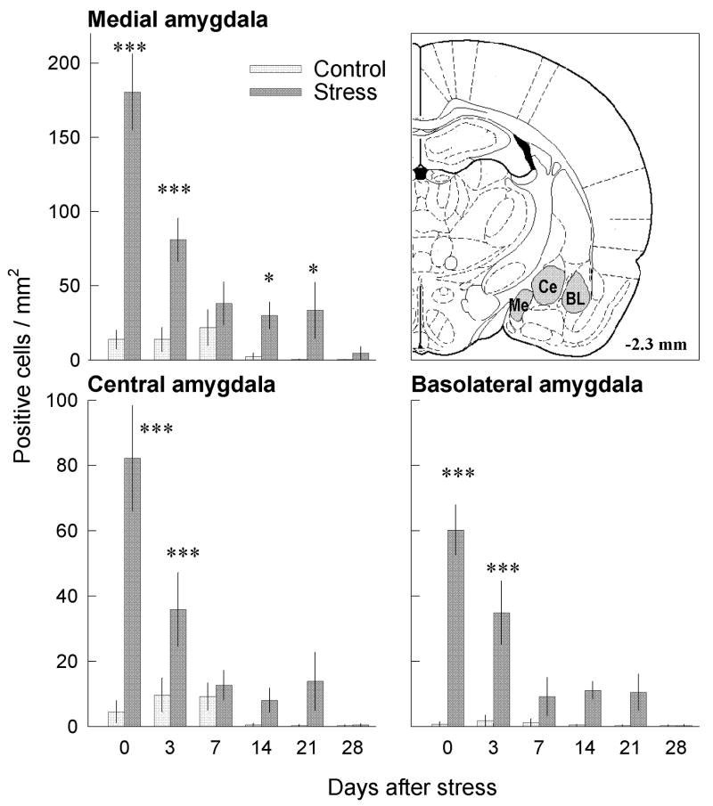
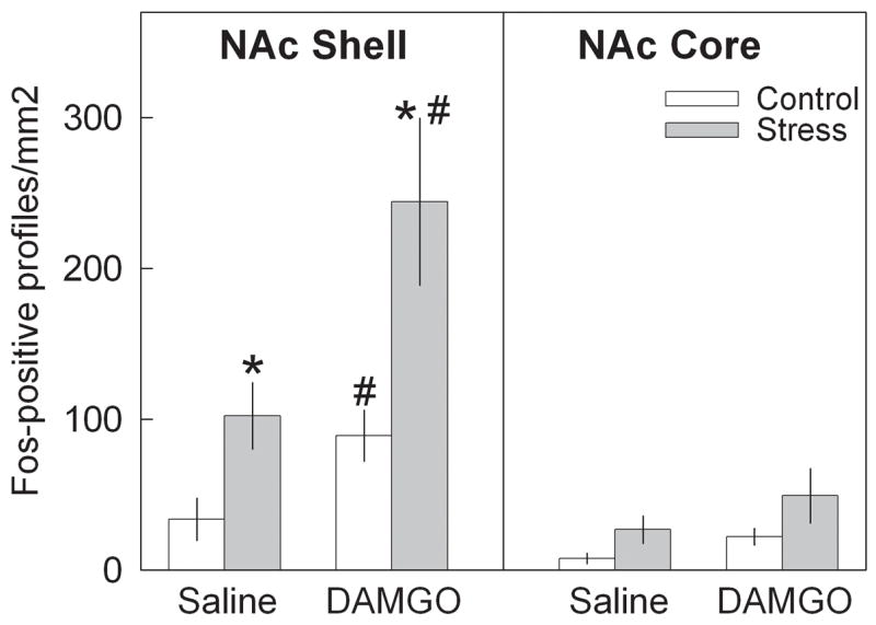
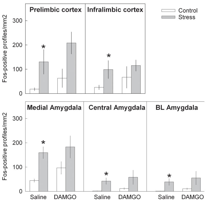
Social defeat stress is a salient stressor that induces neuroadaptive changes in the mesocorticolimbic dopaminergic system. Substantial evidence indicates that mu-opioid receptors (MORs) modulate dopamine transmission in the ventral tegmental area (VTA). FosB/DeltaFosB protein accumulation in dopaminergic projections during repeated treatments is thought to be involved in long-term neuroplasticity. In this study we characterize the magnitude and time-course of MOR mRNA expression and FosB/DeltaFosB immunoreactivity in mesocorticolimbic regions following repeated social defeat stress. Effects of brief repeated social defeat stress or control handling procedures were studied in rats either 2 h after the last exposure, or 3, 7, 14, 21 and 28 days later. We found that MOR mRNA expression in the VTA doubled after the last stress compared with handling, and remained 30-70% higher until day 21. The number of FosB/DeltaFosB-labeled neurons in regions of the frontal cortex, nucleus accumbens (NAc) shell and core, and in the medial, central and basolateral amygdala increased significantly immediately after the last stress episode, and remained enhanced for 21 days. Another group of rats received bilateral intra-VTA infusion of the MOR agonist, DAMGO, 7 days after the last stress. Prior social defeat stress augmented DAMGO-induced Fos expression in the NAc shell, suggesting that Fos expression in this region might be the direct result of MOR activity in the VTA. Social defeat stress leads to an increased capacity for MOR activation in the VTA, which may be relevant to enduring FosB/DeltaFosB expression in mesocorticolimbic areas and to the behaviorally sensitized response to psychostimulant drugs.


Figures

Fig 1
Fig 1
Repeated defeat stress significantly increased the expression of MOR mRNA in the VTA 2 h after the last stress episode, and this level of mRNA expression remained high for at least 21 days. (A) optical density in film autoradiographs expressed as μCi/g of calibrated radiostandard (two-way ANOVA F 1,60 = 47.32; p<0.01) (B) number of labeled neurons in the VTA (F 1,60 = 176.82; p<0.001); (C) labeling ratio (number of grains per cell relative to background labeling) at different time-points after repeated defeat stress (F 1,60 = 70.15; p<0.001). *p<0.05; **p<0.01; *** p<0.001 compared with appropriate control group.
Fig 2
Fig 2
Representative film autoradiographs showing the effect of repeated social stress on MOR mRNA expression in the VTA of handled control and stressed rats 3 and 14 days after the last exposure. Handled control rat (A) 3 and (B) 14 days after handling; stressed rat (C) 3 and (D) 14 days after the last stress exposure. Note increased MOR mRNA expression after social defeat stress. SNc – substantia nigra, pars compacta; SNr - substantia nigra, pars reticulata.
Fig. 3
Fig. 3
Effects of repeated social defeat stress on the number of FosB/ΔFosB neurons in the medial prefrontal cortex and NAc shell and core at different time-points after stress. Repeated social defeat stress significantly increased the number of FosB/ΔFosB-positive neurons in the prelimbic (PrL; F1,60 = 32.63; p<0.001) and infralimbic cortical regions (IL; F1,60 = 20.82; p<0.001); NAc shell (Sh; F1,60 = 76.184; p<0.001) and NAc core (Co; F1,60 = 29.61; p<0.001). *p<0.05; **p<0.01; ***p<0.001 compared with appropriate control group.
Fig. 4
Fig. 4
Photomicrographs showing FosB/ΔFosB labeling in prelimbic cortex from representative handled and stressed rats 3 or 14 days after the last exposure. Handled control rat (A) 3 and 14 (B) 14 days after handling; stressed rat (C) 3 and (D) 14 days after the last stress exposure. The number of FosB/ΔFosB profiles in the prelimbic cortical region significantly increased after exposure to repeated social defeat stress.
Fig. 5
Fig. 5
Photomicrographs showing FosB/ΔFosB labeling in NAc shell from representative handled control and repeated social defeat stress rats 3 and 14 days after the last exposure. (A) – Handled control rat (A) 3 and 14 (B) 14 days after handling;stressed rat (C) 3 and (D) 14 days after the last stress exposure. The number of FosB/ΔFosB profiles in the NAc shell significantly increased after exposure to repeated social defeat stress. V – lateral ventricle; ac – anterior commissure.
Fig. 6
Fig. 6
Time–course of repeated social defeat stress effects on FosB/ΔFosB labeling in medial (Me), central (Ce) and basolateral (BL) regions of amygdala. Repeated social defeat stress significantly increased the number of FosB/ΔFosB-positive neurons in Me (F1,60 = 92.8; p<0.001) Ce (F1,60 = 76.18; p<0.001) and BL regions (F1,60 = 44.48; p<0.001). *p<0.05; ***p<0.001 compared with appropriate control group.
Fig. 7
Fig. 7
Effects of intra-VTA saline and DAMGO infusion on number of Fos-positive cells in NAc shell and core regions 7 days after repeated social defeat stress. * − p<0.05 control vs stress: # − p< saline vs DAMGO.
Fig. 8
Fig. 8
Effects of intra-VTA saline and DAMGO infusion on number of Fos-positive cells in prelimbic and infralimbic regions of frontal cortex; medial, central and basolateral regions of amygdala 7 days after repeated social defeat stress. * − p<0.05 control vs stress.

References

    1. Andersson M, Westin JE, Cenci MA. Time course of striatal ΔFosB-like immunoreactivity and prodynorphin mRNA levels after discontinuation of chronic dopaminomimetic treatment. Eur J Neurosci. 2003;17:661–666. - PubMed
    1. Abercrombie E, Keefe K, DiFrishia D, Zigmond M. Differential effects of stress in vivo dopamine release in striatum, nucleus accumbens and medial prefrontal cortex. J Neurochem. 1989;51:1657–1658. - PubMed
    1. Barrot M, Marinelli M, Abrous DN, Rouge-Pont F, Le Moal M, Piazza PV. Functional heterogeneity in dopamine release and in the expression of Fos-like proteins within the rat striatal complex. Eur J Neurosci. 1999;11:1155–1166. - PubMed
    1. Bontempi B, Sharp FR. Systemic morphine-induced Fos protein in the rat striatum and nucleus accumbens is regulated by μ-opioid receptors in the substantia nigra and ventral tegmental area. J Neurosci. 1997;17:8596–8612. - PMC - PubMed
    1. Broekkamp CL, Phillips AG, Cools AR. Stimulant effects of enkephalin microinjection into the dopaminergic A10 area. Nature. 1979;278:560–562. - PubMed

Publication types