. 2008 Apr 29:8:48.
doi: 10.1186/1471-213X-8-48.

In vivo consequences of deleting EGF repeats 8-12 including the ligand binding domain of mouse Notch1

Affiliations

In vivo consequences of deleting EGF repeats 8-12 including the ligand binding domain of mouse Notch1

Changhui Ge et al. BMC Dev Biol. .

Abstract

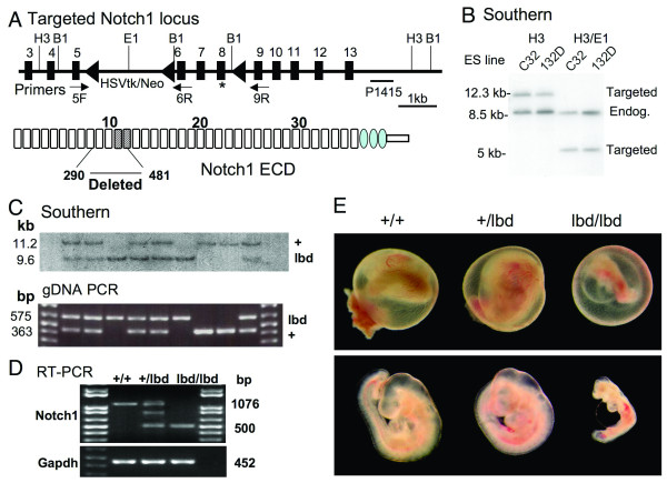
Background: Notch signaling is highly conserved in the metazoa and is critical for many cell fate decisions. Notch activation occurs following ligand binding to Notch extracellular domain. In vitro binding assays have identified epidermal growth factor (EGF) repeats 11 and 12 as the ligand binding domain of Drosophila Notch. Here we show that an internal deletion in mouse Notch1 of EGF repeats 8-12, including the putative ligand binding domain (lbd), is an inactivating mutation in vivo. We also show that maternal and zygotic Notch1(lbd/lbd) mutant embryos develop through gastrulation to mid-gestation.

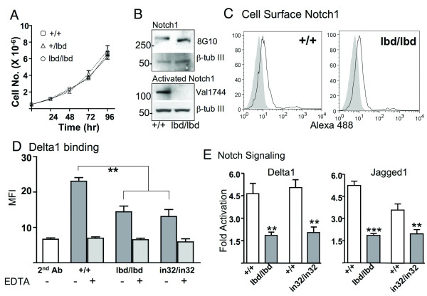
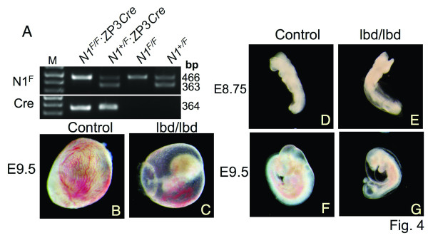
Results: Notch1(lbd/lbd) embryos died at mid-gestation with a phenotype indistinguishable from Notch1 null mutants. In embryonic stem (ES) cells, Notch1(lbd) was expressed on the cell surface at levels equivalent to wild type Notch1, but Delta1 binding was reduced to the same level as in Notch1 null cells. In an ES cell co-culture assay, Notch signaling induced by Jagged1 or Delta1 was reduced to a similar level in Notch1(lbd) and Notch1 null cells. However, the Notch1(lbd/lbd) allele was expressed similarly to wild type Notch1 in Notch1(lbd/lbd) ES cells and embryos at E8.75, indicating that Notch1 signaling is not essential for the Notch1 gene to be expressed. In addition, maternal and zygotic Notch1 mutant blastocysts developed through gastrulation.

Conclusion: Mouse Notch1 lacking the ligand binding domain is expressed at the cell surface but does not signal in response to the canonical Notch ligands Delta1 and Jagged1. Homozygous Notch1(lbd/lbd) mutant embryos die at approximately E10 similar to Notch1 null embryos. While Notch1 is expressed in oocytes and blastocysts, Notch1 signaling via canonical ligands is dispensable during oogenesis, blastogenesis, implantation and gastrulation.


Figures

Figure 1
Figure 1
Targeting of the Notch1 gene. (A) Schematic representation of the floxed region of the mouse Notch1 gene. Exons 6 – 8 (* designates a T466A point mutation termed Notch112f described by [13]) and the HSVTk/Neo cassette were removed by Cre recombinase to generate the Notch1lbd allele. The diagram of Notch1 ECD shows EGF repeats as rectangles and LIN repeats as ovals. The ligand binding domain in EGF repeats 11 and 12 is striped. Amino acids 290–481 were removed by the Notch1lbd mutation. PCR primers 5F, 6R and 9R and the P1415 probe are indicated. B1: BamHI; E1: EcoRI; H3: HindIII. (B) Southern blot analysis of two targeted ES clones (C32 and 132D) by hybridization with probe P1415 after digestion with HindIII or HindIII and EcoRI. (C) Southern blot analysis and PCR genotyping of yolk sac genomic DNA from E9.5 embryos from a Notch1+/lbd heterozygous cross. Genomic DNA was digested with BamHI and probed with P1415. (D) RT-PCR analysis of total RNA from ES cells of the genotypes shown. A hybrid band was obtained from Notch1+/lbd cDNA. (E) E9.5 embryos exhibited defective vascularization of yolk sac and retarded development of Notch1lbd/lbd embryos, but no apparent differences between wild type and heterozygous progeny.
Figure 2
Figure 2
Notch1 lacking the ligand binding domain is expressed on the cell surface but does not signal. (A) Growth curves of ES cells isolated from E3.5 Notch1+/+, Notch1+/lbd and Notch1lbd/lbd blastocysts. Bars represent mean ± SD. (B) Western blot analysis of ES cell lysates (50 μg protein). Full length Notch1 was detected by antibody 8G10 and activated Notch1 was detected by antibody Val1744. Blots were stripped and reprobed using anti-β-Tubulin III. Data are representative of 3 experiments. (C) Flow cytometry of cell surface Notch1 in Notch1lbd/lbd and Notch1+/+ ES cells using anti-Notch1 ECD antibody 8G10 followed by Alexa-488 conjugated anti-hamster IgG. Grey profiles are secondary antibody alone. Profiles are representative of two experiments. (D) Notch ligand binding. ES cells were incubated with soluble Delta1-Fc followed by PE-conjugated anti-human IgG and analyzed by flow cytometry. Notch1in32/in32 null ES cells were line 290-2. EDTA in the binding buffer inhibited binding to all Notch receptors. Bars represent mean ± SEM; n = 5 for Notch1+/+ and Notch1lbd/lbd; n = 3 for Notch1in32/in32 ES cells. (E) ES cells were assayed for Notch signaling after transfection of the Notch TP-1 reporter construct by co-culturing with L cells expressing Delta1 or Jagged1 compared to control L cells. Bars represent fold-activation ± SEM for Notch1+/+ (white), Notch1lbd/lbd and Notch1in32/in32 (gray) (n = 4; ** P < 0.01, *** P < 0.001).
Figure 3
Figure 3
Whole mount in situ hybridization of Notch1 pathway and somitogenic genes. Control (Notch1+/+) embryos (left) and mutant Notch1lbd/lbd denoted lbd/lbd (right) embryos were probed together. Arrows point to highest expression. (A) Notch1 expression in E8.75 control and Notch1lbd/lbd embryos was similar (~9.7 kb probe). (B) At E9.0 Notch1 expression was reduced in Notch1lbd/lbd compared to control embryos (~4.7 kb probe). (C) At E9.5 Notch1 expression was barely detectable in Notch1lbd/lbd embryos (~9.7 kb probe). (D) Expression of the Notch target gene Hes5 was reduced in mutant embryos at E9.5, with residual expression in brain. (E) Myogenin was poorly and diffusely expressed in E9.5 Notch1lbd/lbd embryos. (F) Uncx4.1 was expressed in the caudal compartment of formed somites of control but was missing from the somitic region of E9.5 Notch1lbd/lbd embryos. Uncx4.1 was up-regulated in the midbrain (asterisk) of E9.5 Notch1lbd/lbd embryos (n ≥ 3 for mutant embryos for each probe).
Figure 4
Figure 4
Notch1lbd/lbd maternal and zygotic null embryos. (A) PCR of genomic DNA using primers 5F and 6R (Fig. 1A), and ZP3Cre primers. N1F: Notch1 floxed allele; Cre: ZP3Cre transgene. (B) – (C) E9.5 yolk sac with embryos from crosses between Notch1F/F:ZP3Cre females and Notch1+/lbd males. (D) – (E) E8.75 Notch1+/lbd and Notch1lbd/lbd embryos. (F) – (G) E9.5 Notch1+/lbd and Notch1lbd/lbd embryos. Representative results from a total of 14 E8.75 embryos and 29 E9.5 embryos.

References

    1. Lai EC. Notch signaling: control of cell communication and cell fate. Development (Cambridge, England) 2004;131:965–973. - PubMed
    1. Schweisguth F. Regulation of notch signaling activity. Curr Biol. 2004;14:R129–38. - PubMed
    1. Rebay I, Fleming RJ, Fehon RG, Cherbas L, Cherbas P, Artavanis-Tsakonas S. Specific EGF repeats of Notch mediate interactions with Delta and Serrate: implications for Notch as a multifunctional receptor. Cell. 1991;67:687–699. doi: 10.1016/0092-8674(91)90064-6. - DOI - PubMed
    1. Xu A, Lei L, Irvine KD. Regions of Drosophila Notch That Contribute to Ligand Binding and the Modulatory Influence of Fringe. 2005;280:30158–30165. - PubMed
    1. Hartmann D, de Strooper B, Serneels L, Craessaerts K, Herreman A, Annaert W, Umans L, Lubke T, Lena Illert A, von Figura K, Saftig P. The disintegrin/metalloprotease ADAM 10 is essential for Notch signalling but not for alpha-secretase activity in fibroblasts. Hum Mol Genet. 2002;11:2615–2624. doi: 10.1093/hmg/11.21.2615. - DOI - PubMed

Publication types

LinkOut - more resources