

Splenic rupture as the presenting manifestation of primary splenic angiosarcoma in a teenage woman: a case report
- PMID: 18445294
- PMCID: PMC2387157
- DOI: 10.1186/1752-1947-2-133
Splenic rupture as the presenting manifestation of primary splenic angiosarcoma in a teenage woman: a case report
Abstract
Introduction: Primary splenic angiosarcoma is a rare neoplasm of vascular origin carrying a very poor prognosis, partly due to its high metastatic potential. This disease presents frequently with splenic rupture and hemorrhage. We report the case of a 17-year-old woman who presented with rupture of a primary splenic angiosarcoma.
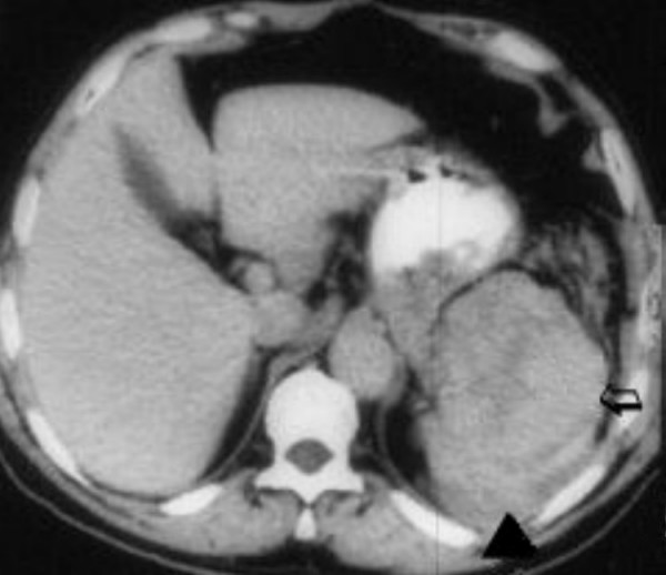
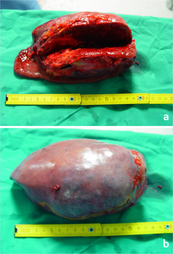
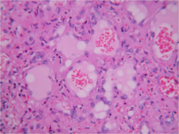
Case presentation: The patient presented with diffuse abdominal pain and distention. Clinical examination revealed severe tenderness in the left upper abdominal quadrant, a palpable abdominal mass, and hemodynamic instability with a systolic arterial blood pressure of 75 mmHg and heart rate of 135 beats per minute. Blood tests revealed anemia (hemoglobin 7.0 g/dl) and thrombocytopenia (platelets 70 x 109/liter). After initial fluid resuscitation and stabilization, abdominal ultrasound and computed tomography were performed, revealing a large quantity of intraperitoneal free fluid, an enlarged spleen, and a heterogeneous low-density signal within the splenic parenchyma, which showed varying degrees of contrast enhancement. At laparotomy a huge (weight 1530 g, diameter 19 cm) actively bleeding spleen was identified and splenectomy was performed. Histopathology showed a primary splenic angiosarcoma. After an uneventful recovery, the patient was discharged on the sixth postoperative day.
Conclusion: Primary splenic angiosarcoma is rare. Although this malignancy is usually encountered in advanced age, there have been a few reported cases among younger patients. The case reported here presented with splenic rupture, was treated by laparotomy and splenectomy, and the patient is disease free 16 months after surgery.
Figures
References
-
- Buckner JW, III, Porterfield G, Williams GR. Spontaneous splenic rupture secondary to angiosarcoma. J Okla State Med Assoc. 1990;83:211–213. - PubMed
-
- Montemayor P, Caggiano V. Primary hemangiosarcoma of the spleen associated with leukocytosis and abnormal spleen scan. Int Surg. 1980;65:369–373. - PubMed
-
- McGinley K, Googe P, Hanna W, Bell J. Primary angiosarcoma of the spleen: a case report and review of the literature. South Med J. 1995;88:873–875. - PubMed
-
- Falk S, Krishnan J, Meis JM. Primary angiosarcoma of the spleen. A clinicopathologic study of 40 cases. Am J Surg Pathol. 1993;17:959–970. - PubMed
LinkOut - more resources
Full Text Sources
