. 2008 Jul;17(7):1175-87.
doi: 10.1110/ps.034892.108. Epub 2008 Apr 29.

Toward chaperone-assisted crystallography: protein engineering enhancement of crystal packing and X-ray phasing capabilities of a camelid single-domain antibody (VHH) scaffold

Affiliations

Toward chaperone-assisted crystallography: protein engineering enhancement of crystal packing and X-ray phasing capabilities of a camelid single-domain antibody (VHH) scaffold

Valentina Tereshko et al. Protein Sci. 2008 Jul.

Abstract

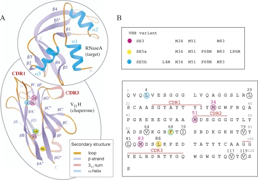
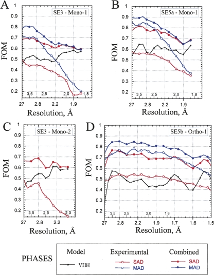
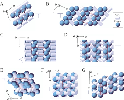
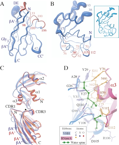
A crystallization chaperone is an auxiliary protein that binds to a target of interest, enhances and modulates crystal packing, and provides high-quality phasing information. We critically evaluated the effectiveness of a camelid single-domain antibody (V(H)H) as a crystallization chaperone. By using a yeast surface display system for V(H)H, we successfully introduced additional Met residues in the core of the V(H)H scaffold. We identified a set of SeMet-labeled V(H)H variants that collectively produced six new crystal forms as the complex with the model antigen, RNase A. The crystals exhibited monoclinic, orthorhombic, triclinic, and tetragonal symmetry and have one or two complexes in the asymmetric unit, some of which diffracted to an atomic resolution. The phasing power of the Met-enriched V(H)H chaperone allowed for auto-building the entire complex using single-anomalous dispersion technique (SAD) without the need for introducing SeMet into the target protein. We show that phases produced by combining SAD and V(H)H model-based phases are accurate enough to easily solve structures of the size reported here, eliminating the need to collect multiple wavelength multiple-anomalous dispersion (MAD) data. Together with the presence of high-throughput selection systems (e.g., phage display libraries) for V(H)H, the enhanced V(H)H domain described here will be an excellent scaffold for producing effective crystallization chaperones.


Figures

Figure 1.
Figure 1.
(A) Ribbon diagrams of cAb-RN5 VHH and RNase A topology. The termini and the secondary structure elements are labeled. CDR1 and CDR3 residues are labeled in red. The position of the SeMet residues that were used for phasing are labeled and represented as small colored spheres. The Cα positions of the three native Met residues—34, 51, and 83—are shown in magenta. The Cα positions of two additional Met residues introduced into the SE5a variant (F68M and L86M) are shown in yellow, and those in the SE5b variant (L4M and F68M) are in cyan. (B) The amino acid sequence of cAb-RN05 VHH. Ten positions that were selected as potential sites for Met incorporation are circled. Positions at which SeMet is present in the proteins used in this study (shown in the upper panel) are colored in the same manner as in A.
Figure 2.
Figure 2.
FOM for phasing based on SeMet-labeled VHH chaperones in the crystals of cAb-RN05 VHH–RNase A complex. Two-wavelength MAD (in blue) and SAD (in red) phasing statistics are plotted as a function of X-ray resolution bins. The value of the average resolution in each bin is indicated along the horizontal axis. Open circles represent the experimental phases derived by SOLVE. Filled circles represent the experimental phases combined with the VHH model-based phases. The phasing was performed for X-ray resolution range indicated in the parentheses in each case. The FOM for VHH model-based phases are included for reference and shown as a gray line. (A) SE3-Mono-1 (50–1.8 Å); (B) SE5a-Mono-1 (50–1.8 Å); (C) SE3-Mono-2 (50–2.0 Å); (D) SE5b-Ortho-1 (50–1.5 Å).
Figure 3.
Figure 3.
Schematics of the packing modes found in the crystals of the cAb-RN05 VHH–RNase A complexes. The unit cell is indicated in red. The orientation of the crystal axes is shown in black. The cyan arrow indicates the view shown in Supplemental Figure S2. The VHH and RNase A molecules are represented as white cylinders and cyan spheres, respectively. This representation is based on the calculation of molecular ellipsoids performed in MOLREP. (A) Monoclinic Mono-1 crystal form with one complex in ASU. This form was observed for the SE3 and SE5a complexes, crystallized isomorphously. (B) Monoclinic SE3-Mono-2 crystal form with two complexes in ASU. (C) Orthorhombic SE5b-Ortho-1 crystal form with one complex in ASU. (D) Orthorhombic SE5b-Ortho-2 crystal form with two complexes in ASU. (E) Trigonal SE5b-Tri crystal form with one complex in ASU. (F) Tetragonal SE5b-Tetra crystal form with one complex in ASU. (G) Original 1BZQ crystal structure crystallized in triclinic space group with four complexes in ASU.
Figure 4.
Figure 4.
Favorable crystal contacts formed by dimerization of VHH through H-bonding in N-terminal β-strands. (A) VHH–VHH alignment in the SE5b dimer. Two VHH domains interact via their N-terminal βA′- and βA″-strands with the residues E6 (βA′), G8, G9, and L11 (βA″) being involved in the H-bonds (2.6–3.3 Å range). The SE5b-Tetra structure was selected for presentation. (Inset) Four H-bonding pairs found in SE5b dimers. The main-chain N- and O-atoms involved in H-bonds are shown as small circles colored in blue and red, respectively. There are four H-bonds in Se5b-Tri and Se5b-Tetra crystals (shown as filled circles). In Se5b-Ortho-2, the residues E6 and L11 contribute both N- and O-atoms in intermolecular H-bonding network, resulting in two additional H-bonds (shown as open circles). (B). VHH–VHH alignment in the 1BZQ dimer. Two VHH domains interact via their N-terminal βA″-strands with the residues G9, L11, and Q13 being involved in the H-bonds. (Inset) Three H-bonding pairs found in 1BZQ. L11 contributes both N- and O-atoms in H-bonding network, resulting in a total of four H-bonds.
Figure 5.
Figure 5.
A comparison of different X-ray structures of the cAb-RN05 VHH–RNase A complexes. Fourteen crystallographically independent copies (those in Table 2 and the 1BZQ structure) were used for superposition and analysis. (A) Worm representations of VHH. The thickness of the worm is proportional to the deviations between the positions of corresponding Cα-atoms in the VHH structures superimposed using their main-chain atoms. The CC′- and DE-loops centered at the residues 42 and 75 exhibit the highest flexibility. CDR1 and CDR3 residues are colored in red. (B) Worm representations of RNase A structures. The thickness of the worm is proportional to the deviations between the positions of corresponding Cα-atoms in RNase A structures superimposed using their main-chain atoms. The residues involved in the interaction with VHH in the complex are indicated in red. (Inset) Six copies of RNase A molecule in the free form from four different crystal forms (PDB codes 7RSA, 1RTB, 1XPS, 1XPT) were superimposed using their main-chain atoms with rmsds <0.7 Å. (C) Superposition of VHH–RNase A complexes. The structures were superimposed using the main-chain atoms of RNase A. Two complexes where the VHH positions differ the most are shown: SE5b-Ortho-2 complex 2 (in blue) and SE3-Mono-2 complex 1 (in red). The VHH domains from two structures may be superimposed by ∼8° rotation around the axis running through the binding interface (close to CDR3) approximately perpendicularly to the page and represented as the black dot. (D) VHH–RNase A interface. Two spines of H-bonded water molecules are represented as connected green bonds. The H-bonds involving the protein residues are shown as dotted lines: water–protein, green; protein–protein, gray. The water spines are connected to the VHH residues G26(O), A28(N), Y31(OH), A98(O), G108(O), G109(O), Q110(NE2) and RNase A residues C58(O), S59(O, OG), Y73(OH), Y76(OH), D105(O), G112(O), Y115(O, N). The H-bond lengths are in 2.6–3.3 Å range. These H-bonds are preserved in all four high-resolution (1.8–1.1 Å) structures studied in this work (Table 2). The 1.1 Å X-ray structure SE5b-Ortho-1 was selected for presentation.

References

    1. Adams, P.D., Pannu, N.S., Read, R.J., Brunger, A.T. Cross-validated maximum likelihood enhances crystallographic simulated annealing refinement. Proc. Natl. Acad. Sci. 1997;94:5018–5023. - PMC - PubMed
    1. Barthelemy, P.A., Raab, H., Appleton, B.A., Bond, C.J., Wu, P., Wiesmann, C., Sidhu, S.S. Comprehensive analysis of the factors contributing to the stability and solubility of autonomous human VH domains. J. Biol. Chem. 2008;283:3639–3654. - PubMed
    1. Boder, E.T., Wittrup, K.D. Yeast surface display for directed evolution of protein expression, affinity, and stability. Methods Enzymol. 2000;328:430–444. - PubMed
    1. Bradbury, A.R.M., Marks, J.D. Antibodies from phage antibody libraries. J. Immunol. Methods. 2004;290:29–49. - PubMed
    1. Brekke, O.H., Loset, G.A. New technologies in therapeutic antibody development. Curr. Opin. Pharmacol. 2003;3:544–550. - PubMed

Publication types