. 2008 May 6;105(18):6578-83.
doi: 10.1073/pnas.0801097105. Epub 2008 Apr 29.

Design of protein function leaps by directed domain interface evolution

Affiliations

Design of protein function leaps by directed domain interface evolution

Jin Huang et al. Proc Natl Acad Sci U S A. .

Abstract

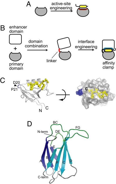
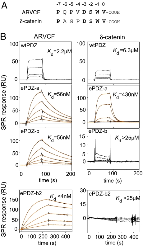
Most natural proteins performing sophisticated tasks contain multiple domains where an active site is located at the domain interface. Comparative structural analyses suggest that major leaps in protein function occur through gene recombination events that connect two or more protein domains to generate a new active site, frequently occurring at the newly created domain interface. However, such functional leaps by combination of unrelated domains have not been directly demonstrated. Here we show that highly specific and complex protein functions can be generated by joining a low-affinity peptide-binding domain with a functionally inert second domain and subsequently optimizing the domain interface. These directed evolution processes dramatically enhanced both affinity and specificity to a level unattainable with a single domain, corresponding to >500-fold and >2,000-fold increases of affinity and specificity, respectively. An x-ray crystal structure revealed that the resulting "affinity clamp" had clamshell architecture as designed, with large additional binding surface contributed by the second domain. The affinity clamps having a single-nanomolar dissociation constant outperformed a monoclonal antibody in immunochemical applications. This work establishes evolutionary paths from isolated domains with primitive function to multidomain proteins with sophisticated function and introduces a new protein-engineering concept that allows for the generation of highly functional affinity reagents to a predefined target. The prevalence and variety of natural interaction domains suggest that numerous new functions can be designed by using directed domain interface evolution.


Conflict of interest statement

Conflict of interest statement: J.H., A.K., and S.K. are named as the inventors of a patent application on the technology described in this article.

Figures

Fig. 1.
Fig. 1.
The concept of directed domain interface evolution and building blocks used in this work. (A and B) Comparison of domain interface engineering with conventional protein engineering. In the conventional engineering that mimics gene duplication and sequence divergence (A), the interface predefined in the starting scaffold is altered/refined, which tends to produce incremental changes in function. In contrast, domain interface engineering that mimics gene combination and sequence divergence (B) produces a new functional site at the interface between two domains, which can result in a major leap in protein function. (C) The structure of the Erbin PDZ bound to a peptide (PDB entry 1MFG). The N and C termini are indicated. The positions for the new termini of the circularly permutated PDZ (cpPDZ) are shown with a triangle and residue numbers. Right shows the surface of the PDZ domain with the peptide as a stick model, illustrating the shallow binding pocket. (D) The structure of FN3 (PDB entry 1FNF). The loops that are diversified to construct combinatorial libraries are labeled. The termini are also labeled. Note that the N terminus and the recognition loops are located on the same side of the FN3 protein.
Fig. 2.
Fig. 2.
Target binding properties of affinity clamps. (A) Amino acid sequences of the target peptides. Residue numbers are according to the convention for PDZ ligands. Identical residues between the two peptides are shown in bold. (B) SPR sensorgrams for the interaction of the ARVCF peptide (Left) and the δ-catenin peptide (Right) to wild-type PDZ, ePDZ-a, ePDZ-b, and ePDZ-b2. The target peptides were fused to SUMO for SPR measurements. Sensorgrams with 0, 10, 20, 40, 80, and 160 nM peptide are in black, and the global fittings of the 1:1 Langmuir binding model are in orange. Kd values are also shown. Kd values for wild-type PDZ were determined from equilibrium analyses using a broader range of target concentration than shown here.
Fig. 3.
Fig. 3.
The x-ray crystal structure of ePDZ-a in complex with the ARVCF peptide. (A) Ribbon representations of the overall structure. The cpPDZ and FN3 portions and the peptide are shown in gray, cyan, and yellow, respectively. Missing residues for the linker segment are indicated with dashed lines. (B) Clamping of the peptide by ePDZ-a. Only the region in the dashed box in A is shown. The surfaces originating from the cpPDZ and FN3 portions are shown in gray and cyan, respectively, and the peptide is shown as a yellow stick model. (C) Interactions of the FN3 loops with the cpPDZ/peptide complex. The three FN3 loops (BC, DE, and FG loops) are shown as sticks in blue, cyan, and red, respectively. The surface of the PDZ portion is shown in gray, and the peptide is shown as yellow spheres. In Lower, the surfaces of the PDZ and peptide portions in contact with the BC, DE, and FG loops (within 5 Å are shown in blue, cyan, and red, respectively, and those in contact with both BC and FG loops are in magenta. The peptide surfaces without FN3 contact are shown in yellow, and the green line encloses the bound peptide. (D) Superposition of wild-type PDZ (PDB entry 1MFG, green) and cpPDZ (gray). The original and new termini are indicated. The rmsd for the equivalent 97 Cα atoms was 0.54 Å. The structures are in the same orientation as in A.
Fig. 4.
Fig. 4.
Stability and applications of affinity clamps. (A) Gel-filtration chromatograms of ePDZ-a before (dashed line) and after (solid line) heat treatment (2 h at 50°C). Gel filtration was performed by using a Superdex75 column (Amersham Biosciences) in PBS (pH 7.4). (B) The SPR sensorgrams of ePDZ-a before (dashed line) and after (solid line) the same heat treatment as in A. (C) Western blotting of wild-type ARVCF in mammalian cell lysates. Lysates of an MDCK cell line stably expressing human ARVCF (denoted as +) (41) and the parent cell line (−) were detected with ePDZ-b2 fused with alkaline phosphatase. mAb indicates a positive control with anti-ARVCF monoclonal antibody 4B1 (41). Left shows Coomassie brilliant blue staining, and Right shows Western blotting. (D) Pull-down (immunoprecipitation) of SUMO tagged with the ARVCF peptide from E. coli lysate by affinity clamps. The lysate was mixed with an affinity clamp immobilized to streptavidin magnetic beads. SDS/PAGE of the input (I), unbound (U), wash (W), and bound (B) fractions visualized with Coomassie brilliant blue staining are shown. “Beads” indicates a control experiment without an immobilized affinity clamp. “cpPDZFN” indicates a control experiment using cpPDZ fused to the unmodified FN3 scaffold. The position of the captured target is marked with the triangle for ePDZ-b2 (lane B), and the equivalent position is also marked for cpPDZFN. “Binder” indicates the position of ePDZ-b2 and cpPDZFN, and “SA” indicates the position of streptavidin.

References

    1. Todd AE, Orengo CA, Thornton JM. Sequence and structural differences between enzyme and nonenzyme homologs. Structure. 2002;10:1435–1451. - PubMed
    1. Pluckthun A, Mayo SL. The design of evolution and the evolution of design. Curr Opin Struct Biol. 2007;17:451–453.
    1. Kortemme T, Baker D. Computational design of protein-protein interactions. Curr Opin Chem Biol. 2004;8:91–97. - PubMed
    1. Winter G, Griffiths AD, Hawkins RE, Hoogenboom HR. Making antibodies by phage display technology. Annu Rev Immunol. 1994;12:433–455. - PubMed
    1. Binz HK, Amstutz P, Pluckthun A. Engineering novel binding proteins from nonimmunoglobulin domains. Nat Biotechnol. 2005;23:1257–1268. - PubMed

Publication types

Associated data