. 2008 May 15;121(Pt 10):1661-70.
doi: 10.1242/jcs.020149. Epub 2008 Apr 29.

PAI1 stimulates assembly of the fibronectin matrix in osteosarcoma cells through crosstalk between the alphavbeta5 and alpha5beta1 integrins

Affiliations

PAI1 stimulates assembly of the fibronectin matrix in osteosarcoma cells through crosstalk between the alphavbeta5 and alpha5beta1 integrins

Daniel Vial et al. J Cell Sci. .

Abstract

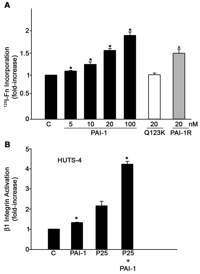
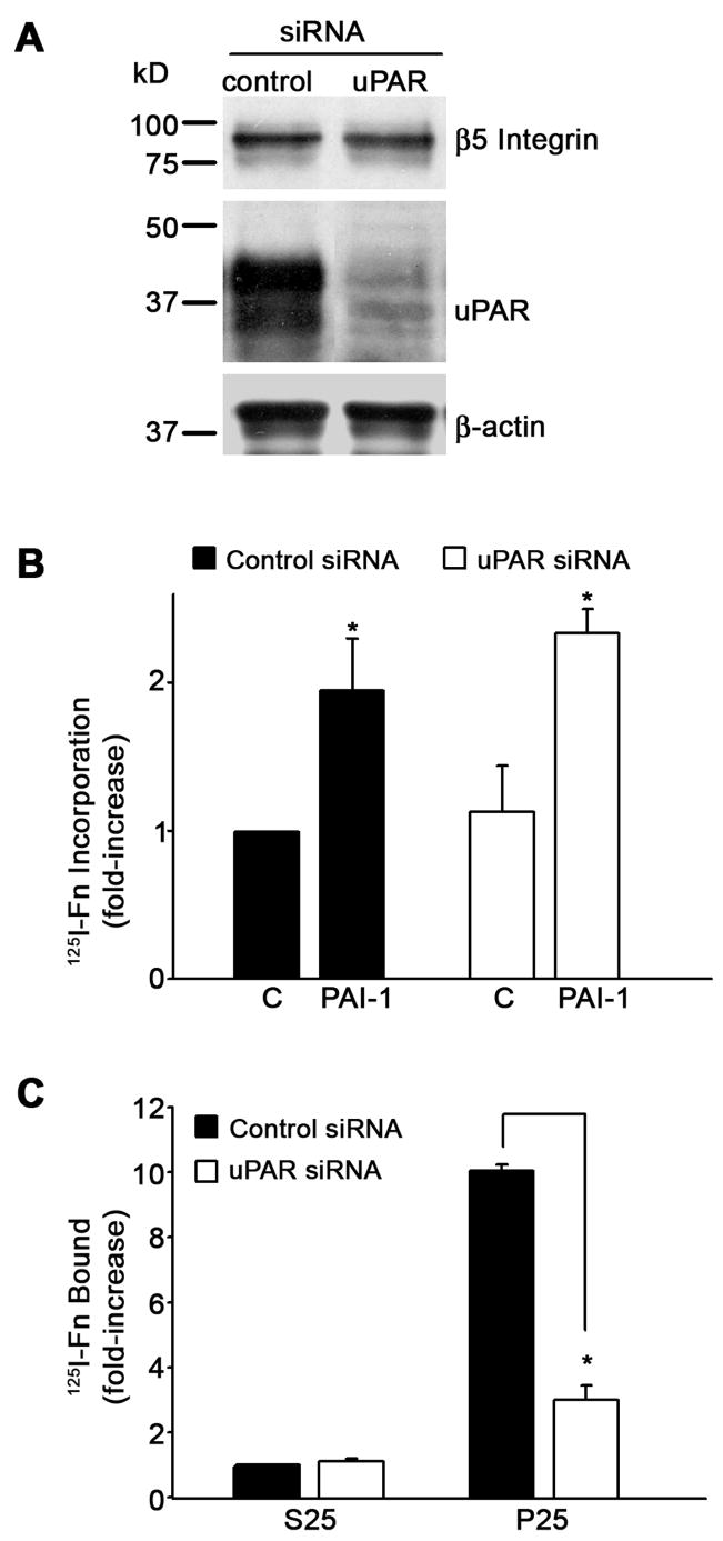
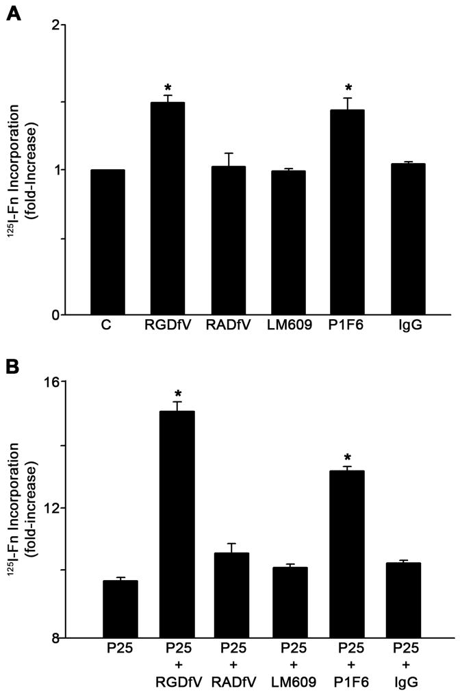
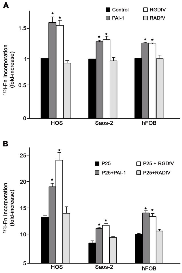
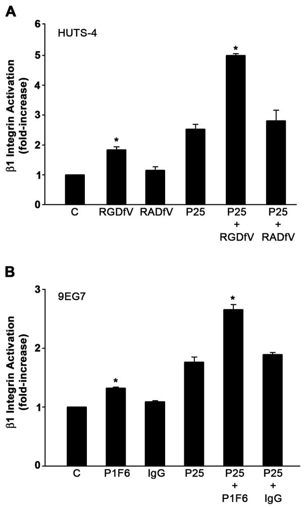
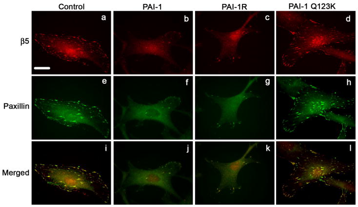
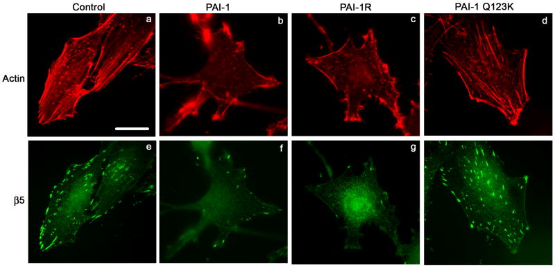
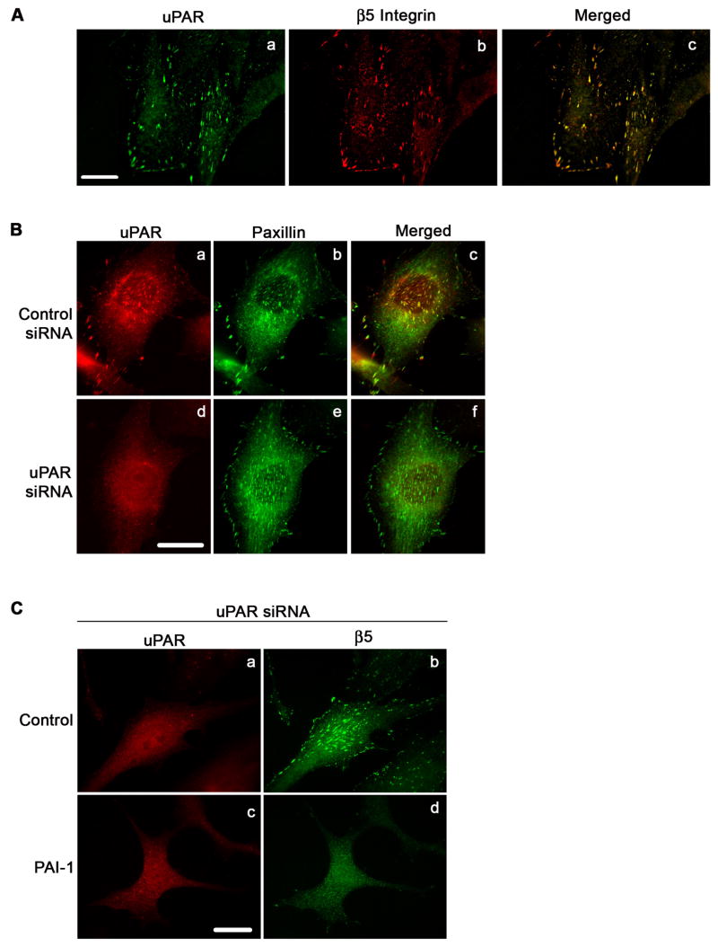
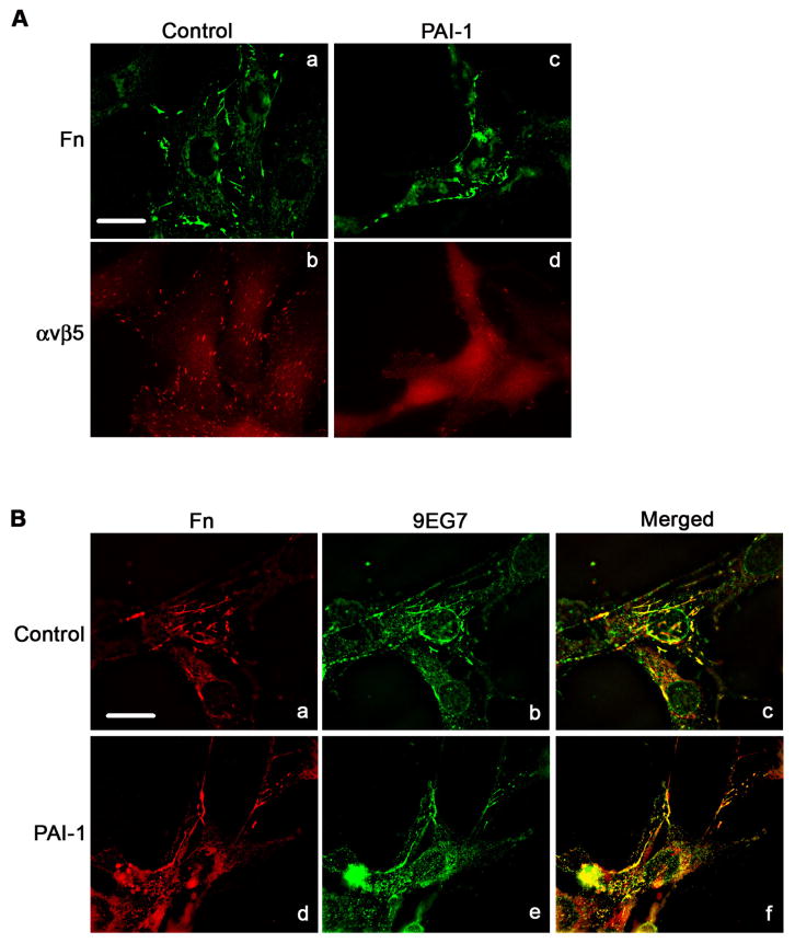
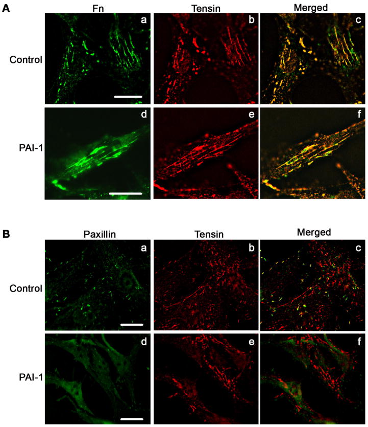
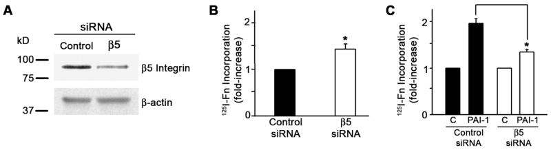
The plasminogen activation system regulates matrix remodeling through both proteolytic and non-proteolytic mechanisms. Studies were undertaken to determine the effects of the plasminogen activator inhibitor 1 (PAI1) on the assembly of the fibronectin matrix. The addition of PAI1 to MG-63 cells caused a 1.5- to threefold increase in the rate of fibronectin matrix assembly which was associated with an increase in beta integrin activation. PAI1 treatment led to a marked decrease in focal contacts and stress fibers, whereas tensin-containing matrix contacts remained unaffected. The effects of PAI1 on matrix assembly were independent of both urokinase-type plasminogen activator (uPA) and urokinase-type plasminogen activator receptor (uPAR), indicating that the stimulation of matrix assembly by PAI1 does not depend on its anti-proteolytic activity or on the association of uPAR with integrin receptors. Antagonists of the alphavbeta5 integrin mimicked the effect of PAI1 on cell morphology and fibronectin matrix deposition, indicating that stimulation of matrix assembly by PAI1 required disruption of the interaction between the alphavbeta5 integrin and vitronectin. Consistent with this conclusion, the Q123K PAI1 mutant which does not bind vitronectin had no effect on matrix assembly. Our data identify PAI1 as a novel regulator of fibronectin matrix assembly, and indicate that this regulation occurs through a previously undescribed crosstalk between the alphavbeta5 and alpha5beta1 integrins.


Figures

Fig. 1
Fig. 1
PAI-I increases fibronectin matrix assembly and β1 integrin activation in MG-63 cells. A - MG-63 cells were incubated with 125I-fibronectin for 6 hours in the presence of the indicated concentrations of PAI-1, PAI-1R or PAI-1 (Q123K) in DMEM+BSA 0.02% + 20 mM Hepes. Cell layers were extracted with 1% DOC, and 125I-fibronectin that was incorporated into the detergent-insoluble matrix was recovered by centrifugation and measured by gamma scintillation. This experiment is representative of three separate experiments. Data are the mean ± S.E. of two experiments performed in duplicate. *Significantly different than control cells, t test, p < 0.05 (n=4). B - MG-63 cells were pretreated with 20 nM PAI-1 for 20 minutes and incubated with 50 μM of uPAR agonist P25 or S25 for 1 hour in DMEM. Activation of β1 integrin was assessed by ELISA using the HUTS-4 antibody. Total β1 integrin was measured using the P5D2 antibody against α5β1. The graph shows the levels of β1 integrin activation after normalization to total β1 levels. Neither P25 nor PAI-1 had any effect on total β1 integrin. Data represent one of three different experiments performed in triplicate. *Significantly different than control or P25-treated cells, respectively, t test, p < 0.05 (n=3).
Fig. 2
Fig. 2
The PAI-1 mediated increase in fibronectin matrix assembly does not require uPAR. A - Cell lysates were prepared from control siRNA or uPAR siRNA transfected MG-63 cells. uPAR and β5 integrin levels were analyzed by Western blotting. The membrane was stripped and reprobed with anti β-actin antibody as a loading control. B - Cells layers from control siRNA or uPAR siRNA transfected MG-63 cells were incubated with 125I-fibronectin for 6 hours in the absence or presence of PAI-1 (20 nM). Cell layers were extracted with 1% DOC, and 125I-fibronectin that was incorporated into the detergent-insoluble matrix was recovered by centrifugation and measured by gamma scintillation. Data are the mean ± S.E. of two experiments performed in duplicate. *Significantly different than non treated cells, t test, p < 0.05 (n=4). C - Cells layers from control siRNA or uPAR siRNA transfected MG-63 cells were incubated with 125I-fibronectin for 6 hours in the presence of the uPAR agonist P25 or the control peptide, S25. Cell layers were washed and scraped into 1% DOC and cell layer associated 125I-fibronectin was measured by gamma scintillation. Data are the mean ± S.E. of two experiments performed in duplicate. *Significantly different than control cells, t test, p < 0.05 (n=4).
Fig. 3
Fig. 3
Inhibition of αvβ5 integrin interaction with vitronectin increases fibronectin matrix assembly. A - MG-63 cells were pre-incubated for 45 minutes with the cyclic peptide RGDfV (20 μM) or its inactive analog RADfV (20 μM) or for 2 hours with 20 μg/ml of normal mouse IgG, β3 integrin blocking antibody LM609 or β5 integrin blocking antibody P1F6 and then incubated with 125I-fibronectin (Fn) for 6 hours. This experiment is representative of three separate experiments. Data are the mean ± S.E. of two different experiments performed in duplicate. *Significantly different than control cells, t test, p < 0.05 (n=4). B - Same as A only under conditions of uPAR stimulation with P25. Cell layers were extracted in 1% DOC, and 125I-fibronectin that was incorporated into the detergent-insoluble matrix was recovered by centrifugation and measured by gamma scintillation.. This experiment is representative of three separate experiments. Data are the mean ± S.E. of two experiments performed in duplicate. *Significantly different than P25-treated cells, t test, p < 0.05 (n=4).
Fig. 4
Fig. 4
PAI-1 stimulates fibronectin matrix assembly in osteosarcoma and osteoblast cells. A - Cells were pre-incubated for 20 minutes with PAI-1 (20 nM) or for 45 minutes with the cyclic peptide RGDfV (20 μM) or its inactive analog RADfV (20 μM) and then incubated with 125I-fibronectin (Fn) for 6 hours. Cell layers were extracted in 1% DOC, and 125I-fibronectin that was incorporated into the detergent-insoluble matrix was recovered by centrifugation and measured by gamma scintillation. This experiment is representative of three separate experiments. Data are the mean ± S.E. of two different experiments performed in duplicate. *Significantly different than control cells, t test, p < 0.05 (n=4). B - Same as A only under conditions of uPAR stimulation with 50 μM P25. This experiment is representative of three separate experiments. Data are the mean ± S.E. of two experiments performed in duplicate. *Significantly different than P25-treated cells, t test, p < 0.05 (n=4).
Fig. 5
Fig. 5
Activation of β1 integrin by β5 integrin blocking agents. A - MG-63 cells were pretreated for 45 minutes with 20 μM cyclic peptides RGDfV or RADfV and incubated with 50 μM of P25 or S25 in DMEM. Activation of β1 integrin was assessed by ELISA using the HUTS-4 antibody. B - MG-63 cells were preincubated for 2 hours with 20 μg/ml of normal mouse IgG, or β5 integrin blocking antibody P1F6 before treatment with P25 or S25. Integrin activation was assessed using monoclonal antibody 9EG7. All graphs show the levels of β1 integrin activation after normalization to total β1 levels as described in Fig. 1. Total β1 integrin did not change. Data represent one of three different experiments performed in triplicate. *Significantly different than control or P25-treated cells, respectively, t test, p < 0.05 (n=3).
Fig. 6
Fig. 6
PAI-1 disrupts focal adhesions. MG-63 cell monolayers were plated overnight onto coverslips in complete media and then incubated for 3 hours with 20 nM of PAI-1 or PAI-1 mutants (PAI-1R and Q123K). Cells were subsequently fixed, permeabilized and stained for 1 hour with the clone 15F11 antibody for β5 integrin (panels a-d). After1 hour staining with the AlexaFluor594 derivatized anti-mouse secondary antibody, cells were washed and labeled for an additional hour with a paxillin-FITC antibody (panels e-h). Panels i-l show merged images indicating colocalization of β5 integrin with paxillin. Scale Bar, 20 μm.
Fig. 7
Fig. 7
PAI-1 disrupts actin stress fibers. MG-63 cell monolayers were plated overnight onto coverslips in complete media and then incubated for 3 hours with 20 nM of PAI-1 or PAI-1 mutants (PAI-1R and Q123K). Cells were subsequently fixed, permeabilized and stained for 1 hour with the clone 15F11 antibody for β5 integrin. Cells were then washed and labeled for an additional hour with the AlexaFluor488 derivatized anti-mouse secondary antibody and the AlexaFluor594-phalloidin. Actin and β5 integrin were visualized by indirect immunofluorescence. Scale Bar, 20 μm.
Fig. 8
Fig. 8
PAI-1 mediated disruption of focal adhesions does not require uPAR. A - MG-63 cells were plated overnight in complete medium. Cells were subsequently fixed, permeabilized and stained with uPAR polyclonal antibody (panel a) and 15F11 monoclonal antibody for β5 integrin (Panel b). uPAR and β5 integrin were visualized by indirect immunofluorescence. Panel c shows the merged image. Scale Bar, 20 μm. B - Cells stained for uPAR and paxillin. Panels a and b show uPAR and paxillin staining in control siRNA treated cells. Panels d and e show uPAR and paxillin staining in uPAR knock down cells. Panels c and f are the merged images. Yellow indicates colocalization of uPAR and α5 integrin. Scale Bar, 20 μm. C - uPAR siRNA treated cells were plated overnight in complete medium and then incubated for 3 hours in DMEM in the presence or absence of PAI-1. Scale Bar, 20 μm. Cells were subsequently fixed, permeabilized and stained. uPAR and β5 integrin were visualized by indirect immuno-fluorescence in non-treated cells (panels a and b) and PAI-1 treated cells (panels c and d).
Fig. 9
Fig. 9
PAI-1 does not disrupt the association of α5β1 with fibronectin matrix. Cells were incubated for 3 hours in DMEM in the presence or absence of PAI-1, subsequently fixed, permeabilized and immunostained. A - Fibronectin matrix and β5 integrin were visualized by indirect immunofluorescence in non-treated cells (panels a and b) and PAI-1 treated cells (panels c and d). Scale Bar, 20 μm. B - Fibronectin and activated β1 integrin (detected with 9EG7 antibody) were visualized by indirect immunofluorescence in non-treated cells (panels a and b) and PAI-1 treated cells (panels d and e). Panels c and f shows merged pictures. Yellow staining indicates areas of colocalization of β1 integrin with fibronectin matrix. Scale Bar, 20 μm.
Fig. 10
Fig. 10
PAI-1 does not disrupt matrix adhesions. Cells were incubated overnight onto coverslips in complete media and then incubated for 3 hours in DMEM in the presence or absence of PAI-1(20 nM). Cells were subsequently fixed, and permeabilized. A - Fibronectin and tensin were visualized by indirect immunofluorescence in non-treated cells (panels a and b) and PAI-1 treated cells (panels d and e). Panels c and e show the merged pictures. Yellow staining represents area of colocalization of tensin and fibronectin. Scale Bar, 20 μm. B – Paxillin and tensin were visualized by direct and indirect immunofluorescence in non-treated cells (panels a and b) and PAI-1 treated cells (panels d and e). Panels c and f shows merged pictures. Scale Bar, 20 μm.
Fig. 11
Fig. 11
β5 knockdown inhibits PAI-1 stimulation of matrix assembly. A - Lysates from MG-63 cells treated with either control siRNA or β5 siRNA were analyzed for β5 integrin and actin by Western blot. B, C - MG-63 cells treated with either control or β5 siRNA were incubated with 125I-fibronectin for 6 hours in the presence of 20 nM PAI. Cells were extracted with 1% DOC and 125I-fibronectin incorporated into detergent insoluble matrix was recovered by centrifugation and measured by gamma scintillation. Experiments are representative of separate experiments performed in triplicate. Data are the mean ± S.E. of one experiment performed in triplicate. t test, p<0.05 (n=3). All values were normalized to protein content. No significant difference in protein content was observed between control siRNA and β5 siRNA treated cells.

References

    1. Aguirre-Ghiso JA, Liu D, Mignatti A, Kovalski K, Ossowski L. Urokinase receptor and fibronectin regulate the ERK(MAPK) to p38(MAPK) activity ratios that determine carcinoma cell proliferation or dormancy in vivo. Mol Biol Cell. 2001;12:863–879. - PMC - PubMed
    1. Arnaout MA, Goodman SL, Xiong J. Structure and mechanics of integrin-based cell adhesion. Current Opin Cell Biol. 2007;19:495–507. - PMC - PubMed
    1. Brenner KA, Corbett SA, Schwarzbauer J. Regulation of fibronectin matrix assembly by activated Ras in transformed cells. Oncogene. 2000;19:3156–3163. - PubMed
    1. Chambers SK, Irvins CM, Carcamgiu ML. Plasminogen activator inhibitor-1 is an independent poor prognostic factor for survival in advanced stage epithelial ovarian cancer patients. Int J Cancer. 1998;79:449–454. - PubMed
    1. Czekay RP, Aertgeerts K, Curriden SA, Loskutoff D. Plasminogen activator inhibitor-1 detaches cells from extracellular matrices by inactivating integrins. J Cell Biol. 2003;160:781–791. - PMC - PubMed

Publication types

MeSH terms