Comparative Study
. 2008 Aug;74(2):360-70.
doi: 10.1124/mol.108.046813. Epub 2008 Apr 29.

Postsynaptic density-95 (PSD-95) and calcineurin control the sensitivity of N-methyl-D-aspartate receptors to calpain cleavage in cortical neurons

Affiliations
Comparative Study

Postsynaptic density-95 (PSD-95) and calcineurin control the sensitivity of N-methyl-D-aspartate receptors to calpain cleavage in cortical neurons

Eunice Y Yuen et al. Mol Pharmacol. 2008 Aug.

Abstract

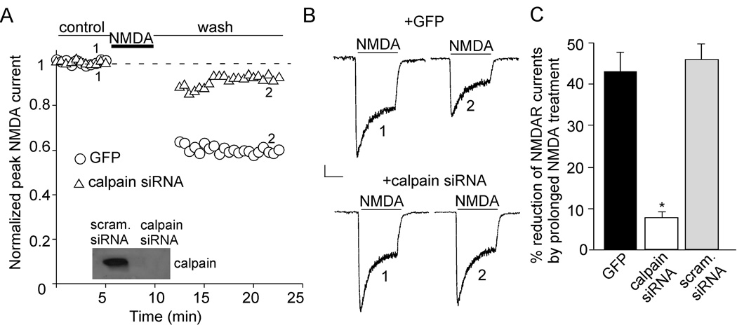
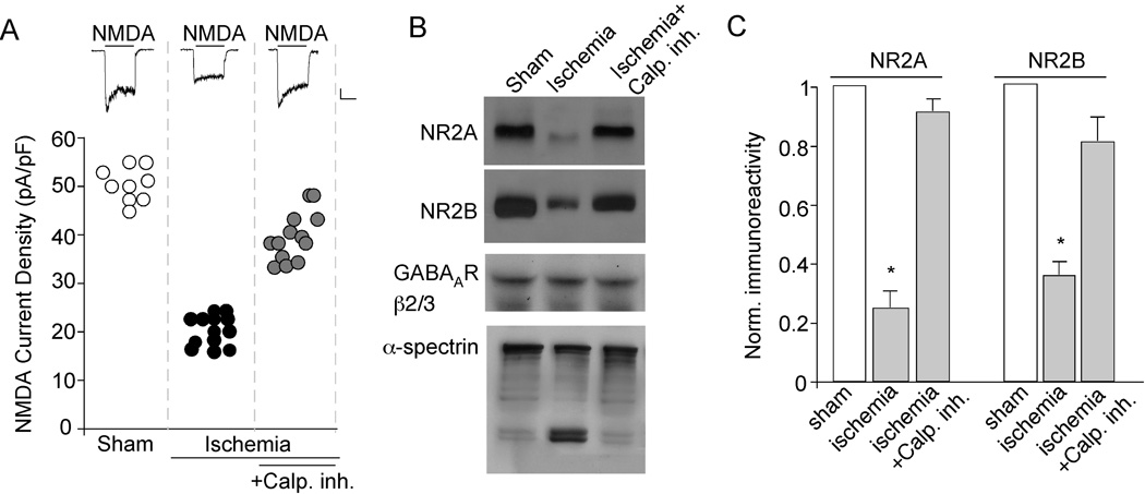
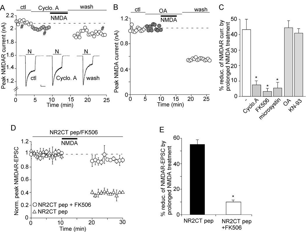
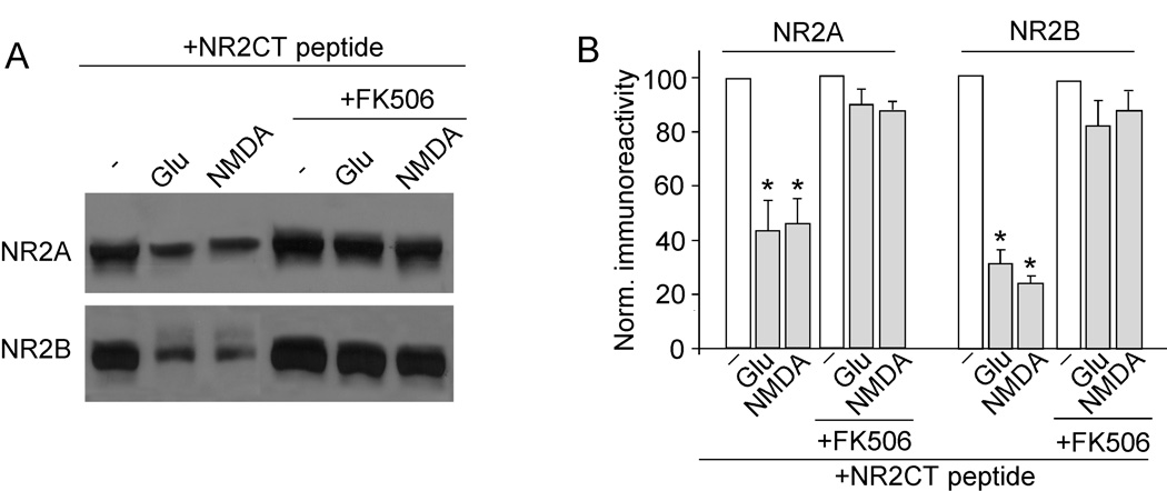
The N-methyl-D-aspartate receptor (NMDAR) is a Ca(2+)-permeable glutamate receptor mediating many neuronal functions under normal and pathological conditions. Ca(2+) influx via NMDARs activates diverse intracellular targets, including Ca(2+)-dependent protease calpain. Biochemical studies suggest that NR2A and NR2B subunits of NMDARs are substrates of calpain. Our physiological data showed that calpain, activated by prolonged NMDA treatment (100 microM, 5 min) of cultured cortical neurons, irreversibly decreased the whole-cell currents mediated by extrasynaptic NMDARs. Animals exposed to transient forebrain ischemia, a condition that activates calpain, exhibited the reduced NMDAR current density and the lower full-length NR2A/B level in a calpain-dependent manner. Disruption of the association between NMDARs and the scaffolding protein postsynaptic density (PSD)-95 facilitated the calpain regulation of synaptic NMDAR responses and NR2 cleavage in cortical slices, whereas inhibition of calcineurin activity blocked the calpain effect on NMDAR currents and NR2 cleavage. Calpain-cleaved NR2B subunits were removed from the cell surface. Moreover, cell viability assays showed that calpain, by targeting NMDARs, provided a negative feedback to dampen neuronal excitability in excitotoxic conditions. These data suggest that calpain activation suppresses NMDAR function via proteolytic cleavage of NR2 subunits in vitro and in vivo, and the susceptibility of NMDARs to calpain cleavage is controlled by PSD-95 and calcineurin.


Figures

Figure 1
Figure 1. Prolonged NMDA treatment reduces NMDAR-mediated currents via calpain activation in cultured cortical pyramidal neurons
A, Plot of normalized peak NMDAR currents (INMDA) with a prolonged NMDA application (100 µM, 5 min) in GFP-positive neurons transfected with or without calpain siRNA. INMDA was elicited by NMDA pulses (100 µM, 2 sec). Inset: Immunoblot of calpain regulatory subunit in cultured cortical neurons transfected with calpain siRNA or a scrambled siRNA. B, Representative current traces taken from the records used to construct A (at time points denoted by numbers). Scale bars: 100 pA, 1 sec. C, Cumulative data (mean ± SEM) showing the percentage reduction of NMDAR currents by prolonged NMDA treatment in GFP-positive cells transfected without or with calpain siRNA or a scrambled siRNA. *: p < 0.001. ANOVA.
Figure 2
Figure 2. Transient forebrain ischemia reduces NMDAR current density via calpain activation
A, Scatterplot depicting the NMDAR current density in cortical pyramidal neurons acutely dissociated from sham-operated vs. ischemic animals injected with or without calpain inhibitor III (3 mg/kg). Inset: Representative current traces (evoked by 100 µM NMDA) taken from various experimental groups. Scale bars: 100 pA, 1 sec. B, Western blot analysis of NR2A and NR2B (detected with C-terminal antibodies), GABAAR β2/3 and α-spectrin in cortical slices from sham-operated vs. ischemic animals with or without calpain inhibitor III. C, Quantitive analysis (means ± SEM) showing the levels of NR2A and NR2B in cortical slices from sham-operated vs. ischemic animals with or without calpain inhibitor III. *: p < 0.001, ANOVA.
Figure 3
Figure 3. Disruption of the PSD-95/NMDAR interaction facilitates calpain regulation of NMDAR-EPSC
A, Effect of TAT-NR2CT peptide (25 µM, 30 min treatment) on the interaction of NMDA receptors with PSD-95. A scrambled peptide was used as a control. After treatment, cell lysates from cortical slices were immunoprecipitated with anti-PSD-95 and Western blotted with anti-NR2A, anti-NR2B or PSD-95. B, Bar graphs showing levels of NR2A and NR2B bound to PSD-95 in the absence or presence of TAT-NR2CT peptide or a scrambled peptide. *: p < 0.001, ANOVA. C, Plot of normalized peak NMDAR-EPSC showing the effect of prolonged NMDA treatment (100 µM, 5 min) in neurons dialyzed with or without TAT-NR2CT peptide (10 µM) in the absence or presence of calpain inhibitor ALLN (25 µM). Inset: Representative traces (average of 3 trials) taken from the recordings used to construct C (at time points denoted by numbers). Scale bars: 100 pA, 100 ms. D, Cumulative data (mean ± SEM) summarizing the percentage reduction of NMDAR-EPSC amplitude by prolonged NMDA treatment under different conditions. *: p < 0.001, ANOVA.
Figure 4
Figure 4. Calpain cleavage of NR2A and NR2B subunits requires dissociation with PSD-95, and cleaved NMDARs are removed from the surface
A, Immunoblots of NR2A, NR2B and NR1 subunits (detected with C-terminal antibodies) in lysates of cortical slices following prolonged glutamate (500 µM, 5 min) or NMDA (100 µM, 5 min) treatment in the absence or presence of TAT-NR2CT peptide (10 µM, added 30 min before glutamate/NMDA treatment). Cells were collected after 10 min of washing following glutamate/NMDA treatment. B, Quantitive analysis (means ± SEM) showing the levels of NR2A and NR2B with glutamate or NMDA treatment in cortical slices in the absence or presence of TAT-NR2CT peptide. *: p < 0.001, ANOVA. C, Immunoblots of the total and surface NR2B subunit in lysates of cortical slices treated with glutamate (500 µM, 10 min) in the absence or presence of TAT-NR2CT peptide (10 µM, 30 min). Cells were collected after 10 min of washing. NR2B was detected with an antibody against the extracellular N-terminal, which labeled both the cleaved and uncleaved subunit. The total and surface GABAAR β subunits were also measured as a control. Similar results were obtained from four experiments. D, Quantitive analysis (means ± SEM) showing the level of cleaved NR2B fragment (115KDa) in total lysate or cell surface with glutamate treatment in cortical slices in the absence or presence of TAT-NR2CT peptide. #: p < 0.05, *: p < 0.01, ANOVA.
Figure 5
Figure 5. Disruption of NR2/PSD-95 reduces NMDA-induced cell death via calpain activation
A–E, Immunocytochemical images showing the co-staining of MAP2 (green) and propidium iodide (PI, a nuclear marker, red). Cortical cultures (DIV 14) were treated with NMDA (100 µM, 10 min) in the absence or presence of calpain inhibitor III (20 µM, added 30 min prior to NMDA treatment) or/and TAT-NR2CT peptide (10 µM, added 30 min prior to NMDA treatment). Neurons were collected 24 hrs later for staining. Surviving neurons are positive for MAP2 staining. Apoptotic neuronal death was indicated by shrunk and condensed nucleus in PI staining. F, G, Cumulative data (mean ± SEM) showing the percentage of surviving neurons (F) or neuronal death (G) under various treatments. Data were summarized from 5–7 experiments with each condition. *: p < 0.001, ANOVA.
Figure 6
Figure 6. Inhibition of calcineurin attenuates the effect of prolonged NMDA treatment on NMDAR currents
A, B, Plot of INMDA with a prolonged NMDA treatment (100 µM, 5 min) in the presence of cyclosporine A (20 µM, calcineurin inhibitor, A) or okadaic acid (1 µM, PP1/2A inhibitor, B) in acutely dissociated cortical pyramidal neurons. C, Cumulative data (mean ± SEM) summarizing the percentage reduction of NMDAR currents by prolonged NMDA treatment with different agents that affect calcineurin, PP1/2A or CaMKII. *: p < 0.001, ANOVA. D, Plot of normalized peak NMDAR-EPSC with a prolonged NMDA treatment (100 µM, 5 min) in cells injected with TAT-NR2CT peptide (10 µM) in the presence or absence of FK506 (5 µM). Inset: Representative traces taken from the recordings at indicated times. Scale bars: 100 pA, 100 ms. E, Cumulative data (mean ± SEM) showing the percentage reduction of NMDAR-EPSC by prolonged NMDA treatment in the presence of TAT-NR2CT peptide with or without FK506. *: p < 0.001, ANOVA.
Figure 7
Figure 7. Calpain cleavage of NR2A and NR2B subunits requires calcineurin activity
A, Western blot analysis of NR2A and NR2B (detected with C-terminal antibodies) in lysates of cortical slices following glutamate (500 µM, 5 min) or NMDA (100 µM, 5 min) treatment in the absence or presence of FK506 (5 µM, added 10 min before glutamate/NMDA treatment). Note that slices were incubated with TAT-NR2CT peptide (10 µM) throughout the experiments. Slices were collected after 10 min of washing. B, Quantitive analysis (mean ± SEM) showing the levels of NR2A and NR2B with glutamate or NMDA treatment in cortical slices in the absence or presence of FK506. *: p < 0.001, ANOVA.

References

    1. Aarts M, Liu Y, Liu L, Besshoh S, Arundine M, Gurd JW, Wang YT, Salter MW, Tymianski M. Treatment of ischemic brain damage by perturbing NMDA receptor- PSD-95 protein interactions. Science. 2002;298:846–850. - PubMed
    1. Abe K, Takeichi M. NMDA-receptor activation induces calpain-mediated beta-catenin cleavages for triggering gene expression. Neuron. 2007;53:387–397. - PubMed
    1. Adamec E, Beermann ML, Nixon RA. Calpain I activation in rat hippocampal neurons in culture is NMDA receptor selective and not essential for excitotoxic cell death. Brain Res Mol Brain Res. 1998;54:35–48. - PubMed
    1. Amadoro G, Ciotti MT, Costanzi M, Cestari V, Calissano P, Canu N. NMDA receptor mediates tau-induced neurotoxicity by calpain and ERK/MAPK activation. Proc Natl Acad Sci U S A. 2006;103:2892–2897. - PMC - PubMed
    1. Ankarcrona M, Dypbukt JM, Bonfoco E, Zhivotovsky B, Orrenius S, Lipton SA, Nicotera P. Glutamate-induced neuronal death: a succession of necrosis or apoptosis depending on mitochondrial function. Neuron. 1995;15:961–973. - PubMed

Publication types

LinkOut - more resources