Review
. 2008 Jul;233(7):779-91.
doi: 10.3181/0711-MR-308. Epub 2008 Apr 29.

An unexpected role for ion channels in brain tumor metastasis

Affiliations
Review

An unexpected role for ion channels in brain tumor metastasis

Harald Sontheimer. Exp Biol Med (Maywood). 2008 Jul.

Abstract

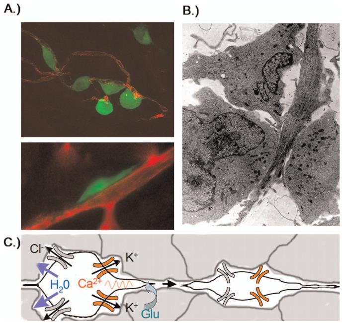
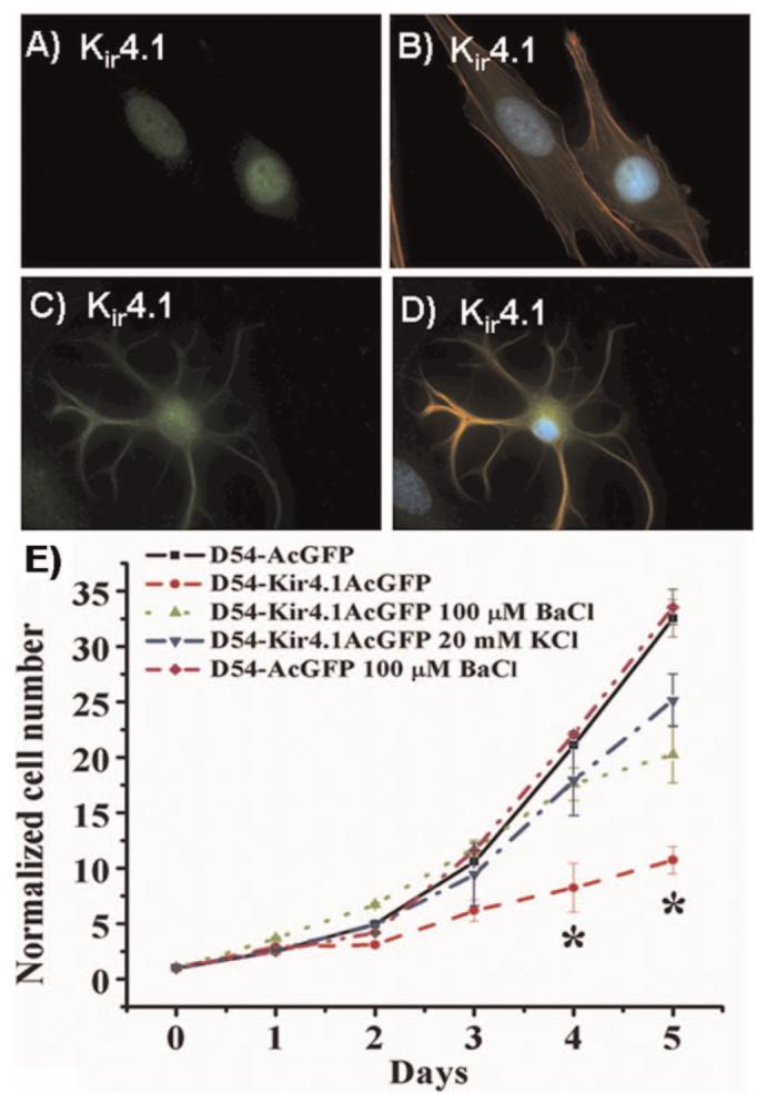
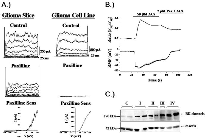
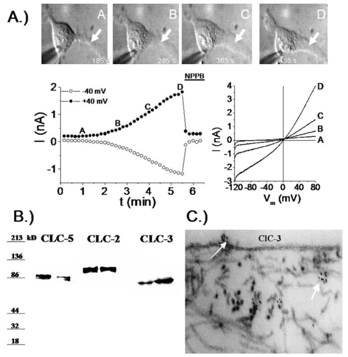
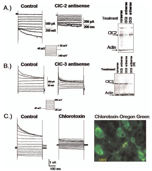
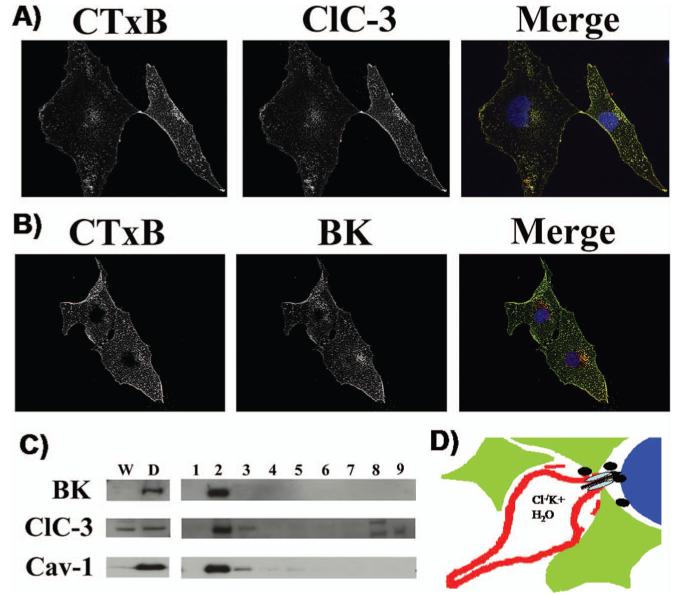
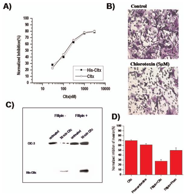
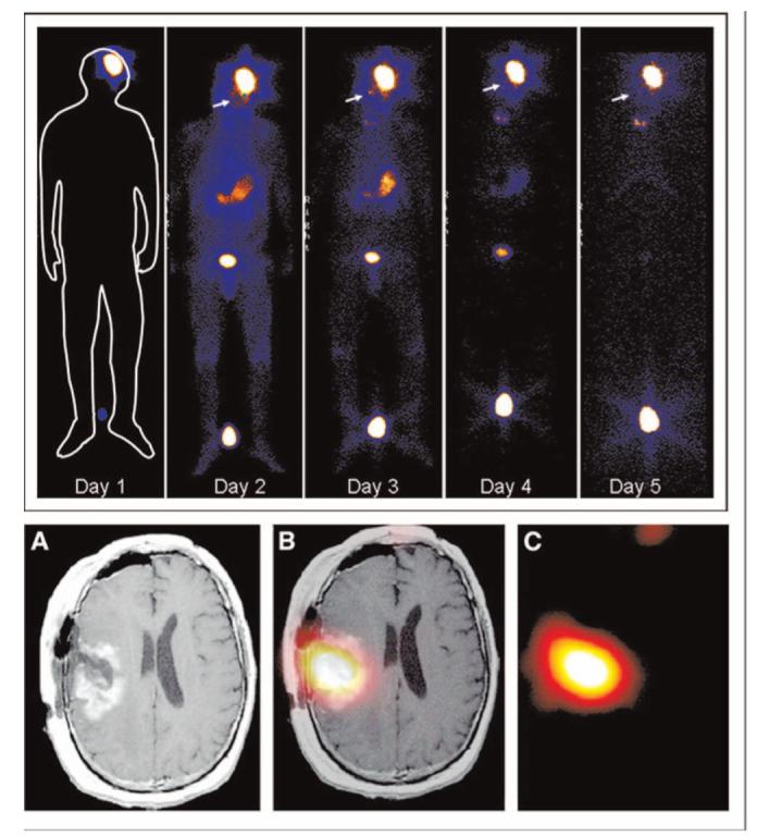
Over the past two decades it has become apparent that essentially all living cells express voltage-activated ion channels. While the role of ion channels for electrical signaling between excitable cells is well known, their function in non-excitable cells is somewhat enigmatic. Research on cancer cells suggests that certain ion channels, K+ channels in particular, may be involved in aberrant tumor growth and channel inhibitors often lead to growth arrest. An unsuspected role for K+ and Cl(-) channels has now been documented for primary brain tumors, glioma, where the concerted activity of these channels promotes cell invasion and the formation of brain metastasis. Specifically, Ca2+-activated K+ (BK) channels colocalize with ClC-3 Cl(-) channels to the invading processes of these tumor cells. Upon a rise in intracellular Ca2+, these channels activate and release K+ and Cl(-) ions together with obligated water causing a rapid shrinkage of the leading process. This in turn facilitates the invasion of the cell into the narrow and tortuous extracellular brain spaces. The NKCC1 cotransporter accumulates intracellular Cl(-) to unusually high concentrations, thereby establishing an outward directed gradient for Cl(-) ions. This allows glioma cells to utilize Cl(-) as an osmotically active anion during invasion. Importantly, the inhibition of Cl(-) channels retards cell volume changes, and, in turn, compromises tumor cell invasion. These findings have led to the clinical evaluation of a Cl(-) channel blocking peptide, chlorotoxin, in patients with malignant glioma. Data from this clinical trial shows remarkable tumor selectivity for chlorotoxin. The experimental therapeutic was well tolerated and is now evaluated in a multi-center phase II clinical trial. A similar role for Cl(-) and K+ channels is suspected in other metastatic cancers, and lessons learned from studies of gliomas may pave the way towards the development of novel therapeutics targeting ion channels.


Figures

Figure 1
Figure 1
Invading glioma cells in situ. (A) Confocal images of invading D54MG cell stably expressing EGFP adhere to blood vessels. Cells often show an elongated wedge-shaped appearance, A, bottom. (B) The elongated shape is quite apparent in an electronmicrograph that captured an invading glioma cell extending between normal brain cells. (C) Cell shrinkage requires efflux of water which is energetically driven out of the cell through the concerted secretion of Cl- and K+ through ion channels. Glutamate is shown as possible motogenic stimulus acting via AMPA-R to raise intracellular Ca2+ which may in turn activate Ca2+-activated BK channels. A color version of this figure is available in the online version of the journal.
Figure 2
Figure 2
Glioma cells sequester Kir4.1 channels to the nucleus as they otherwise suppress cell growth. (A-B) Immunolabeling of cultured STTG1 glioma cells and spinal cord astrocytes (C-D) with Kir4.1 antibodies (green), phalloidin (red) and DAPI (blue). While both cell types show Kir4.1 channel expression, only astrocytes have them on the plasma membrane. (E) Cell growth over a 5-days period comparing wild type D54-MG glioma cells to cell expressing GFP tagged Kir4.1 and vector only D54-MG cells. Kir expression inhibited growth unless channel activity was blocked with Ba2+ or cells were depolarized by high K+. [(A-D) reproduced with permission from (17); (E) reproduced with permission from (22).] A color version of this figure is available in the online version of the journal.
Figure 3
Figure 3
Glioma cells express Ca2+-activated K+ channels. (A) Representative recordings from glioma cells in a brain section or isolated cell. Outward currents were sensitive to paxilline, a specific inhibitor of Ca2+-activated K+ channels. Block was incomplete in brain slices because drug perfusion was limited in tissue. (B) Application of Acetylcholin (Ach) to a cultured glioma cell recorded using amphotericin perforated patch, hence without disturbing intracellular Ca2+ caused a rapid increase in Ca2+ determined by Fura-II recording (top) associated with a large hyperpolarizing voltage shift indicating activation of BK channels. (C) Two biopsies each from patients with gliomas ranging in malignancy grade from WHO-I-IV show an increase in BK channel protein with increasing malignancy on Western blot. [(C) reproduced with permission from (23); (B) reproduced with permission from (25).]
Figure 4
Figure 4
Expression and function of Cl- channels in glioma cells. (A) Extension of a process in a cultured glioma cells correlates with the progressive development of a NPPB sensitive outwardly rectifying Cl- current. (B) Western blots from 2 biopsies each probed for ClC-2, ClC-3 and ClC-5 show prominent expression of all 3 members of this channel family. (C) Using immuno-gold electron microscopy clusters of ClC-3 channels can be seen on the membrane surface of glioma cells. [(A) reproduced with permission from (18).]
Figure 5
Figure 5
Glioma cells express functional ClC-2 and ClC-3 channels. Using specific antisense oligonucleotides to ClC-2 (A) and ClC-3 (B) it was possible to identify currents attributable to these channels respectively. The Western blots illustrate effective reduction in protein after antisense treatment and show specificity. (C) Currents with a biophysical signature of ClC-3 are inhibited by 1 μM Chlorotoxin. OregonGreen-conjugated Chlorotoxin was used to label an acute biopsy section from a glioblastoma patient (right). [(A) reproduced with permission from (27); (C) reproduced with permission from (34).] A color version of this figure is available in the online version of the journal.
Figure 6
Figure 6
ClC-3 and BK channels colocalize to lipid rafts. D54-MG glioma cells were immunostained with the beta subunit of Cholera toxin (CTxB) to identify lipid rafts. (A) ClC-3 antibodies labeled the same domains as indicated in the merged image. (B) Similarly, BK antibodies localize to lipid rafts suggesting a co-localization of ClC-3 and BK channels to lipid rafts. (C) Biochemical isolation of lipid rafts indeed demonstrates that BK and ClC-3 channels are enriched in the lipid raft domain that also contains the structural protein caveolin-1 found in caveolar rafts. (D) We hypothesize that Cl- and K+ channels colocalize to invadipodia to facilitate cell shrinkage and that lipid rafts localize them in close proximity. [Reproduced in modified form with permission from (34).] A color version of this figure is available in the online version of the journal.
Figure 7
Figure 7
Inhibition of Cl- channels with Chlorotoxin retards cell invasion. (A) Dose-dependent inhibition of cell invasion using Transwell migration/invasion assays shows effective inhibition with natural (Cltx) and recombinant (His-Cltx) chlorotoxin. (B) Underside of Transwell filters shows a significant decrease in migrated cells in the presence of 5 μM Cltx. (C) Cltx interferes with trafficking of ClC-3 channels. Isolation of membrane associated ClC-3 channels was achieved after cell surface biotinylation. 30 min incubation with Cltx causes a significant loss of ClC-3 from the plasma membrane presumably by internalization of channels into caveolar vesicles. Disruption of caveoli with the sterol binding drug Filipin eliminated the effect of Cltx and retained all ClC-3 protein on the cell surface. (D) The inhibitory effects of Cltx on cell migration/invasion were also overcome by treatment of cells with Filipin which would prevent internalization of ClC-3 channels. [(B) reproduced with permission from (32); (A, C, D) reproduced with permission from (33).] A color version of this figure is available in the online version of the journal.
Figure 8
Figure 8
Chlorotoxin in clinical trial: (top) Whole body gamma scan shows specific localization and retention of 131I-ClTx to a glioblastoma in a patient from a recent Phase I study [reproduced with permission from (38)]. Hot spot between the subject’s feet is a radiation standard. (Bottom) Axial view of T1-Wc (A), coregistered (B), and SPECT (day 8) images of intrathecal-delivered peptide. [Reproduced with permission from (39).] A color version of this figure is available in the online version of the journal.

References

    1. Maher EA, Furnari FB, Bachoo RM, Rowitch DH, Louis DN, Cavenee WK, DePinho RA. Malignant glioma: genetics and biology of a grave matter. Genes Dev. 2001;15:1311–1333. - PubMed
    1. Schwartzbaum JA, Fisher JL, Aldape KD, Wrensch M. Epidemiology and molecular pathology of glioma. Nat Clin Pract Neurol. 2006;2:494–503. - PubMed
    1. Kleihues P, Soylemezoglu F, Schaueble B, Scheithauer BW, Burger PC. Histopathology, classification and grading of gliomas. Glia. 1995;15:211–221. - PubMed
    1. Kleihues P, Burger PC, Scheithauer BW. The new who classification of brain tumours. Brain Pathol. 1993;3:255–268. - PubMed
    1. Jemal A, Siegel R, Ward E, Murray T, Xu J, Smigal C, Thun MJ. Cancer statistics, 2006. CA Cancer J Clin. 2006;56:106–130. - PubMed

Publication types

MeSH terms