

Neurogenesis after primary intracerebral hemorrhage in adult human brain
- PMID: 18446166
- PMCID: PMC2575114
- DOI: 10.1038/jcbfm.2008.37
Neurogenesis after primary intracerebral hemorrhage in adult human brain
Abstract
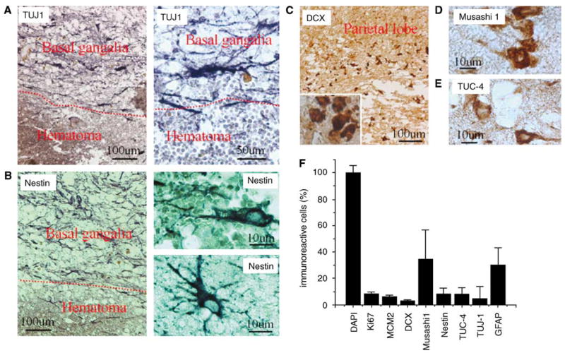
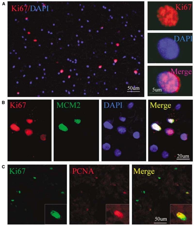
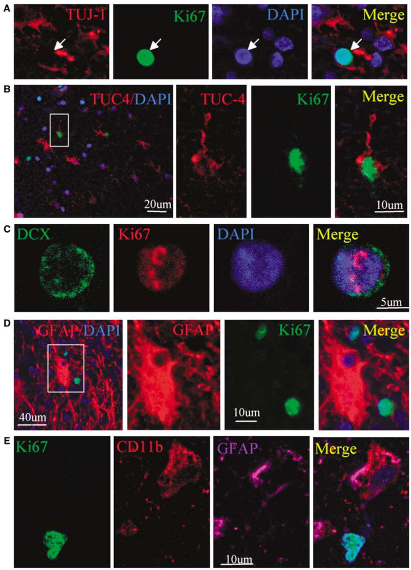
Neurogenesis occurs in discrete regions of normal brains of adult mammals including humans, and is induced in response to brain injury and neurodegenerative disease. Whether intracerebral hemorrhage can also induce neurogenesis in human brain is unknown. Specimens were obtained from patients with primary intracerebral hemorrhage undergoing surgical evacuation of an intracerebral hematoma, and evaluated by two-photon laser scanning confocal microscopy. We found that neural stem/progenitor cell-specific protein markers were expressed in cells located in the perihematomal regions of the basal ganglia and parietal lobe of the adult human brain after primary intracerebral hemorrhage (n=5). Cells in this region also expressed cell proliferation markers, which colocalized to the same cells that expressed neural stem/progenitor cell-specific proteins. Our data suggest that intracerebral hemorrhage induces neurogenesis in the adult human brain.
Figures


Similar articles
-
Activation of endogenous neural stem cells in the adult human brain following subarachnoid hemorrhage.J Neurosci Res. 2007 Jun;85(8):1647-55. doi: 10.1002/jnr.21303. J Neurosci Res. 2007. PMID: 17455304
-
Endogenous neurogenesis after intracerebral hemorrhage.Histol Histopathol. 2012 Mar;27(3):303-15. doi: 10.14670/HH-27.303. Histol Histopathol. 2012. PMID: 22237708
-
Predictors of hematoma volume in deep and lobar supratentorial intracerebral hemorrhage.JAMA Neurol. 2013 Aug;70(8):988-94. doi: 10.1001/jamaneurol.2013.98. JAMA Neurol. 2013. PMID: 23733000 Free PMC article.
-
Progressive bleeding in spontaneous thalamic hemorrhage.Neurologia. 1994 Oct;9(8):364-7. Neurologia. 1994. PMID: 7803055 Review.
-
Progenitor cells and adult neurogenesis in neurodegenerative diseases and injuries of the basal ganglia.Clin Exp Pharmacol Physiol. 2007 May-Jun;34(5-6):528-32. doi: 10.1111/j.1440-1681.2007.04609.x. Clin Exp Pharmacol Physiol. 2007. PMID: 17439428 Review.
Cited by
-
Involvement of Notch1 signaling in neurogenesis in the subventricular zone of normal and ischemic rat brain in vivo.J Cereb Blood Flow Metab. 2009 Oct;29(10):1644-54. doi: 10.1038/jcbfm.2009.83. Epub 2009 Jun 17. J Cereb Blood Flow Metab. 2009. PMID: 19536070 Free PMC article.
-
Role of low-level laser therapy in neurorehabilitation.PM R. 2010 Dec;2(12 Suppl 2):S292-305. doi: 10.1016/j.pmrj.2010.10.013. PM R. 2010. PMID: 21172691 Free PMC article. Review.
-
Role of neural precursor cells in promoting repair following stroke.Acta Pharmacol Sin. 2013 Jan;34(1):78-90. doi: 10.1038/aps.2012.107. Epub 2012 Oct 15. Acta Pharmacol Sin. 2013. PMID: 23064725 Free PMC article. Review.
-
White matter repair and treatment strategy after intracerebral hemorrhage.CNS Neurosci Ther. 2019 Oct;25(10):1113-1125. doi: 10.1111/cns.13226. Epub 2019 Oct 2. CNS Neurosci Ther. 2019. PMID: 31578825 Free PMC article. Review.
-
Acquired Brain Injury in Adults: A Review of Pathophysiology, Recovery, and Rehabilitation.Perspect ASHA Spec Interest Groups. 2021 Aug;6(4):714-727. doi: 10.1044/2021_persp-21-00013. Epub 2021 Aug 20. Perspect ASHA Spec Interest Groups. 2021. PMID: 34746412 Free PMC article.
References
-
- Alvarez-Buylla A, Lois C. Neuronal stem cells in the brain of adult vertebrates. Stem Cells. 1995;13:263–72. - PubMed
-
- Arvidsson A, Collin T, Kirik D, Kokaia Z, Lindvall O. Neuronal replacement from endogenous precursors in the adult brain after stroke. Nat Med. 2002;8:963–70. - PubMed
-
- Broderick JP, Adams HP, Jr, Barsan W, Feinberg W, Feldmann E, Grotta J, Kase C, Krieger D, Mayberg M, Tilley B, Zabramski JM, Zuccarello M. Guidelines for the management of spontaneous intracerebral hemorrhage: a statement for healthcare professionals from a Special Writing Group of the Stroke Council, American Heart Association. Stroke. 1999;30:905–15. - PubMed
-
- Eriksson PS, Perfilieva E, Bjork-Eriksson T, Alborn AM, Nordborg C, Peterson DA, Gage FH. Neurogenesis in the adult human hippocampus. Nat Med. 1998;4:1313–7. - PubMed
-
- Fanarraga ML, Avila J, Zabala JC. Expression of unphosphorylated class III beta-tubulin isotype in neuroepithelial cells demonstrates neuroblast commitment and differentiation. Eur J Neurosci. 1999;11:516–27. - PubMed
Publication types
MeSH terms
Grants and funding
LinkOut - more resources
Full Text Sources
Other Literature Sources
Medical
