

A new concept for medical imaging centered on cellular phone technology
- PMID: 18446199
- PMCID: PMC2312332
- DOI: 10.1371/journal.pone.0002075
A new concept for medical imaging centered on cellular phone technology
Abstract
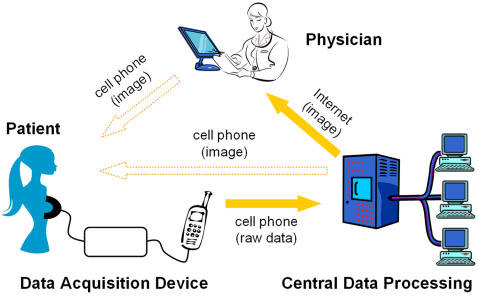
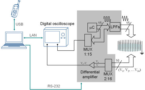
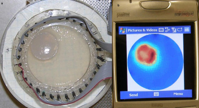
According to World Health Organization reports, some three quarters of the world population does not have access to medical imaging. In addition, in developing countries over 50% of medical equipment that is available is not being used because it is too sophisticated or in disrepair or because the health personnel are not trained to use it. The goal of this study is to introduce and demonstrate the feasibility of a new concept in medical imaging that is centered on cellular phone technology and which may provide a solution to medical imaging in underserved areas. The new system replaces the conventional stand-alone medical imaging device with a new medical imaging system made of two independent components connected through cellular phone technology. The independent units are: a) a data acquisition device (DAD) at a remote patient site that is simple, with limited controls and no image display capability and b) an advanced image reconstruction and hardware control multiserver unit at a central site. The cellular phone technology transmits unprocessed raw data from the patient site DAD and receives and displays the processed image from the central site. (This is different from conventional telemedicine where the image reconstruction and control is at the patient site and telecommunication is used to transmit processed images from the patient site). The primary goal of this study is to demonstrate that the cellular phone technology can function in the proposed mode. The feasibility of the concept is demonstrated using a new frequency division multiplexing electrical impedance tomography system, which we have developed for dynamic medical imaging, as the medical imaging modality. The system is used to image through a cellular phone a simulation of breast cancer tumors in a medical imaging diagnostic mode and to image minimally invasive tissue ablation with irreversible electroporation in a medical imaging interventional mode.
Conflict of interest statement
Figures

References
-
- WHO report. Essential Health Technologies Strategy 2004–2007. World Health Organization. 2003 http://www.who.int/eht/en/EHT_strategy_2004-2007.pdf.
-
- WHO report, Health Technologies- the backbone of Health Services. World Health Organization. http://www.who.int/eht/en/Backbone.pdf.
-
- WHO report, Essential Diagnostic Imaging. World Health Organization. http://www.who.int/eht/en/DiagnosticImaging.pdf.
-
- WHO report, About diagnostic imaging. World Health Organization. http://www.who.int/diagnostic_imaging/about/en/
-
- WHO report, Diagnostic imaging. World Health Organization. http://www.who.int/diagnostic_imaging/en/
Publication types
MeSH terms
Grants and funding
LinkOut - more resources
Full Text Sources
Other Literature Sources
Medical
