. 2008 Apr 30;3(4):e2084.
doi: 10.1371/journal.pone.0002084.

Muscle dystroglycan organizes the postsynapse and regulates presynaptic neurotransmitter release at the Drosophila neuromuscular junction

Affiliations

Muscle dystroglycan organizes the postsynapse and regulates presynaptic neurotransmitter release at the Drosophila neuromuscular junction

Laurent Bogdanik et al. PLoS One. .

Abstract

Background: The Dystrophin-glycoprotein complex (DGC) comprises dystrophin, dystroglycan, sarcoglycan, dystrobrevin and syntrophin subunits. In muscle fibers, it is thought to provide an essential mechanical link between the intracellular cytoskeleton and the extracellular matrix and to protect the sarcolemma during muscle contraction. Mutations affecting the DGC cause muscular dystrophies. Most members of the DGC are also concentrated at the neuromuscular junction (NMJ), where their deficiency is often associated with NMJ structural defects. Hence, synaptic dysfunction may also intervene in the pathology of dystrophic muscles. Dystroglycan is a central component of the DGC because it establishes a link between the extracellular matrix and Dystrophin. In this study, we focused on the synaptic role of Dystroglycan (Dg) in Drosophila.

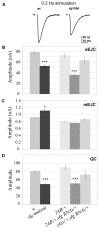
Methodology/principal findings: We show that Dg was concentrated postsynaptically at the glutamatergic NMJ, where, like in vertebrates, it controls the concentration of synaptic Laminin and Dystrophin homologues. We also found that synaptic Dg controlled the amount of postsynaptic 4.1 protein Coracle and alpha-Spectrin, as well as the relative subunit composition of glutamate receptors. In addition, both Dystrophin and Coracle were required for normal Dg concentration at the synapse. In electrophysiological recordings, loss of postsynaptic Dg did not affect postsynaptic response, but, surprisingly, led to a decrease in glutamate release from the presynaptic site.

Conclusion/significance: Altogether, our study illustrates a conservation of DGC composition and interactions between Drosophila and vertebrates at the synapse, highlights new proteins associated with this complex and suggests an unsuspected trans-synaptic function of Dg.


Conflict of interest statement

Competing Interests: The authors have declared that no competing interests exist.

Figures

Figure 1
Figure 1. Localization of Dg at the NMJ.
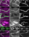
A-D) Dg (A2, B2, C2, D2) and HRP (A3, B3, C3, D3) immunoreactivity at the NMJ of muscle 4. Merge of both images with Dg in magenta and HRP in green (A1, B1, C1, D1). The LG5 antibody was used to analyze Dg localization. (A) In wild-type (ywCS) flies, Dg concentration is clearly visible at type Ib boutons. Dg immunoreactivity is much larger than motoneuron-specific HRP immunoreactivity, indicating that Dg is present in the muscle cell, i.e. postsynaptically. (B) In larvae expressing dg-directed RNAi in the muscle using the 24B Gal4 driver, the NMJ LG5 staining was decreased in intensity. (C) In larvae mutant for dg (dge01554/dg323), the NMJ LG5 staining was more strongly affected. (D) In larvae overexpressing Dg-C isoform in the muscles using the 24B Gal4 driver, a clear increase in LG5 immunoreactivity was observed (laser intensity was decreased to avoid too much signal saturation in this genotype) both at the NMJ, and also in distant patches (arrows). (E) Two serial sections (every 0.7 µm) of a synaptic varicosity of a larva expressing a Dg-C-GFP construct in the muscle cells (with the 24B Gal4 driver). GFP fluorescence (E2) is present in the subsynaptic reticulum (SSR) (arrow) and is partially excluded from the sensus-stricto synapses (arrowhead). Synapses are labelled with the active zone marker Bruchpilot (BRP)(E3). Merge of both images with Dg-GFP in green and BRP in magenta is shown in E1. (F) Dg staining with anti-Dgex8 antibody, specific of the Dg-C isoform (F2) and HRP (F3). The Dgex8 immunoreactivity is similar to the LG5 immunoreactivity (A2). Again, Dgex8 immunoreactivity is much larger than motoneuron-specific HRP immunoreactivity (F1), indicating that Dg-C protein is present at the postsynapse. In all panels, a muscle 4 NMJ is shown. Scale bar is 10 µm in A–D, 5 µm in E and 10 µm in F.
Figure 2
Figure 2. Laminin localization at the drosophila NMJ varicosities is influenced by dg.
A) Serial sections of two adjacent varicosities (every 0.7 µm) labelled for BRP (green) and Lam (red) to analyze Lam localization. There is no colocalization between the active zone marker BRP and the Lam staining, indicating that Lam is mainly perisynatically localized. B–D) Lower magnification of NMJ stained for Lam (B2,C2,D2) and HRP (B3,C3,D3). The merge image is shown in B1,C1,D1 (Lam in red and HRP in blue). (B) In wild-type (ywCS) larvae, Lam is present both in varicosities and inter-varicosities connectives. (C) In larvae expressing dg-directed RNAi in the muscle using the 24B Gal4 driver (dg-RNAi/+; 24B Gal4/dg-RNAi), the varicosity Lam staining was decreased in intensity, whereas the connective staining remained unchanged. (D) In larvae mutant for dg (dge01554/dg323), the Lam varicosity staining was strongly affected, but not the connective staining. (E) In larvae overexpressing Dg-C isoform with the 24B Gal4 driver, no clear increase in Lam immunoreactivity was observed at the NMJ. However, larger Lam stretches were observed (see arrows in E1, E2 and in the insert showing a double BRP (green), Lam (red) staining of a synaptic bouton in this genotype). In addition Lam patches (arrowhead) appeared in this genotype. N.B. an immunoreactive trachea is visible in E1 and E2 (asterisk). In all panels, a muscle 4 NMJ is shown. Scale bar is 10 µm.
Figure 3
Figure 3. Dg controls synaptic Dys localization.
Triple staining for HRP (blue), Dys (red) and Dlg (green) in control (ywCS) (A), dg mutant (B) and Dg-C overexpressing (C) larvae. Merge images are shown in A1, B1, C1. Single channel stainings are shown in A2, B2, C2 for HRP, A3, B3, C3 for Dys and A4, B4, C4 for Dlg. In dge01554/dg323 mutant larvae, the Dys staining (B3) is almost absent compared to wild-type larvae (A3), although the Dlg staining is still present (compare A4 and B4). When Dg-C is overexpressed with the 24B Gal4 driver, Dys staining is strongly enhanced around HRP positive boutons, and also in the entire muscle cell (compare C3 and A3). In all panels, a muscle 4 NMJ is shown. Scale bar is 10 µm.
Figure 4
Figure 4. Dys controls Dystroglycan postsynaptic concentration.
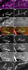
(A–C) Double staining for Fas2 (A green, B), Dys (A magenta, C) on control (1) and UAS-dys-RNAi flies crossed with 24B Gal4 (2). In larvae expressing the dys-RNAi (dys-RNAi/+; 24B Gal4/+), there is almost no Dys immunoreactivity detectable at the NMJ. (D–G) Triple staining for Fas2 (D blue, E), Dystroglycan (D green, F) and Coracle (D red, G) in the same genotypes. Dg postsynaptic labelling is reduced in absence of postsynaptic Dystrophin. Coracle immunoreactivity is also reduced, but to a much lower extent compared to the loss of postsynaptic Dg. (H) alpha-Spectrin immunostaining on the same genotypes. Scale bar is 10 µm. In all panels, a muscle 4 NMJ is shown.
Figure 5
Figure 5. Dg controls postsynaptic Cora concentration.
Double staining for Cora (Magenta) and HRP (green)(1) in (A) control (ywCS) larvae, (B) larvae expressing muscle dg-RNAi (dg-RNAi/+; 24B Gal4/dg-RNAi), (C) dge01554/dg323 mutant larvae and (D) larvae overexpressing Dg-C in muscles (24B-Gal4/UAS-DgC). Arrows indicate patches of Cora protein. Single stainings for Cora and HRP are shown respectively in (2) and (3). In all panels, a muscle 4 NMJ is shown. Scale bar is 10 µm. 3D views of the preparations shown in A1 and C1 are shown in E and F: we look at the NMJ from the inside of the muscle cell. Cora is in magenta and HRP in green.
Figure 6
Figure 6. Dg controls postsynaptic Spectrin concentration and both Cora and Spectrin co-immunoprecipitate with Dg.
Double staining for alpha-Spectrin (Magenta) and HRP (green)(1) in (A) control (ywCS) larvae, (B) larvae expressing muscle dg-RNAi (dg-RNAi/+; 24B Gal4/dg-RNAi), (C) dge01554/dg323 mutant larvae and (D) larvae overexpressing Dg-C isoform in the muscles (24B-Gal4/UAS-DgC). Single stainings for alpha-Spectrin are shown in (2). Scale bar is 10 µm. (E) Co-immunoprecipitation was performed with a polyclonal anti-GFP antibody on protein extracts from flies expressing Dg-C-GFP. S corresponds to the supernatant and P to the pellet. Cora and alpha-Spectrin co-immunoprecipitate with Dg-C-GFP, but not Shaggy (Sgg), a cytoplasmic protein kinase.
Figure 7
Figure 7. Cora controls Spectrin, Dystroglycan, Dystrophin but not Dlg postsynaptic concentration.
(A–C) Triple staining for HRP (A), Cora (B) and Dlg (C) in control (ywCS) (1) and cora14/corak08713 larvae (2). Cora concentration at the NMJ is largely decreased in the cora mutant, whereas synaptic HRP and Dlg stainings show the same signal intensity. This indicates that Cora synaptic loss in the cora mutant is not the direct consequence of a total disruption of NMJ structure. (D–F) Stainings for alpha-Spectrin (D), Dgex8 (E) and Dystrophin (F) in control (1) and cora14/corak08713 larvae (2). (D) In cora mutants, alpha-Spectrin postsynaptic concentration decreases, but does not completely disappear, like in dg mutants. (E) Dystroglycan staining appears thinner in cora mutants. Inserts show higher magnification of a synaptic bouton with presynaptic HRP in magenta and Dg staining in green. The thinner appearance of the NMJ in cora mutants is due to a decrease in postsynaptic Dg staining compared to WT larvae. (F) In cora mutants, Dys labelling is reduced, but is still visible, contrarily to dg mutants, in which it disappears more strongly. Scale bar is 20 µm.
Figure 8
Figure 8. Dg influences glutamate receptor subunit composition.
Triple staining for HRP (1), DGluRIIC (2) and DGluRIIA (3) in control (ywCS) (A) and dge01554/dg323 mutant larvae (B). DGluRIIA immunoreactivity is reduced in the dg mutant whereas DGluRIIC and HRP immunoreactivities are unchanged. (C) Quantification of the ratio of DGluRIIA versus DGluRIIC staining intensities (n = 6 for ywCS and dg mutant, and n = 8 for dg-RNAi/+ and dg-RNAi/+; 24B Gal4/+). (* p<0.5; ** p<0.01). Error bars represent SEM.
Figure 9
Figure 9. NMJ electrophysiological defects due to the loss of dg function.
A) Example traces of evoked postsynaptic currents (EJCs) from NMJs of wild type (w1) and dg mutant dge01554 larvae. B) Mean EJC amplitude for wild type (w1 larvae), dg mutant dge01554, Canton S crossed with 24B Gal4 (24B Gal4/+), dg-RNAi crossed with 24B Gal4 (dg-RNAi/+;24B Gal4/+) and dg-RNAi crossed with elavC155 Gal4 (elav Gal4/+; dg-RNAi/+). B) mini EJC amplitudes and C) quantal content for the same genotypes. The number of measured larvae is indicated within each histogram bar. (* p<0.5; *** p<0.001). Error bars represent SEM.

Similar articles

Cited by

References

    1. Ervasti JM, Campbell KP. A role for the dystrophin-glycoprotein complex as a transmembrane linker between laminin and actin. J Cell Biol. 1993;122:809–823. - PMC - PubMed
    1. Ibraghimov-Beskrovnaya O, Ervasti JM, Leveille CJ, Slaughter CA, Sernett SW, et al. Primary structure of dystrophin-associated glycoproteins linking dystrophin to the extracellular matrix. Nature. 1992;355:696–702. - PubMed
    1. Ibraghimov-Beskrovnaya O, Milatovich A, Ozcelik T, Yang B, Koepnick K, et al. Human dystroglycan: skeletal muscle cDNA, genomic structure, origin of tissue specific isoforms and chromosomal localization. Hum Mol Genet. 1993;2:1651–1657. - PubMed
    1. Ohlendieck K, Campbell KP. Dystrophin-associated proteins are greatly reduced in skeletal muscle from mdx mice. J Cell Biol. 1991;115:1685–1694. - PMC - PubMed
    1. Pasternak C, Wong S, Elson EL. Mechanical function of dystrophin in muscle cells. J Cell Biol. 1995;128:355–361. - PMC - PubMed

Publication types

MeSH terms

LinkOut - more resources