. 2008 Apr 30;3(4):e2119.
doi: 10.1371/journal.pone.0002119.

The Nod-like receptor (NLR) family: a tale of similarities and differences

Affiliations

The Nod-like receptor (NLR) family: a tale of similarities and differences

Martina Proell et al. PLoS One. .

Abstract

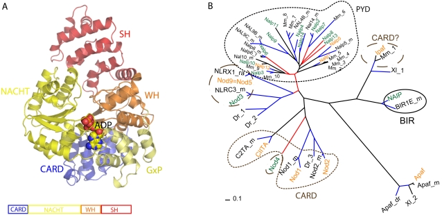
Innate immunity represents an important system with a variety of vital processes at the core of many diseases. In recent years, the central role of the Nod-like receptor (NLR) protein family became increasingly appreciated in innate immune responses. NLRs are classified as part of the signal transduction ATPases with numerous domains (STAND) clade within the AAA+ ATPase family. They typically feature an N-terminal effector domain, a central nucleotide-binding domain (NACHT) and a C-terminal ligand-binding region that is composed of several leucine-rich repeats (LRRs). NLRs are believed to initiate or regulate host defense pathways through formation of signaling platforms that subsequently trigger the activation of inflammatory caspases and NF-kB. Despite their fundamental role in orchestrating key pathways in innate immunity, their mode of action in molecular terms remains largely unknown. Here we present the first comprehensive sequence and structure modeling analysis of NLR proteins, revealing that NLRs possess a domain architecture similar to the apoptotic initiator protein Apaf-1. Apaf-1 performs its cellular function by the formation of a heptameric platform, dubbed apoptosome, ultimately triggering the controlled demise of the affected cell. The mechanism of apoptosome formation by Apaf-1 potentially offers insight into the activation mechanisms of NLR proteins. Multiple sequence alignment analysis and homology modeling revealed Apaf-1-like structural features in most members of the NLR family, suggesting a similar biochemical behaviour in catalytic activity and oligomerization. Evolutionary tree comparisons substantiate the conservation of characteristic functional regions within the NLR family and are in good agreement with domain distributions found in distinct NLRs. Importantly, the analysis of LRR domains reveals surprisingly low conservation levels among putative ligand-binding motifs. The same is true for the effector domains exhibiting distinct interfaces ensuring specific interactions with downstream target proteins. All together these factors suggest specific biological functions for individual NLRs.


Conflict of interest statement

Competing Interests: The authors have declared that no competing interests exist.

Figures

Figure 1
Figure 1. Multiple sequence alignment of NLR NACHT-WH-SH domains and the Apaf-1 NACHT-WH-SH domain.
Degree of conservation is shown as blue shading. The secondary structure of Apaf-1 is shown above the Apaf-1 sequence. Arrows underneath the alignment indicate domain boundaries. Conserved sequence features important for catalytic activity are shown in black boxes. Orange and magenta boxes depict interfaces residues while the orange ones contribute to interactions with the left partner and the magenta residues are thought to interact with the right partner in the oligomer. Green boxes indicate additional motifs as described in the text.
Figure 2
Figure 2
A Apaf-1 structure and domain organization. Apaf-1ΔWD40 (PDB id: 1z6t) is shown in ribbon representation color-coded according to domain boundaries (CARD aa 1–101, NACHT-GxP aa108–365, WH aa366–450, and SH aa451–586. ADP molecule bound to the active site is shown in sphere representation. B Evolutionary tree of human NLR NACHT-WH domain generated for NLR proteins and orthologues. Labels correspond to the accession numbers (Uniprot) Mm_1: Q3TAU8, Mm_2: Q2LKU8, Mm_3: Q2LKU9, Mm_4: Q2LKV8, Mm_5: A1Z198; NALP5_m: NALP5_MOUSE, Mm_6: Q4PLSO, Mm_7: Q66JP4; Mm_8: NAL4C_MOUSE, Mm_9:Q08EE9, Mm_10: Q8R4B8. Dr_1: A5PEZ1; Dr_2: A3KQD4, Dr_3: Q1AMZ9, Xl_1: Q28DS5, Xl_2: Q6GNU6. Black lines indicate a probability of more than 50%, blue lines indicate a 95% confidence in the grouping, and red lines indicate a confidence below the 40%. Green fonts indicate human proteins, black fonts alternative organisms. Mm and _m is Mus musculus (mouse), Dr and _Dr is Danio rerio (fish), Xl and _Xl is Xenopus leavis (frog). Orange fonts indicate an “S” in the Walker A motif in human sequences. Circles and modified ovals over the clades indicate the type of domain present at the N-terminal region of the NACHT domain. PYD is Pyrin domain, CARD is Card domain, Card? indicates CARD-like and BIR are BIR repeats. Apaf-1 is used to root the tree.
Figure 3
Figure 3. Model of the NOD2 nucleotide-binding site with an ADP molecule and conserved sequence motifs Walker A, Walker B, Sensor 1, GxP, and WH-His shown in sticks.
Figure 4
Figure 4
A NLR oligomerization interface: Apaf-1 oligomer modeled on the basis of the NtrC1 heptamer crystal structure. For clarity only three NACHT domains are shown in ribbon representation with an ADP molecule depicted in sticks to highlight the nucleotide-binding site in each domain. Side-chains of residues in the oligomerization interfaces are shown in sticks color-coded according to the alignment in Figure 1. The two interfaces form across the nucleotide-binding site of the NACHT domain including the GxP domain. B Model of NLR activation and inflammasome formation based on the Apaf-1 apoptosome.
Figure 5
Figure 5
A Multiple sequence alignment of NLR and Apaf-1 CARD domains. Acidic key residues participating in the CARD-CARD interface are indicated by red borders. Residues that belong to the basic patch of the CARD-CARD interface are indicated by blue borders. Nod2.1 and Nod2.2 refer to NOD2 CARD domain 1 and 2, respectively. B Multiple sequence alignment of NLR PYRIN domains. Patch of negatively charged residues from ASC2 in helices 1 and 4 and their corresponding residues in the PYRIN domain containing NLR proteins (red box). Patch of positively charged residues from ASC2 in helices 2 and 3 and their corresponding residues in the PYRIN containing NLR proteins (blue box).
Figure 6
Figure 6
A Homology model of NOD2 LRR domain based on the ribonuclease inhibitor (pdb id: 1bnh). Predicted structure of NOD2 LRR domains with conserved residues shown in green and non-conserved residues in white. B Position of loss-of-function mutations shown in darkblue. Mutations found in CD patients are depicted in red. (Core forming residues that do not contribute to the ligand-binding patch are highlighted in lightblue). C NOD2 homology model based on templates Apaf-1 (aa1–581, PDB id: 1z6t, seqID 11%) and ribonuclease inhibitor (aa1–413, PDB entry: 1bnh, seqID 33%) (33). NOD2 model depicted as a cartoon color-coded according to domain structure: blue CARD2 aa95–182, green linker aa183–240, purple NACHT aa241–465, orange winged helix and superhelical domain aa466–734, green-yellow LRR aa735–1040). An ADP molecule bound in the ATPase active site is depicted in sticks. The position of the SNP mutations P268S, R702W, and G908R, are shown as red spheres. The truncation due to mutation L1007fsinsC is colored red.

References

    1. Meylan E, Tschopp J, Karin M. Intracellular pattern recognition receptors in the host response. Nature. 2006;442:39–44. - PubMed
    1. Fritz JH, Ferrero RL, Philpott DJ, Girardin SE. Nod-like proteins in immunity, inflammation and disease. Nat Immunol. 2006;7:1250–1257. - PubMed
    1. Werts C, Girardin SE, Philpott DJ. TIR, CARD and PYRIN: three domains for an antimicrobial triad. Cell Death Differ. 2006;13:798–815. - PubMed
    1. Inohara N, Nunez G. NODs: intracellular proteins involved in inflammation and apoptosis. Nat Rev Immunol. 2003;3:371–382. - PubMed
    1. Mariathasan S, Newton K, Monack DM, Vucic D, French DM, et al. Differential activation of the inflammasome by caspase-1 adaptors ASC and Ipaf. Nature. 2004;430:213–218. - PubMed

Publication types

Substances