. 2008 Apr 30;3(4):e2063.
doi: 10.1371/journal.pone.0002063.

Rescue of salivary gland function after stem cell transplantation in irradiated glands

Affiliations

Rescue of salivary gland function after stem cell transplantation in irradiated glands

Isabelle M A Lombaert et al. PLoS One. .

Abstract

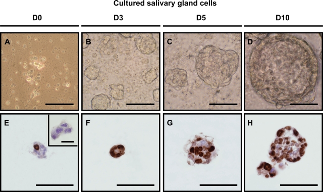
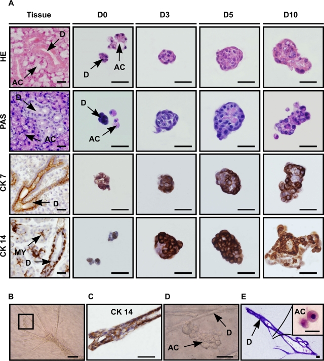
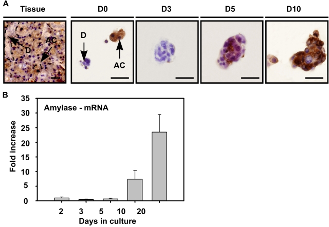
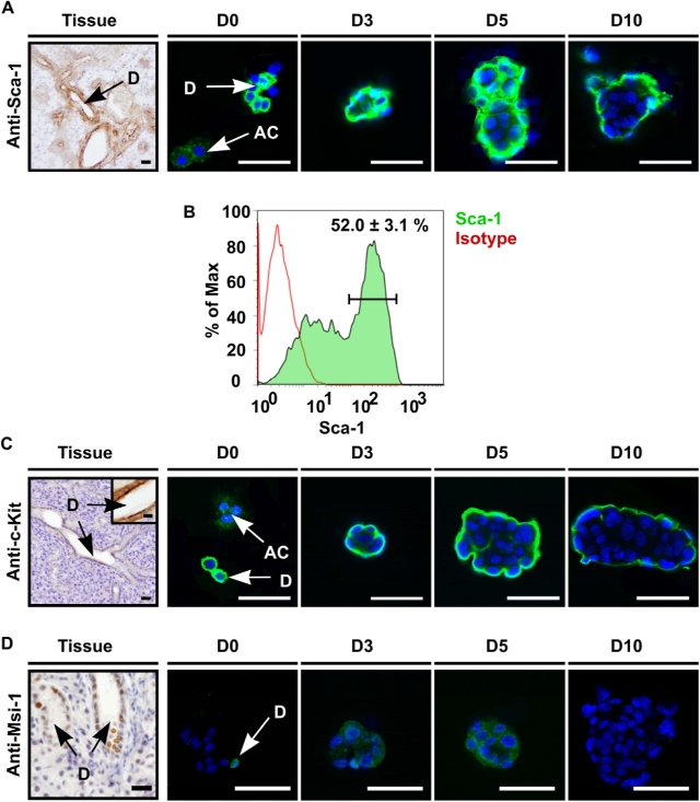
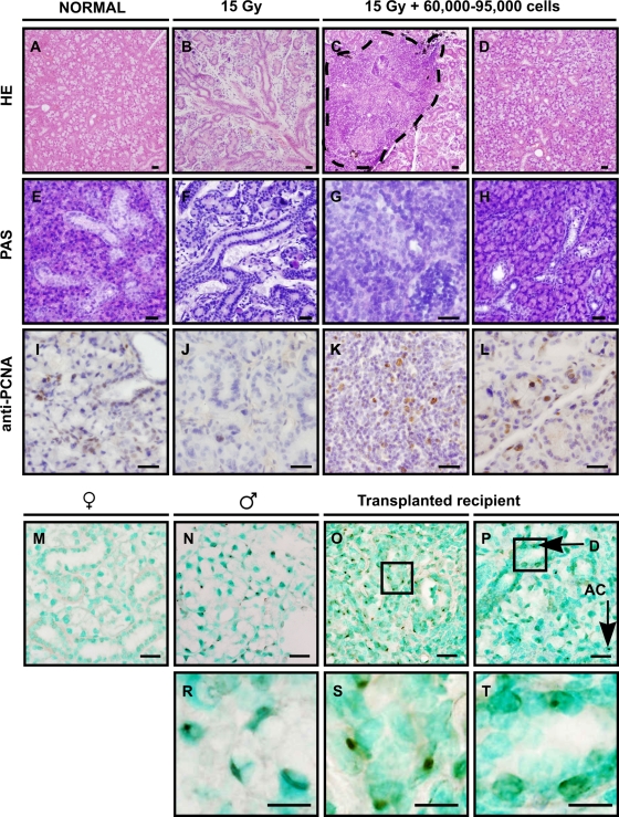
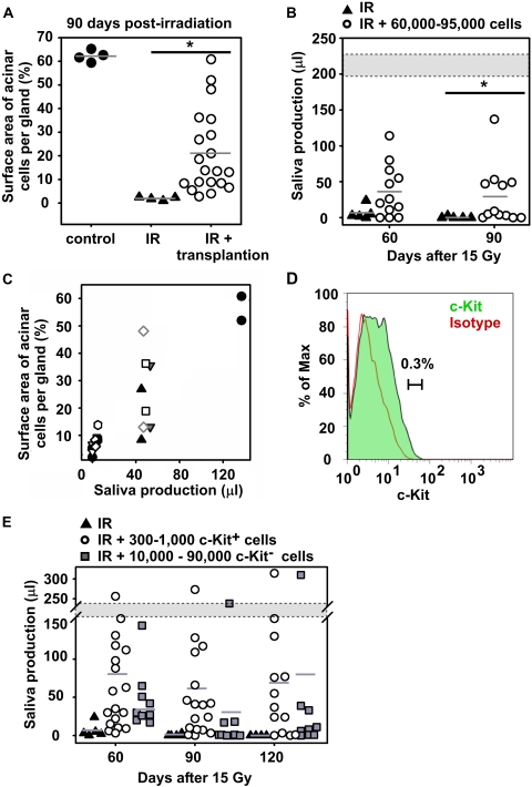
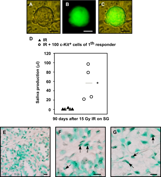
Head and neck cancer is the fifth most common malignancy and accounts for 3% of all new cancer cases each year. Despite relatively high survival rates, the quality of life of these patients is severely compromised because of radiation-induced impairment of salivary gland function and consequential xerostomia (dry mouth syndrome). In this study, a clinically applicable method for the restoration of radiation-impaired salivary gland function using salivary gland stem cell transplantation was developed. Salivary gland cells were isolated from murine submandibular glands and cultured in vitro as salispheres, which contained cells expressing the stem cell markers Sca-1, c-Kit and Musashi-1. In vitro, the cells differentiated into salivary gland duct cells and mucin and amylase producing acinar cells. Stem cell enrichment was performed by flow cytrometric selection using c-Kit as a marker. In vitro, the cells differentiated into amylase producing acinar cells. In vivo, intra-glandular transplantation of a small number of c-Kit(+) cells resulted in long-term restoration of salivary gland morphology and function. Moreover, donor-derived stem cells could be isolated from primary recipients, cultured as secondary spheres and after re-transplantation ameliorate radiation damage. Our approach is the first proof for the potential use of stem cell transplantation to functionally rescue salivary gland deficiency.


Conflict of interest statement

Competing Interests: The authors have declared that no competing interests exist.

Figures

Figure 1
Figure 1. In vitro salisphere formation.
(A) Dissociation of submandibular glands using hyaluronidase and collagenase resulted in clustered cell suspensions. 9723 ± 795 spheres were formed after 2–3 days of culturing (B), which increased in size in time (C,D). BrdU incorporation indicated that the cells in the culture were actively dividing (E–H). BrdU incorporation stained in brown, nuclei in blue. Scale bar = 50 µm. Inset shows negative control for BrdU, scale bar = 20 µm.
Figure 2
Figure 2. Salispheres originate from duct cells.
(A) Cells from salispheres visualized by dye staining and immunohistochemistry in submandibular gland tissue (vertical lane Tissue) and after 0, 3, 5 or 10 days of culture (vertical lanes D0, D3, D5, D10, resp.). Hematoxylin-Eosin (horizontal lane HE) staining emphasized the morphology of typical submandibular gland duct (small) (D) and acinar cells (AC) recognized by their polarized nucleus. Acinar cells were present at the onset of cultivation (HE-D0), but disappeared within 3 days (HE-D3). Horizontal lane PAS: acinar presence was confirmed by PAS staining (pink) which revealed the formation of mucin/mucopolysaccharide containing cells from day 5 on (PAS-D5). Specific ductal markers CK 7 (horizontal lane CK7) and CK 14 (horizontal lane CK 14) showed the ductal origin of the sphere. When three day cultured spheres were transferred into 3D collagen formation, duct-like branches (B) appeared within the next 7 days of culture. The branches contained specific CK 14 positive duct cells (C), whereas acinar-like (D, enlargement of B) structures contained granulae and were PAS positive (E). Antibody labeling is shown in brown, nuclei in blue. Scale bar = 50 µm, inset = 20 µm. (D = ductal cell type, AC = acinar cell, MY = myoepithelial cell).
Figure 3
Figure 3. Differentiation of salisphere into acinar cells.
(A) Amylase expressing cells (AC) in submandibular gland tissue (Tissue) were also present at the onset of culture (A-D0), and were visualized in the sphere at the onset of day 5 (A-D5), whereas granulae-containing spheres appeared in culture at later time-points (A-D10). Antibody labeling is shown in brown, nuclei in blue. Scale bar = 50 µm. (D = duct cells, AC = acinar cells, D0–3–5–10 represent days in culture). (B) Real time RT-PCR confirmed the enhanced expression of amylase during in vitro culturing and differentiation. Error bars represent SEM (N = 2). Amylase mRNA expression levels at 2 days of culture were normalized to one.
Figure 4
Figure 4. Salispheres contain stem cells.
(A) Sca-1 is present in the mouse submandibular gland on endothelial cells as well as on excretory and striated duct cells (D), and was clearly present at the onset of cultivation (D0) whereas acinar cells (AC) did not express Sca-1. At day 3 and later time-points, nearly all cells at the periphery of the salispheres showed high Sca-1 expression which decreased in time (D10). (B) Approximately 52.0+/−3.1% (Mean+/−STDEV) of cells in D3 cultured spheres expressed Sca-1, as quantified by flow cytometry. (C) c-Kit is only expressed by excretory duct cells (Tissue). Salispheres showed similar c-Kit staining patterns as for Sca-1. (D) Most nuclei in excretory duct compartments and few nuclei in striated duct cells showed Musashi-1 presence (Tissue). Whereas Musashi-1 was still present in the nuclei of some cells at the onset of culturing (D0-arrow), most cells showed cytoplasmic localization which diminished in time (D3–5–10). Cells were visualized with DAPI (blue). Scale bar = 50 µm, inset = 20 µm. D = duct cells, AC = acinar cell, D0–3–5–10 represent days in culture.
Figure 5
Figure 5. Restored morphology after injection of cultured stem cells.
Single cells from day 3 salispheres were intra-glandularly injected 30 days post-irradiation and glands were analyzed 90 days post-irradiation. From left to right, normal unirradiated glands (A,E,I), irradiated non-transplanted glands (B,F,J), irradiated transplanted glands (C,G,K) at the site of injection and irradiated transplanted glands away from the injection site (D,H,L). Irradiated glands hardly contained acinar cells (B,F) as visualized by HE or PAS. In contrast, in transplanted glands, ductal cells at the site of injection (dashed line) (C,G), and acinar cells further in the tissue were readily detectable (D,H). In normal (I) and irradiated glands (J) hardly any cells stained for PCNA. In contrast, PCNA labeled proliferating cells were present at and outside the site of injection (K,L). In situ hybridization to detect Y chromosomes of male transplanted cells in female recipient mice. (M) No staining in female control tissue and specific staining in male control mice (N, enlargement in R). Cells at the site of injection (O, enlargement in S), as well as distant ductal cells (P, enlargement in T) and acinar cells were donor derived. Nuclei are visualized in blue or green. Scale bar = 50 µm, enlarged pictures (R,S,T) = 20 µm.
Figure 6
Figure 6. Restored organ function by transplanted progenitor/stem cells.
(A) Quantifying the surface area occupied by acinar cells at 90 days post-irradiation revealed that transplanted glands contain significantly more acinar cells (○) compared to irradiated glands (▴). Saliva production measured on 90 days post-irradiation (B) was significantly higher enhanced in transplanted mice (○) compared to irradiated mice (▴). Gland function was restored in successfully transplanted mice to 23% up to 70% of normal production (gray area). (C) Saliva production correlated strongly with acinar cell restoration. Each symbol indicates a unique animal, at each time point two glands per animal were evaluated. (D) After 3 days in culture, the c-Kit brightest cells were FACS purified based on the indicated gate. (E) Injection of c-Kit+ cells (300 to 1,000 cells per gland) restored gland function in 9 out of 13 recipients until 120 days post-irradiation, whereas 3/9 responded when 10,000–90,000 c-Kit cells were transplanted.
Figure 7
Figure 7. Restored organ function by transplantation of c-kit+ cells from secondary spheres in secondary recipients.
Primary recipients were sacrificed 120 days after transplantation and secondary spheres were cultured from isolated glands. (A) Bright field image of secondary sphere, (B) GFP expression of this sphere, (C) overlay. (D) c-Kit+ cells were re-isolated from secondary salispheres and 100 cells were serially transplanted into irradiated gland of secondary recipients. Saliva production of secondary recipients was significantly (P<0.01) increased compared to irradiated control mice. (E,F,G) In situ hybridization to detect precence of Y chromosomes in male secondary transplanted cells in female recipient mice. Both ductal cells (F, arrows) and acinar cells (G, arrows) were Y chromosome+. Nuclei are visualized in green. Scale bar is 50 µm (E), or 20 µm (F,G).

Similar articles

Cited by

References

    1. Vissink A, Burlage FR, Spijkervet FK, Jansma J, Coppes RP. Prevention and treatment of the consequences of head and neck radiotherapy. Crit Rev Oral Biol Med. 2003;14:213–225. - PubMed
    1. Vissink A, Jansma J, Spijkervet FK, Burlage FR, Coppes RP. Oral sequelae of head and neck radiotherapy. Crit Rev Oral Biol Med. 2003;14:199–212. - PubMed
    1. Gresik EW. The granular convoluted tubule (GCT) cell of rodent submandibular glands. Microsc Res Tech. 1994;27:1–24. - PubMed
    1. Denny PC, Ball WD, Redman RS. Salivary glands: a paradigm for diversity of gland development. Crit Rev Oral Biol Med. 1997;8:51–75. - PubMed
    1. Burford-Mason AP, Cummins MM, Brown DH, MacKay AJ, Dardick I. Immunohistochemical analysis of the proliferative capacity of duct and acinar cells during ligation-induced atrophy and subsequent regeneration of rat parotid gland. J Oral Pathol Med. 1993;22:440–446. - PubMed

Publication types

MeSH terms

Substances