. 2008 Apr 29;6(4):e103.
doi: 10.1371/journal.pbio.0060103.

Bergmann glia and the recognition molecule CHL1 organize GABAergic axons and direct innervation of Purkinje cell dendrites

Affiliations

Bergmann glia and the recognition molecule CHL1 organize GABAergic axons and direct innervation of Purkinje cell dendrites

Fabrice Ango et al. PLoS Biol. .

Abstract

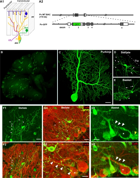
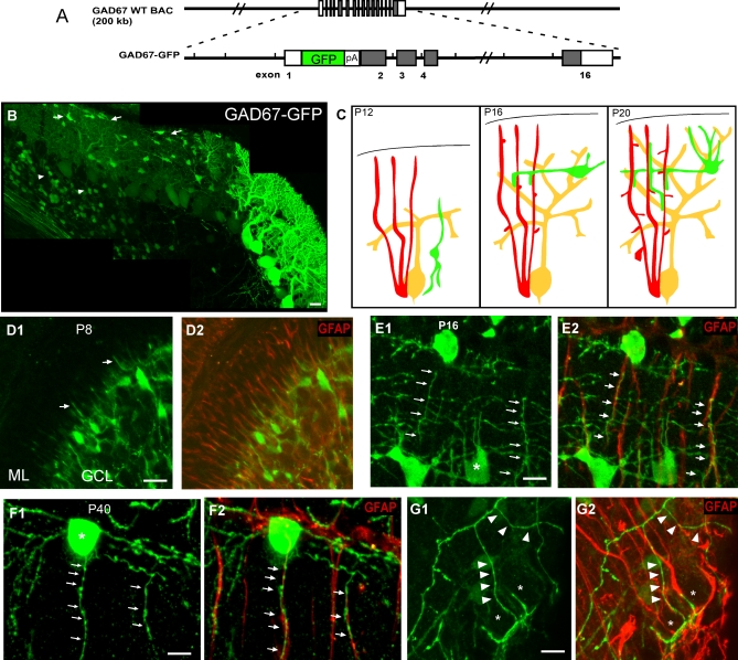
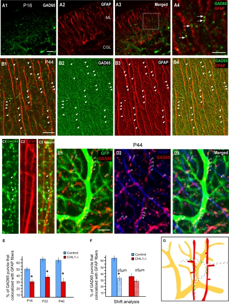
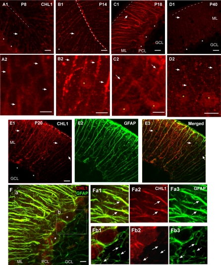
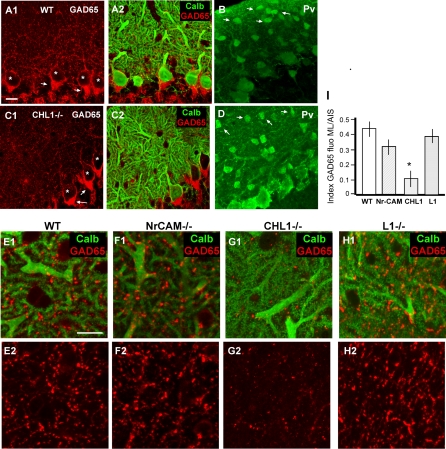
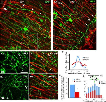
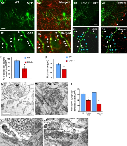
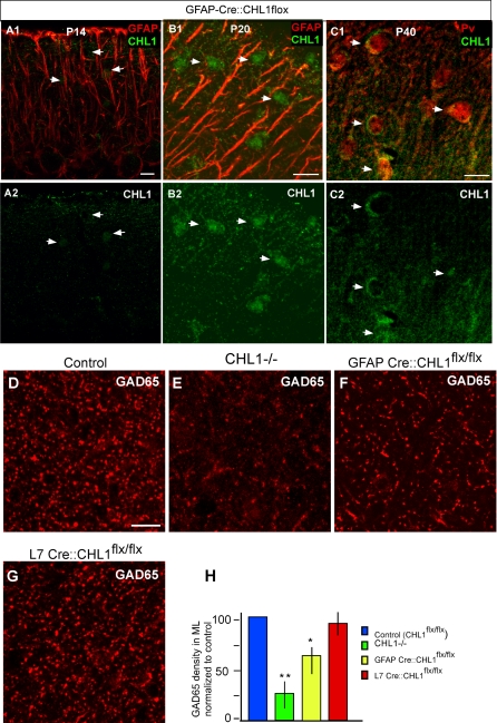
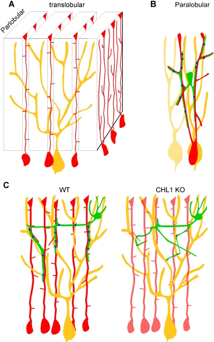
The geometric and subcellular organization of axon arbors distributes and regulates electrical signaling in neurons and networks, but the underlying mechanisms have remained elusive. In rodent cerebellar cortex, stellate interneurons elaborate characteristic axon arbors that selectively innervate Purkinje cell dendrites and likely regulate dendritic integration. We used GFP BAC transgenic reporter mice to examine the cellular processes and molecular mechanisms underlying the development of stellate cell axons and their innervation pattern. We show that stellate axons are organized and guided towards Purkinje cell dendrites by an intermediate scaffold of Bergmann glial (BG) fibers. The L1 family immunoglobulin protein Close Homologue of L1 (CHL1) is localized to apical BG fibers and stellate cells during the development of stellate axon arbors. In the absence of CHL1, stellate axons deviate from BG fibers and show aberrant branching and orientation. Furthermore, synapse formation between aberrant stellate axons and Purkinje dendrites is reduced and cannot be maintained, leading to progressive atrophy of axon terminals. These results establish BG fibers as a guiding scaffold and CHL1 a molecular signal in the organization of stellate axon arbors and in directing their dendritic innervation.


Conflict of interest statement

Competing interests. The authors have declared that no competing interests exist.

Figures

Figure 1
Figure 1. Stellate and Basket Cells Use Different Cellular Mechanisms to Innervate Purkinje Cells
(A1) A schematic of the major neuronal components of the cerebellar cortex organized along the translobular and parlobular plane. Bsk, basket cell; CF, climbing fiber; PF, parallel fiber; Pj, Purkinje cell; St, stellate cell. (A2) A schematic of the PV-GFP BAC reporter construct. Long black lines indicate the PV BAC clone; shaded boxes, coding exons; open boxes, noncoding exons. Sequences coding for EGFP and polyadenylation signal (pA) were inserted at the translation initiation site (see Materials and Methods). (B) Low-magnification view of a cerebellar sagital section from an adult PV-GFP reporter mouse (B20 line). Note the low density of cells expressing GFP. (C–H) Individual Purkinje (C), stellate (D), and basket cells (E) can be resolved with synaptic resolution to their entirety. Arrows in (D) and (E) indicate dendrites. The basket axons ([E] and [H], arrowheads) are smooth, grow along Purkinje proximal dendrite labeled by calbindin (H2), soma (star), AIS, and form pinceau synapses (yellow arrowheads) at AIS. The stellate axons ([D], [F], and [G], arrowheads) are beaded, send ascending and descending collaterals that intercept with Purkinje dendrites ([F2] and [G2], labeled by calbindin immunofluorescence) at sharp angles. (G2) is a higher magnification of (G1). Note a straight and descending stellate axon branch (arrowheads) that reaches a Purkinje cell soma (star) but, in contrast to basket axons, does not grow along the Purkinje cell and terminates abruptly. Bk, basket cell; St, stellate cell. Scale bars indicate 20 μm.
Figure 2
Figure 2. Developing Stellate Axons Extend along Bergmann Glial Fibers
(A) A schematic of the GAD67-GFP BAC reporter construct. Long black lines indicate the GAD67 BAC clone; shaded boxes, coding exons; open boxes, noncoding exons. Sequences coding for EGFP and polyadenylation signal (pA) were inserted at the translation initiation site. (B) During the second postnatal week, stellate neurons migrate into the ML, reaching their laminar position between P12 and P16. At P14, some stellate cells already settled in the upper ML close to the pia (arrows), while other interneurons were still migrating in the white matter (arrowheads). (C) Schematic representations of the development of stellate axons (green) and their relationship to Purkinje cells (yellow) and Bergmann glia (red) between P12 and P20. (D) At P8, BG fibers (GFAP immunofluorescence in [D2]) were already prominent in the ML while interneurons were migrating towards and into the ML ([D1], arrows indicate neurites). (E) At P16, stellate cells (E1) extended their axons with ascending and descending branches that strictly adhered to the GFAP-labeled BG fibers (E2, arrows). (F) At more-mature ages (P40), stellate cell axons (F1) were still associated with BG fibers (F2). (G) Basket axons at P40 (G1, arrowheads) did not associate with GFAP-labeled BG fibers (G2) but outlined Purkinje cell soma (stars). Scale bars indicate 20 μm
Figure 3
Figure 3. GABAergic Synapses Develop at the Intersection between Purkinje Dendrites and GFAP Fibers
(A) At P16, double labeling of GAD65 (A1) and GFAP (A2) revealed a significant colocalization of GABAergic presynaptic boutons and BG fibers in the ML (A3). Note the association of GAD65 with BG fibers in (A4) (arrows), a higher magnification of the boxed area in (A3). (B and C) Prominent colocalization of GAD65 along GFAP fibers at P44. Strings of GAD65 puncta can be discerned that align to the radial GFAP fibers (arrowheads) and also to some lateral appendages (star). (B2–B4) was obtained using the “sharper” filter function of the LSM510 confocal software. (D) Triple labeling of GABAergic boutons (GAD65 in red), BG fibers (GFAP in blue), and a Purkinje dendrite (PV-GFP expression in B20 line; green). GAD65 puncta colocalized with the dendritic shaft of this Purkinje cell (arrowhead in [D1]). Note that the same clusters of GAD65 punta also aligned with GFAP fiber (D2), and GAD65 labeled boutons localized to the intersection between Purkinje dendritic shaft and GFAP positive BG fibers (arrowheads, [D3]). (E) Quantification of GAD65 association with GFAP fibers at P16, P20, and P40 in WT and CHL1−/− mice. Note that there is a significant reduction of GAD65 association with GFAP at all ages (an asterisk [*] indicates p < 0.001). (F) An artificial shift of BG fibers (see Materials and Methods) by 5 μm to the left or to the right induced a 55 ± 7% reduction of their colocalization with GAD65. In contrast, shift analysis performed in image stacks from CHL1−/− mice did not change the percentage of this association. (An asterisk [*] indicates p < 0.001.) (G) A schematic representation of the relationship between stellate axons (dashed lines), BG fibers (red), and Purkinje dendrite (yellow). Stellate axons extend along BG fibers to reach Purkinje dendrite, and synaptic boutons are preferentially formed or stabilized at their interceptions. Scale bars indicate 20 μm.
Figure 4
Figure 4. CHL1 Expression in the Developing Cerebellar Cortex
(A1–D1) CHL1 was prominently expressed in radial stripe patterns (arrows; corresponding largely to BG fibers) in the ML at P8 (A1) and P14 (B1), reaching higher levels and becoming more diffuse at P18 (C1), and declining in adult (P40, [D1]) mice. Pia is indicated by dotted lines. White stars indicate somata of Purkinje cells. (A2–D2) Images are higher magnification of (A1–D1) and are taken from the upper ML, which contained stellate cell somata. CHL1 expression in stellate cells was undetectable at P8 (A2), appeared around P14 ([B2], yellow star), and remained at P18 (C2) and in adult (D2) mice. (E) CHL1 (E1) closely colocalized with GFAP (E2) in developing cerebellar cortex (E3); only P20 data are shown. Note that CHL1 signals became more diffuse in the ML at this age. (Also see Figure S4.) (F) GFAP (green) labeled both the apical BG fibers in the ML (a) and basal lamellae (b); the latter enwrap Purkinje soma and AIS. At P18, the colocalization of CHL1 and GFAP was restricted to the ML and PCL (inset a, arrows), but not to the basal lamellae in the granule cell layer ([GCL], inset b, arrows). CHL1 (F2a and F2b) is colabeled with GFAP (F3a and F3b). (F1a) and (F1b) are the merged pictures of (F2a and F3a) and (F2b and F3b), respectively Scale bars indicate 20 μm.
Figure 5
Figure 5. Deficient GABAergic Innervation in the ML of CHL1−/− Mice
(A) In WT mice, GAD65 staining revealed GABAergic boutons in the ML and the pinceau synapses at Purkinje cell AISs (arrows). (B–D) In CHL1−/− mice (C), GAD65 labeling in the ML was significantly decreased, whereas the pinceau synapses remained intact (arrows). Stars indicate Purkinje soma. GABAergic interneurons (arrows in [B] and [D]) labeled with Pv were present in normal density and locations in the ML of CHL1−/− (D), compared to those of WT (B) mice. (E–I) GAD65 expression in the upper ML of WT (E), NrCAM−/− (F), CHL1−/− (G), and L1−/− (H) mice were quantified as a ratio of fluorescence intensity in ML and at Purkinje AIS. GAD65 (E2, F2, G2, and H2) is colabeled with Calbindin (E1, F1, G1, and H2). (I) There is a 3-fold decreased in this ratio only in CHL1−/− mice ([I], p ≤ 0.001). Purkinje dendrites appeared normal in all phenotypes. Scale bars indicate 20 μm.
Figure 6
Figure 6. Aberrant Stellate Axonal Arborization in CHL1−/− Mice
(A) (A1) single stellate axon arbor labeled by PV-GFP in the B20 reporter line at P40. Even though the axon arbor was complex, close correlation between axon branches and GFAP fibers can be readily detected ([A2 and A3], arrows). Axon branches often switched between neighboring GFAP fibers (stars). Large arrows in (A1) indicate basket cell dendrites. (A2) and (A3) are higher magnification of the white box in (A1). (B) In CHL1−/−::PV-GFP mice, stellate axons appeared thinner and disoriented, with much reduced association to GFAP fibers (arrows in [B3]). Axon branches often crossed over BG fibers at sharp angles (arrowheads in [B3]). (B2) and (B3) are higher magnification of the white box in (B1). (C) Quantification of the orientation of stellate axon branches in WT mice revealed a normal distribution towards the pia surface, with 55% of branches oriented at angles between 80°–100° towards the pia. In CHL1−/− mice, only 32% axon branches were oriented in this range. (D) Quantification of the association of vertically oriented stellate axon branches with GFAP fibers revealed a significant reduction in CHL1−/− mice (29.6 ± 6.2% ) compared to that in WT mice (69.7 ± 11.1%). (E) Quantification of the association of stellate axon branches oriented at multiple angles with GFAP fibers showed significant reductions in CHL1−/− compared to WT mice. Note that in the WT, more than 60% of axonal branches colocalized with GFAP, regardless of their angle with the pia (n = 400, an asterisk [*] indicates p < 0.001 from 10 single cells in each phenotype). Scale bars indicate 20 μm.
Figure 7
Figure 7. Deficient and Decreased Number of Stellate Synapses in CHL1−/− Mice.
(A) Single stellate cell axons (A1) labeled in WT PV-GFP mice showed large and distinct boutons that colocalized with GAD65 (A2).. (B) Higher magnification view showed that nearly all of these boutons (B1) contained GAD65 ([B2], arrowheads). (C and D) In CHL1−/−::PV-GFP mice, stellate boutons appeared smaller (white arrowheads), and many of them did not contain detectable levels of GAD65 (blue arrowheads). Note the normal GAD65 signal at pinceau synapses in (C2) (arrows). (E) Quantification of the percentage of stellate boutons colocalization with GAD65 showed a 43% reduction in CHL1−/− compared to WT mice (p ≤ 0.001. (F) Quantification of stellate bouton size revealed a 40% reduction in CHL1−/− compared to WT mice (p ≤ 0.01). (G and H) Ultrastructural analysis showed stellate cell synapses along Purkinje dendritic shafts in the ML of CHL1−/− mice (H), with largely normal morphology and organelle organization as compared to those in WT mice (G). (I) Quantification of stellate cell synapse density along Purkinje dendritic surface in the upper ML of CHL1 −/− mice revealed a approximately 40% reduction compared to WT mice at P30 (p < 0.03), and a 60% reduction compared to WT mice at P40 (p ≤ 0.001). (J) Ultrastructural analysis showed atrophy of stellate axon terminals in 3-mo-old CHL1−/− mice. Purkinje dendrites (Pd) were often apposed by degenerating profiles exhibiting electron-dense membrane accumulations (arrows) and electron-lucent empty spaces (stars). (J1) and (J2) are two different examples. Note a symmetrical synapse from most likely a stellate cell axon terminal (St).
Figure 8
Figure 8. Conditional Deletion of CHL1 in Bergmann Glial Cells Results in Significant Reduction of GAD65 Expression in the Upper ML
(A–C) BG-restricted CHL1 deletion was achieved by breeding CHL1flx/flx and GFAP-Cre mice. At both P14 (A) and P20 (B), CHL1 (A2 and B2) was no longer present as stripe patterns that colocalize with GFAP (red in [A1] and [B1]) in the ML as in WT mice (Figure 4); but CHL1 was detected as somata profiles (arrows) and diffuse signals in the ML, especially at P20 and P40 (C2). (C1) showed CHL1 colabeled with calbindin (red). (D–G) CHL1 expression remained at low levels in the ML at P40 both as diffuse signals and also outlined stellate cell somata, which were positive for Pv ([D], Pv in red). GAD65 expression in the upper ML of CHL1flx/flx control (D), germline CHL1−/− (E), GFAP-Cre::CHL1flx/flx (F), and L7-Cre::CHL1flx/flx (G) mice. (H) Compared to GAD65 labeling in the ML in control mice, mice lacking CHL1 in BG showed a 27% reduction (an asterisk [*] indicates p ≤ 0.01, n = 4), germline CHL1−/− mice showed a 65% reduction (double asterisks [**] indicate p ≤ 0.001, n = 4), and L7-Cre::CHL1flx/flx mice showed no reduction. Scale bars indicate 20 μm.
Figure 9
Figure 9. Schematic Representation of the Bergmann Glial Fiber Scaffold and CHL1 in Directing Stellate Axons to Innervate Purkinje Dendrites
(A) In the cerebellar cortex, Purkinje dendrites (yellow) are restricted in the translobular plane. Each BG cell (red) gives rise to several ascending BG fibers, which extend in both the translobular and parlobular plane [14,15]. These largely radial fibers from neighboring BG cells further aligned into thin walls, or palisades, in the parlobular plane, perpendicular to the Purkinje dendrites. As a consequence, several BG palisades cut across and impinge upon an individual Purkinje dendrite in a largely vertical orientation. (B) A stellate axon likely contacts segments of multiple intercalated Purkinje dendrites (parlobular view; neighboring Purkinje dendrites are represented in different shades of yellow). Stellate synapses (blue dots) are formed or stabilized at the interception of BG fibers and Purkinje dendrites. (C) In the translobular plane in WT mice (left panel), stellate axons (green) associate with and extend along BG fibers, and are thus organized into characteristic orientations and trajectories towards Purkinje dendrites. In CHL1-deficient mice (right panel), stellate axons can no longer associate with BG fibers, show aberrant orientation and trajectory, and are deficient in synapse formation and/or stability.

Comment in

Similar articles

Cited by

References

    1. Hausser M, Spruston N, Stuart GJ. Diversity and dynamics of dendritic signaling. Science. 2000;290:739–744. - PubMed
    1. Larkum ME, Zhu JJ, Sakmann B. A new cellular mechanism for coupling inputs arriving at different cortical layers. Nature. 1999;398:338–341. - PubMed
    1. Larkum ME, Zhu JJ, Sakmann B. Dendritic mechanisms underlying the coupling of the dendritic with the axonal action potential initiation zone of adult rat layer 5 pyramidal neurons. J Physiol. 2001;533:447–466. - PMC - PubMed
    1. Schaefer AT, Larkum ME, Sakmann B, Roth A. Coincidence detection in pyramidal neurons is tuned by their dendritic branching pattern. J Neurophysiol. 2003;89:3143–3154. - PubMed
    1. London M, Hausser M. Dendritic computation. Annu Rev Neurosci. 2005;28:503–532. - PubMed

Publication types