. 2008 Apr 29;6(4):e111.
doi: 10.1371/journal.pbio.0060111.

A competition between stimulators and antagonists of Upf complex recruitment governs human nonsense-mediated mRNA decay

Affiliations

A competition between stimulators and antagonists of Upf complex recruitment governs human nonsense-mediated mRNA decay

Guramrit Singh et al. PLoS Biol. .

Abstract

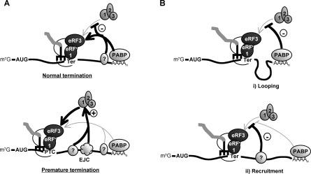
The nonsense-mediated decay (NMD) pathway subjects mRNAs with premature termination codons (PTCs) to rapid decay. The conserved Upf1-3 complex interacts with the eukaryotic translation release factors, eRF3 and eRF1, and triggers NMD when translation termination takes place at a PTC. Contrasting models postulate central roles in PTC-recognition for the exon junction complex in mammals versus the cytoplasmic poly(A)-binding protein (PABP) in other eukaryotes. Here we present evidence for a unified model for NMD, in which PTC recognition in human cells is mediated by a competition between 3' UTR-associated factors that stimulate or antagonize recruitment of the Upf complex to the terminating ribosome. We identify cytoplasmic PABP as a human NMD antagonizing factor, which inhibits the interaction between eRF3 and Upf1 in vitro and prevents NMD in cells when positioned in proximity to the termination codon. Surprisingly, only when an extended 3' UTR places cytoplasmic PABP distally to the termination codon does a downstream exon junction complex enhance NMD, likely through increasing the affinity of Upf proteins for the 3' UTR. Interestingly, while an artificial 3' UTR of >420 nucleotides triggers NMD, a large subset of human mRNAs contain longer 3' UTRs but evade NMD. We speculate that these have evolved to concentrate NMD-inhibiting factors, such as PABP, in spatial proximity of the termination codon.


Conflict of interest statement

Competing interests. The authors have declared that no competing interests exist.

Figures

Figure 1
Figure 1. A 3′ UTR Intron Is Not Sufficient to Trigger NMD
(A–E) Decay assays in human HeLa Tet-off cells for β-globin (A,D) or TPI mRNAs (E) with inserted 3′ UTR introns or β-globin wild-type (B) or PTC-39 mutant (C) mRNAs in the presence of Luciferase (Luc) or hUpf1 siRNAs as indicated (knockdown efficiencies are shown in Figure S1). For each panel, schematics are shown for each tested mRNA below the panels; β-globin or TPI exons are indicated as light-gray bars, introns as lines, AdML exons are shown in black, and TPIi6 exons are patterned. Numbers above the panels indicate time after transcriptional repression. mRNA half-lives were calculated by comparison with the constitutively expressed internal control mRNAs (βUAC-GAP or βwt; top panels in each assay) and are given on the right.
Figure 2
Figure 2. Intron-Less Extended 3′ UTRs Trigger NMD in Human Cells
(A and B) mRNA decay assays showing decay rates of β-globin-derived βGAP, βUAC-GAP, βGFP, and βUAC-GFP mRNAs (see schematics below) in human HeLa Tet-off cells expressing siRNAs against hUpf1, hUpf2, or as a negative control, Luciferase (Luc), as indicated on the left of each panel (knockdown efficiencies are shown in Figure S1). Constitutively expressed βwt mRNA was used as internal controls for quantification. Numbers above the panels indicate time after transcriptional repression. Schematics on the bottom show the used constructs with β-globin exons indicated as light-gray bars, introns as lines, and GAPDH and GFP sequences as dark-gray and dotted bars, respectively. Numbers on the right indicate mRNA half-lives (t1/2; in minutes) calculated from the shown experiment. Numbers in parentheses indicate the fold stabilization with standard deviation (n ≥ 3) upon hUpf knockdown as compared to the Luc control. (C) mRNA decay assays for intron-containing GPx1 mRNA with a PTC (GPx1-UAA) or a GPx1 mRNA with a PTC expressed from an intron-less construct (GPx1Δi-UAA) (see schematics below) in the presence of Luc or hUpf1 siRNAs as indicated on the left. mRNA decay rates for the shown experiments are given on the right and numbers in parentheses indicate the fold stabilization with standard deviation (n ≥ 3) upon hUpf knockdown as compared to the Luc control.
Figure 3
Figure 3. PABPC1 Antagonizes NMD
(A and B) mRNA decay assays for β39–2xMS2-Ex2 and βGAP-4xMS2 mRNAs (see schematics below; MS2 binding sites indicated as black bars and GAPDH sequence in dark gray) in HeLa Tet-off cells expressing the MS2 coat protein fused to PABPC1 (MS2-PABPC1) or PABPN1 (MS2-PABPN1) or individual unfused proteins as indicated on the left of the panels. Constitutively expressed βUAC-GAP or βwt mRNAs were used as internal controls (upper panels). (C and D) Northern blots showing decay rates of β39 and βGAP mRNAs, with or without A30 or N30 sequences inserted downstream of the PTCs (schematic below). For all panels (A–D), numbers on the right indicate mRNA half-lives (t1/2; in minutes) calculated from the shown experiments, and numbers in parentheses indicate the fold stabilization with standard deviation (n ≥ 3) as compared with the control experiments shown in the corresponding top panels.
Figure 4
Figure 4. Normal Long 3′ UTRs Can Evade NMD
mRNA decay assays for βSmg5, βTram1, and βCript1 mRNAs (schematics with the respective 3′ UTR lengths are shown below) in the presence of Luc or hUpf1 siRNAs. mRNA half-lives are given on the right. The number in parentheses for the βSmg5 mRNAs indicates the fold stabilization with standard deviation (n ≥ 3) upon hUpf knockdown as compared to the Luc control.
Figure 5
Figure 5. A 3′ UTR Intron Enhances NMD
Decay assays for βGAP mRNA in the absence or presence of the AdML intron inserted into the GAPDH sequence or exon 3 as indicated. For each panel, schematics are shown for each tested mRNA below the panels; AdML exons are shown in black. mRNA half-lives are given on the right with the fold enhancement of mRNA decay rates and standard deviation (n ≥ 3) as compared with the control experiment in the top panel shown in parentheses.
Figure 6
Figure 6. PABPC1 Antagonizes the Interaction between eRF3 and hUpf1 In Vitro
(A) Co-IP assays showing the co-IP of endogenous hUpf1 and PABPC1 and HuR proteins with an antibody against eRF3 (α-eRF3; 1%, 3%, 10%, 30%, and 100% of pellet loaded in lanes 4–8, respectively), or pre-immune serum (Pre-I, lane 1) as a control. 3% and 0.6% of the total lysate are shown in lanes 2 and 3, respectively. (B) In vitro pull-down assays showing anti-FLAG Western blots of pull-down pellets (upper panels) resulting after myc-tagged eRF3 (lanes 4–7) or no myc-tagged protein (lanes 1–3), immobilized on an anti-myc antibody resin, was incubated with various amounts of FLAG-tagged hUpf1, PABPC1, or hnRNP A1 as indicated. Estimated amounts of FLAG-tagged proteins in each reaction are given in μM. Bottom panels show 5% of input protein for each reaction. The asterisk (*) and dagger (†) on the right indicates likely degradation products of PABPC1 and cross-reacting Myc-eRF3, respectively. (C) Lanes 1–6: Western blot for exogenously expressed Myc-tagged eRF3 (wt, lanes 1–3) or eRF3 KAKA mutant protein (KAKA, lanes 4–6) that co-IP with FLAG-tagged PABPC1, hUpf1, or as a negative control, MS2, as indicated above the lanes. Lanes 7–9: Western blot for exogenously expressed Myc-tagged eRF1 that co-IP with FLAG-tagged eRF3, eRF3 KAKA mutant protein, or as a negative control, MS2, as indicated above the lanes. For all lanes, 5% of total input extracts are shown in the bottom panels.
Figure 7
Figure 7. A Competition between Stimulators and Antagonists of Upf Complex Recruitment in Human NMD
(A) A unified model where NMD is determined by the balance between 3′ UTR–associated factors that stimulate (such as the EJC) or antagonize (such as cytoplasmic PABP) recruitment of the hUpf complex (shown as spheres labeled 1–3) to the terminating ribosome. (B) Mechanisms by which mammalian mRNAs with long 3′ UTRs may evade NMD (see Discussion for details).

References

    1. Amrani N, Sachs MS, Jacobson A. Early nonsense: mRNA decay solves a translational problem. Nat Rev Mol Cell Biol. 2006;7:415–425. - PubMed
    1. Conti E, Izaurralde E. Nonsense-mediated mRNA decay: molecular insights and mechanistic variations across species. Curr Opin Cell Biol. 2005;17:316–325. - PubMed
    1. Isken O, Maquat LE. Quality control of eukaryotic mRNA: safeguarding cells from abnormal mRNA function. Genes Dev. 2007;21:1833–1856. - PubMed
    1. Sharifi NA, Dietz HC. Physiologic substrates and functions for mammalian NMD. In: Maquat LE, editor. Nonsense-mediated mRNA decay. Georgetown, Texas: Landes Bioscience; 2006. pp. 97–106.
    1. Behm-Ansmant I, Gatfield D, Rehwinkel J, Hilgers V, Izaurralde E. A conserved role for cytoplasmic poly(A)-binding protein 1 (PABPC1) in nonsense-mediated mRNA decay. EMBO J. 2007;26:1591–1601. - PMC - PubMed

Publication types

MeSH terms