Review
. 2008 May 15;197 Suppl 3(Suppl 3):S294-306.
doi: 10.1086/533419.

Antiretroviral therapy and central nervous system HIV type 1 infection

Affiliations
Review

Antiretroviral therapy and central nervous system HIV type 1 infection

Richard W Price et al. J Infect Dis. .

Abstract

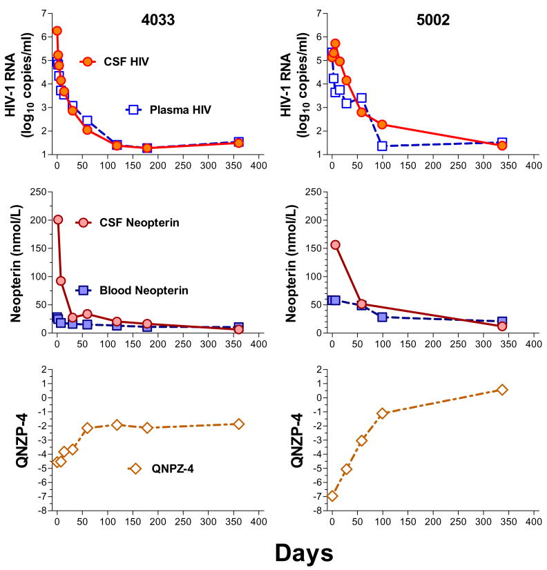
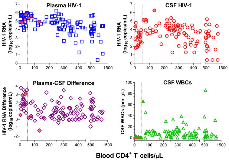
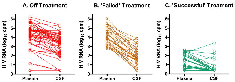
Central nervous system (CNS) human immunodeficiency virus type 1 (HIV-1) infection begins during primary viremia and continues throughout the course of untreated systemic infection. Although frequently accompanied by local inflammatory reactions detectable in cerebrospinal fluid (CSF), CNS HIV-1 infection usually is not clinically apparent. In a minority of patients, CNS HIV-1 infection evolves into encephalitis during the late stages of systemic infection, which compromises brain function and presents clinically as acquired immunodeficiency syndrome dementia complex (ADC). Combination antiretroviral therapy (ART) has had a major impact on all aspects of CNS HIV-1 infection and disease. In those with asymptomatic infection, ART usually effectively suppresses HIV-1 in CSF and markedly reduces the incidence of symptomatic ADC. In those presenting with ADC, ART characteristically prevents neurological progression and leads to variable, and at times substantial, recovery. Similarly, treatment has reduced CNS opportunistic infections. With better control of these severe disorders, attention has turned to the possible consequences of chronic silent infection and the issue of whether indolent, low-grade brain injury might require earlier treatment intervention.


Figures

Figure 1
Figure 1
Effect of HAART in ADC. Graphs show the changes in plasma and CSF HIV-1 RNA concentrations (top panels), blood and CSF (middle panels), and composite quantitative neurological performance Z scores on four tests (QNPZ-4) [22, 127] (bottom panels) for 2 ADC patients after starting HAART. Patient 4033 (left column), a 38 year old man with ADC stage 2, was treated with abacavir, lamivudine, nevirapine and boosted indinavir. This led to a rapid fall in both CSF and plasma HIV and CSF neopterin concentrations from high baseline levels. During this same 3-month period, the patient improved neurologically (as documented by the rise in QNPZ-4) but he plateaued at an ‘impaired’ level of about -2, reflecting continued motor dysfunction including a mild spastic gait. He was able to return to school and to acting. Patient 5002 was 36 years old when he presented with ADC stage 3 and was treated with abacavir, lamivudine, nevirapine and nelfinavir. While there was an initial delay in the fall of his CSF HIV-1 RNA, it eventually fell to an undetectable level. Similarly, his CSF neopterin concentration declined from a high baseline. The QNPZ-4 score rose steadily over the first 3 months and continued further improvement over the remaining year, eventually reaching a normal level as he returned to a nearly asymptomatic functional level (ADC stage 0.5).
Figure 2
Figure 2
CSF changes in HIV-1 infection. These cross-sectional data combine experience from two cohorts [5, 56] and include only untreated HIV-infected subjects. With the exception of 8 patients presenting with ADC (filled symbols), the remainder were neurologically asymptomatic. The graphs show the high prevalence of detectable CSF HIV-1 RNA across blood CD4+ T cell counts and its variable relationship to plasma HIV-1 RNA levels. The frequent presence of CSF pleocytosis is also shown. The vertical dotted line marks a blood CD4+ count of 50 cells per μL and shows the generally high plasma, lower CSF, and high plasma-CSF log10 difference in HIV-1 RNA, along with the lower incidence of pleocytosis.
Figure 3
Figure 3
Effect of treatment on CSF HIV-1 RNA. These data are taken from Spudich et al [56] but are presented to show the relationships of plasma to CSF HIV-1 RNA in individual subjects (connecting lines). In those currently not receiving or those who have discontinued treatment, the relationships are highly variable as shown by the different slopes of the connecting lines. In those defined for this study as ‘failures’ (due to drug resistance) who are on treatment but with plasma HIV-1 RNA levels >500 copies per mL, the CSF HIV-1 RNA levels are consistently lower with a single exception. Successful suppression of plasma HIV-1 to < 500 copies per mL was accompanied by similar suppression of HIV-1 in the CSF with only a few exceptions.

Similar articles

Cited by

References

    1. Davis LE, Hjelle BL, Miller VE, et al. Early viral brain invasion in iatrogenic human immunodeficiency virus infection. Neurology. 1992;42:1736–9. - PubMed
    1. Pilcher CD, Shugars DC, Fiscus SA, et al. HIV in body fluids during primary HIV infection: implications for pathogenesis, treatment and public health. AIDS. 2001;15:837–45. - PubMed
    1. Ritola K, Pilcher CD, Fiscus SA, et al. Multiple V1/V2 env variants are frequently present during primary infection with human immunodeficiency virus type 1. J Virol. 2004;78:11208–18. - PMC - PubMed
    1. Ellis RJ, Hsia K, Spector SA, et al. Cerebrospinal fluid human immunodeficiency virus type 1 RNA levels are elevated in neurocognitively impaired individuals with acquired immunodeficiency syndrome. HIV Neurobehavioral Research Center Group. Ann Neurol. 1997;42:679–88. - PubMed
    1. Spudich SS, Nilsson AC, Lollo ND, et al. Cerebrospinal fluid HIV infection and pleocytosis: Relation to systemic infection and antiretroviral treatment. BMC Infect Dis. 2005;5:98. - PMC - PubMed

Publication types

MeSH terms

Substances