

Relation between myocardial edema and myocardial mass during the acute and convalescent phase of myocarditis--a CMR study
- PMID: 18447954
- PMCID: PMC2396625
- DOI: 10.1186/1532-429X-10-19
Relation between myocardial edema and myocardial mass during the acute and convalescent phase of myocarditis--a CMR study
Abstract
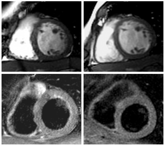
Background: Myocardial edema is a substantial feature of the inflammatory response in human myocarditis. The relation between myocardial edema and myocardial mass in the course of healing myocarditis has not been systematically investigated. We hypothesised that the resolution of myocardial edema as visualised by T2-weighted cardiovascular magnetic resonance (CMR) is associated with a decrease of myocardial mass in steady state free precession (SSFP)-cine imaging.
Methods: 21 patients with acute myocarditis underwent CMR shortly after onset of symptoms and 1 year later. For visualization of edema, a T2-weighted breath-hold black-blood triple-inversion fast spin echo technique was applied and the ratio of signal intensity of myocardium/skeletal muscle was assessed. Left ventricular (LV) mass, volumes and function were quantified from biplane cine steady state free precession images. 11 healthy volunteers served as a control group for interstudy reproducibility of LV mass.
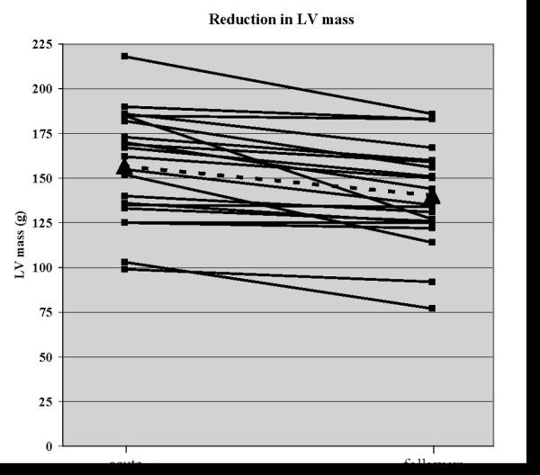
Results: In patients with myocarditis, a significant decrease in LV mass was observed during follow-up compared to the acute phase (156.7 +/- 30.6 g vs. 140.3 +/- 28.3 g, p < 0.0001). The reduction of LV mass paralleled the normalization of initially increased myocardial signal intensity on T2-weighted images (2.4 +/- 0.4 vs. 1.68 +/- 0.3, p < 0.0001). In controls, the interstudy difference of LV mass was lower than in patients (5.1 +/- 2.9 g vs. 16.3 +/- 14.2 g, p = 0.02) resulting in a lower coefficient of variability (2.1 vs 8.9%, p = 0.04).
Conclusion: Reversible abnormalities in T2-weighted CMR are paralleled by a transient increase in left ventricular mass during the course of myocarditis. Myocardial edema may be a common pathway explaining these findings.
Figures
Similar articles
-
Cardiac magnetic resonance monitors reversible and irreversible myocardial injury in myocarditis.JACC Cardiovasc Imaging. 2009 Feb;2(2):131-8. doi: 10.1016/j.jcmg.2008.09.014. JACC Cardiovasc Imaging. 2009. PMID: 19356545
-
Assessment of acute myocarditis by cardiovascular MR: diagnostic performance of shortened protocols.Int J Cardiovasc Imaging. 2013 Jun;29(5):1077-83. doi: 10.1007/s10554-013-0189-7. Epub 2013 Feb 13. Int J Cardiovasc Imaging. 2013. PMID: 23404383
-
Native T1-mapping detects the location, extent and patterns of acute myocarditis without the need for gadolinium contrast agents.J Cardiovasc Magn Reson. 2014 May 23;16(1):36. doi: 10.1186/1532-429X-16-36. J Cardiovasc Magn Reson. 2014. PMID: 24886708 Free PMC article.
-
Myocarditis or "true" infarction by cardiac magnetic resonance in patients with a clinical diagnosis of myocardial infarction without obstructive coronary disease: A meta-analysis of individual patient data.Atherosclerosis. 2015 Jul;241(1):87-91. doi: 10.1016/j.atherosclerosis.2015.04.816. Epub 2015 May 1. Atherosclerosis. 2015. PMID: 25967935 Review.
-
T2 mapping in myocardial disease: a comprehensive review.J Cardiovasc Magn Reson. 2022 Jun 6;24(1):33. doi: 10.1186/s12968-022-00866-0. J Cardiovasc Magn Reson. 2022. PMID: 35659266 Free PMC article. Review.
Cited by
-
Biomarkers in inflammatory and noninflammatory cardiomyopathy.Herz. 2009 Dec;34(8):614-23. doi: 10.1007/s00059-009-3318-2. Herz. 2009. PMID: 20024641
-
Peripartum Cardiomyopathy: Diagnostic and Prognostic Value of Cardiac Magnetic Resonance in the Acute Stage.Diagnostics (Basel). 2022 Feb 1;12(2):378. doi: 10.3390/diagnostics12020378. Diagnostics (Basel). 2022. PMID: 35204469 Free PMC article.
-
Successful Management of Recurrent Pyothorax in a Cat: Clinical Findings with Medical and Surgical Approaches.Animals (Basel). 2025 Apr 29;15(9):1253. doi: 10.3390/ani15091253. Animals (Basel). 2025. PMID: 40362068 Free PMC article.
-
An unusual case of apical myocarditis: a case report.Eur Heart J Case Rep. 2020 Nov 7;4(6):1-5. doi: 10.1093/ehjcr/ytaa347. eCollection 2020 Dec. Eur Heart J Case Rep. 2020. PMID: 33634222 Free PMC article.
-
Transient myocardial thickening associated with acute myocardial injury and congestive heart failure in two Toxoplasma gondii-positive cats.JFMS Open Rep. 2022 Oct 31;8(2):20551169221131266. doi: 10.1177/20551169221131266. eCollection 2022 Jul-Dec. JFMS Open Rep. 2022. PMID: 36339325 Free PMC article.
References
-
- Aherne T, Yee ES, Tscholakoff D, Gollin G, Higgins C, Ebert PA. Diagnosis of acute and chronic cardiac rejection by magnetic resonance imaging: a non-invasive in-vivo study. J Cardiovasc Surg (Torino) 1988;29:587–590. - PubMed
-
- Garcia-Dorado D, Oliveras J, Gili J, Sanz E, Perez-Villa F, Barrabes J, Carreras MJ, Solares J, Soler-Soler J. Analysis of myocardial oedema by magnetic resonance imaging early after coronary artery occlusion with or without reperfusion. Cardiovasc Res. 1993;27:1462–1469. doi: 10.1093/cvr/27.8.1462. - DOI - PubMed
Publication types
MeSH terms
LinkOut - more resources
Full Text Sources
Medical
