

Misdirection of regenerating motor axons after nerve injury and repair in the rat sciatic nerve model
- PMID: 18448099
- PMCID: PMC2967197
- DOI: 10.1016/j.expneurol.2007.12.023
Misdirection of regenerating motor axons after nerve injury and repair in the rat sciatic nerve model
Abstract
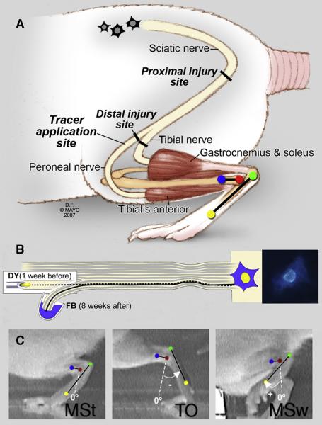
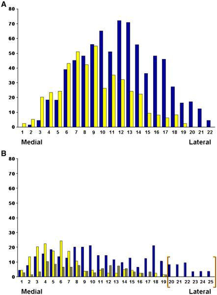
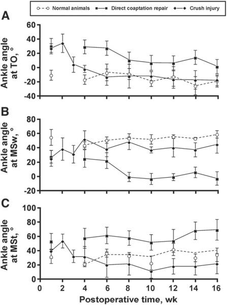
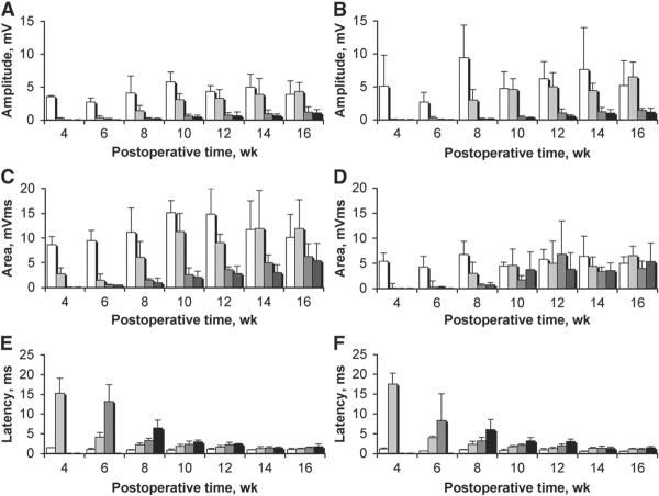
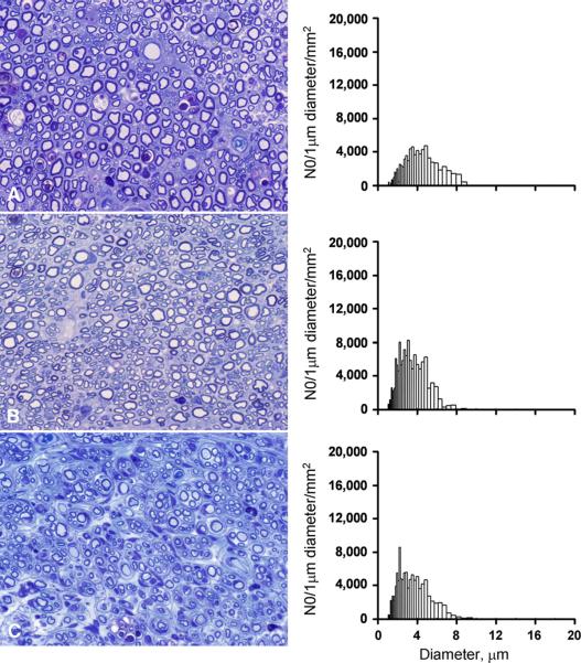
Misdirection of regenerating axons is one of the factors that can explain the poor results often found after nerve injury and repair. In this study, we quantified the degree of misdirection and the effect on recovery of function after different types of nerve injury and repair in the rat sciatic nerve model; crush injury, direct coaptation, and autograft repair. Sequential tracing with retrograde labeling of the peroneal nerve before and 8 weeks after nerve injury and repair was performed to quantify the accuracy of motor axon regeneration. Digital video analysis of ankle motion was used to investigate the recovery of function. In addition, serial compound action potential recordings and nerve and muscle morphometry were performed. In our study, accuracy of motor axon regeneration was found to be limited; only 71% (+/-4.9%) of the peroneal motoneurons were correctly directed 2 months after sciatic crush injury, 42% (+/-4.2%) after direct coaptation, and 25% (+/-6.6%) after autograft repair. Recovery of ankle motion was incomplete after all types of nerve injury and repair and demonstrated a disturbed balance of ankle plantar and dorsiflexion. The number of motoneurons from which axons had regenerated was not significantly different from normal. The number of myelinated axons was significantly increased distal to the site of injury. Misdirection of regenerating motor axons is a major factor in the poor recovery of nerves that innervate different muscles. The results of this study can be used as basis for developing new nerve repair techniques that may improve the accuracy of regeneration.
Figures
Similar articles
-
Accuracy of motor axon regeneration across autograft, single-lumen, and multichannel poly(lactic-co-glycolic acid) nerve tubes.Neurosurgery. 2008 Jul;63(1):144-53; discussion 153-5. doi: 10.1227/01.NEU.0000335081.47352.78. Neurosurgery. 2008. PMID: 18728579 Free PMC article.
-
The influence of motor axon misdirection on muscle contraction force in early nerve repair in a rat sciatic nerve model.Hand Surg. 2004 Dec;9(2):151-7. doi: 10.1142/s0218810404002200. Hand Surg. 2004. PMID: 15810099
-
Misdirection of regenerating axons and functional recovery following sciatic nerve injury in rats.J Comp Neurol. 2011 Jan 1;519(1):21-33. doi: 10.1002/cne.22446. J Comp Neurol. 2011. PMID: 21120925 Free PMC article.
-
Misdirection and guidance of regenerating axons after experimental nerve injury and repair.J Neurosurg. 2014 Feb;120(2):493-501. doi: 10.3171/2013.8.JNS122300. Epub 2013 Oct 11. J Neurosurg. 2014. PMID: 24116727 Review.
-
Role of chronic Schwann cell denervation in poor functional recovery after nerve injuries and experimental strategies to combat it.Neurosurgery. 2009 Oct;65(4 Suppl):A105-14. doi: 10.1227/01.NEU.0000358537.30354.63. Neurosurgery. 2009. PMID: 19927054 Review.
Cited by
-
Brief Electrical Stimulation Promotes Recovery after Surgical Repair of Injured Peripheral Nerves.Int J Mol Sci. 2024 Jan 4;25(1):665. doi: 10.3390/ijms25010665. Int J Mol Sci. 2024. PMID: 38203836 Free PMC article. Review.
-
Comments on "Comparison between normal and reverse orientation of graft in functional and histomorphological outcomes after autologous nerve grafting: An experimental study in the mouse model".Microsurgery. 2022 May;42(4):393-394. doi: 10.1002/micr.30876. Epub 2022 Mar 1. Microsurgery. 2022. PMID: 35229351 Free PMC article. No abstract available.
-
Treatment with Riluzole Restores Normal Control of Soleus and Extensor Digitorum Longus Muscles during Locomotion in Adult Rats after Sciatic Nerve Crush at Birth.PLoS One. 2017 Jan 17;12(1):e0170235. doi: 10.1371/journal.pone.0170235. eCollection 2017. PLoS One. 2017. PMID: 28095499 Free PMC article.
-
Collateral development and spinal motor reorganization after nerve injury and repair.Am J Transl Res. 2016 Jul 15;8(7):2897-911. eCollection 2016. Am J Transl Res. 2016. PMID: 27508011 Free PMC article.
-
The Effect of Electrical Stimulation on Nerve Regeneration Following Peripheral Nerve Injury.Biomolecules. 2022 Dec 12;12(12):1856. doi: 10.3390/biom12121856. Biomolecules. 2022. PMID: 36551285 Free PMC article. Review.
References
-
- Aldskogius H, Thomander L. Selective reinnervation of somatotopically appropriate muscles after facial nerve transection and regeneration in the neonatal rat. Brain Res. 1986;375:126–134. - PubMed
-
- Aldskogius H, Molander C, Persson J, Thomander L. Specific and nonspecific regeneration of motor axons after sciatic nerve injury and repair in the rat. J. Neurol. Sci. 1987;80:249–257. - PubMed
-
- Beer GM, Steurer J, Meyer VE. Standardizing nerve crushes with a non-serrated clamp. J. Reconstr. Microsurg. 2001;17:531–534. - PubMed
-
- Bernstein JJ, Guth L. Nonselectivity in establishment of neuromuscular connections following nerve regeneration in the rat. Exp. Neurol. 1961;4:262–275. - PubMed
Publication types
MeSH terms
Grants and funding
LinkOut - more resources
Full Text Sources
Other Literature Sources
