. 2008 Jul;212(1):14-28.
doi: 10.1016/j.expneurol.2008.01.031. Epub 2008 Feb 20.

Motor cortical stimulation promotes synaptic plasticity and behavioral improvements following sensorimotor cortex lesions

Affiliations

Motor cortical stimulation promotes synaptic plasticity and behavioral improvements following sensorimotor cortex lesions

DeAnna L Adkins et al. Exp Neurol. 2008 Jul.

Abstract

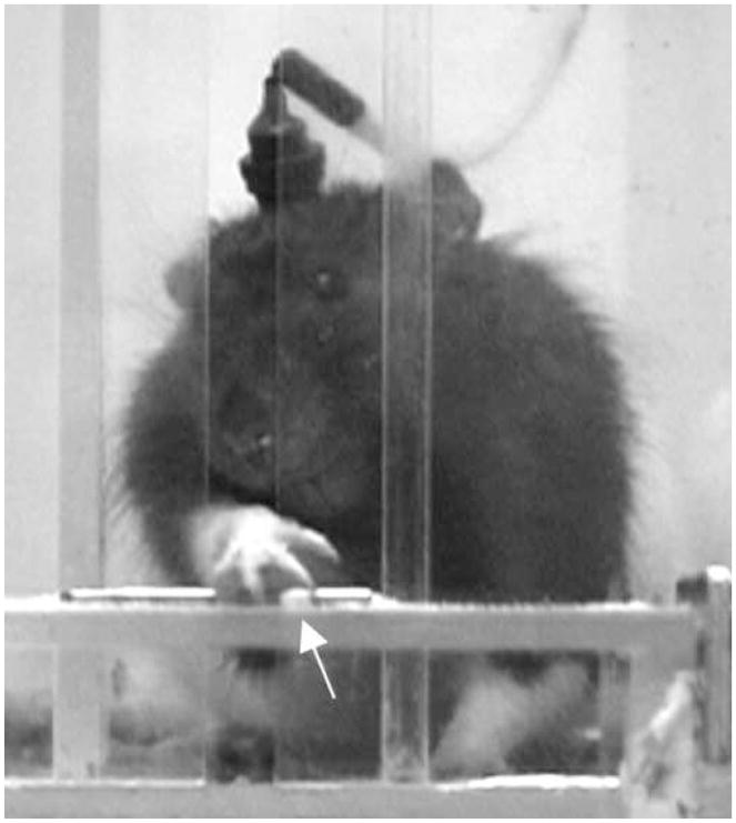
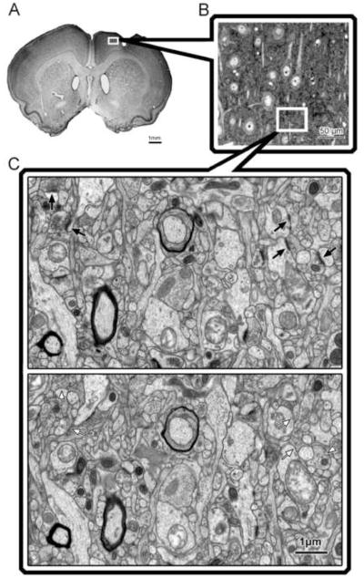
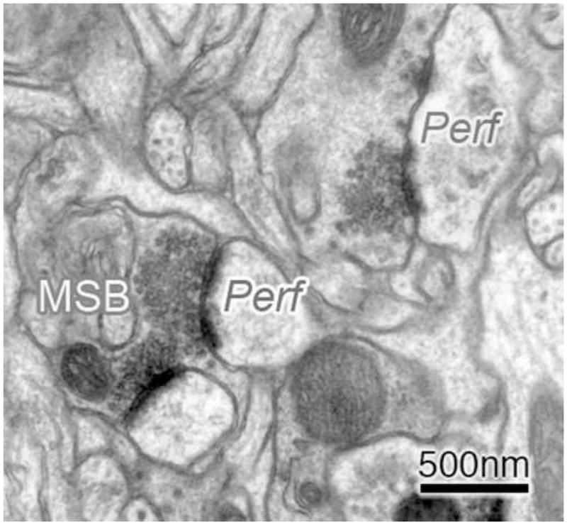
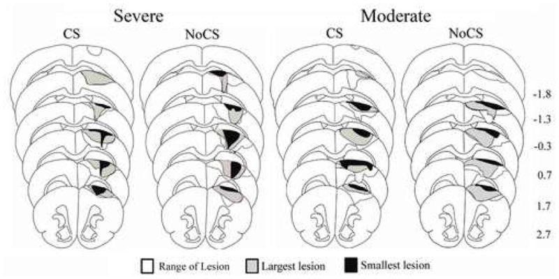
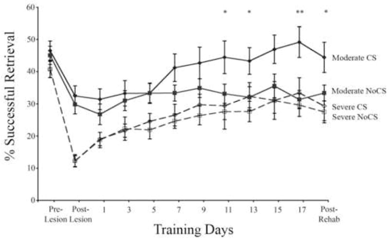
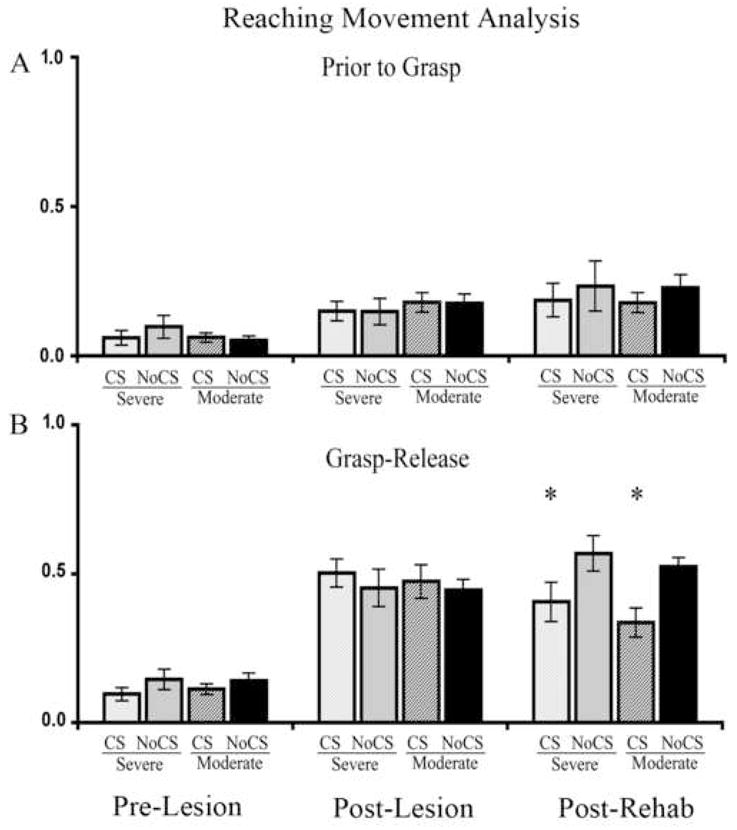
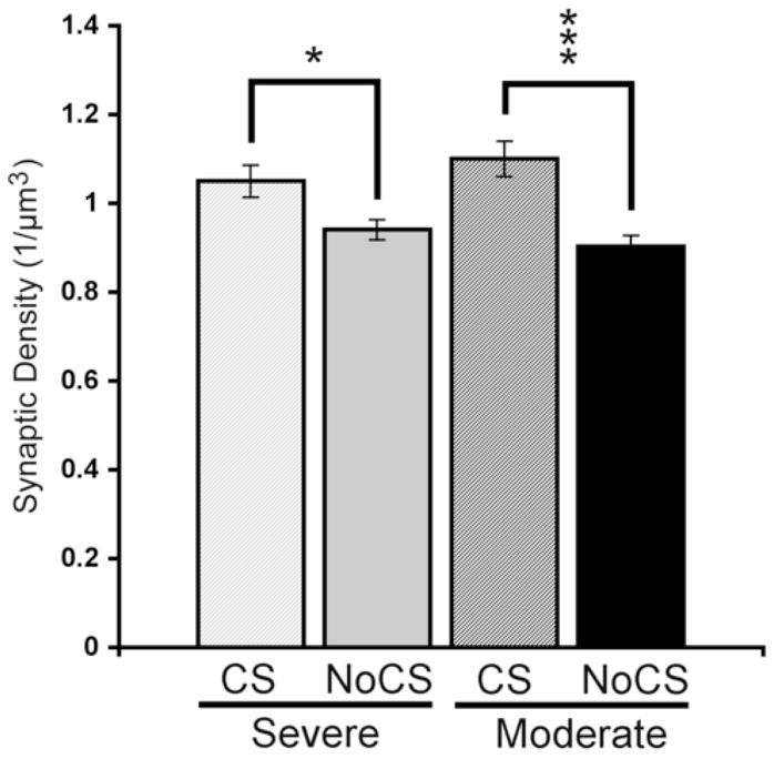
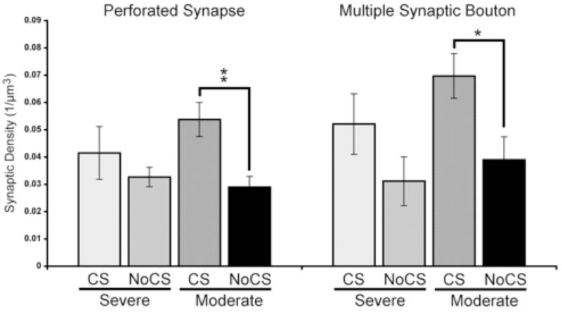
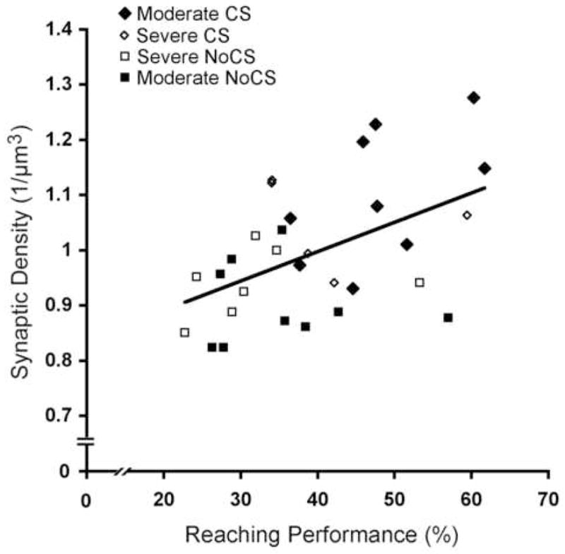
Cortical stimulation (CS) as a means to modulate regional activity and excitability in cortex is emerging as a promising approach for facilitating rehabilitative interventions after brain damage, including stroke. In this study, we investigated whether CS-induced functional improvements are linked with synaptic plasticity in peri-infarct cortex and vary with the severity of impairments. Adult rats that were proficient in skilled reaching received subtotal unilateral ischemic sensorimotor cortex (SMC) lesions and implantation of chronic epidural electrodes over remaining motor cortex. Based on the initial magnitude of reaching deficits, rats were divided into severely and moderately impaired subgroups. Beginning two weeks post-surgery, rats received 100 Hz cathodal CS at 50% of movement thresholds or no-stimulation control procedures (NoCS) during 18 days of rehabilitative training on a reaching task. Stereological electron microscopy methods were used to quantify axodendritic synapse subtypes in motor cortical layer V underlying the electrode. In moderately, but not severely impaired rats, CS significantly enhanced recovery of reaching success. Sensitive movement analyses revealed that CS partially normalized reaching movements in both impairment subgroups compared to NoCS. Additionally, both CS subgroups had significantly greater density of axodendritic synapses and moderately impaired CS rats had increases in presumed efficacious synapse subtypes (perforated and multiple synapses) in stimulated cortex compared to NoCS. Synaptic density was positively correlated with post-rehabilitation reaching success. In addition to providing further support that CS can promote functional recovery, these findings suggest that CS-induced functional improvements may be mediated by synaptic structural plasticity in stimulated cortex.


Figures

Figure 1
Figure 1. Single pellet retrieval task
Image of a rat reaching for and retrieving a pellet (arrow).
Figure 2
Figure 2. Sampling strategy in peri-lesion motor cortex
Changes in synaptic density in layer V of the peri-lesion cortex were assessed using the physical disector method for quantitative transmission electron microscopy. A, The region sampled was the remaining agranular cortex near the rostral portion of the lesion. B, Semithin sections were used for neuronal density estimates and to identify the sampling region for electron microscopy. C, Disector pair of electron micrographs of layer V motor cortex. Arrows indicate postsynaptic densities that disappear from one section to the next and are therefore counted.
Figure 3
Figure 3. Representative electron micrographs of subtypes of layer V motor cortical axodendritic synapses
Synapses with perforated or segmented post-synaptic densities were identified as perforated (Perf) synapses. Boutons forming synaptic contacts with more than one distinct dendritic element (spine or shaft) were identified as multisynaptic boutons (MSBs). These synapse subtypes were chosen for analysis because they have been linked to increased synaptic efficacy.
Figure 4
Figure 4. Reconstruction of sensorimotor cortex lesion extent and placement
Lesions produced damage to the forelimb representation region of the sensorimotor cortex (SMC). There were no major differences in extent or placement between groups nor differences in remaining cortical volume (see text for details). For each group, black outlines the range of all lesions, grey shading represents the largest lesion, and black shading represents the smallest lesion. Numbers to the right indicate approximate coronal section coordinates in mm relative to Bregma.
Figure 5
Figure 5. Cortical stimulation concurrent with rehabilitative training enhances reaching performance in moderately impaired animals
All animals had a decline in reaching performance from their pre-lesion level following SMC lesions (Post-Lesion) and an increase in success level over the 18 days of practice in the single pellet reaching task. In groups with initial success rates of > 20% after the lesions (Moderate), 100Hz epidural cortical stimulation (CS) significantly enhanced reaching success over days of testing compared to all other groups. CS did not produce performance enhancements in the animals with more severe initial deficits (≤ 20% success rates; Severe). Data are means ± S.E.M. *Moderate CS significantly different from Moderate NoCS, p < 0.05.
Figure 6
Figure 6. Cortical stimulation partially normalizes reaching movements following rehabilitative training
Average abnormality scores of movements prior to (“Prior to Grasp”) and after (“Grasp-Release”) pellet contact in the single pellet retrieval task. Movements were scored as normal (0.0), slightly abnormal (0.5), or highly abnormal (1.0) and averaged over 5 trials. A, After SMC lesions (“Post-Lesion”), all animals had a significant, but moderate, increased abnormality prior to grasping the pellet with their impaired forelimb compared to pre-lesion levels (“Pre-Lesion”). There were no significant group differences in these movement abnormalities. B, More severe abnormalities in grasp through the release of the pellet were found after the lesions. CS reduced these abnormalities in groups with both moderate and severe initial post-lesion impairments (Post-Rehab). Data are means ± S.E.M. *p < 0.01 CS versus NoCS of the same severity level.
Figure 7
Figure 7. Cortical stimulation increases axodendritic synaptic density
CS treated rats had greater synaptic densities compared with impairment-matched NoCS controls in both severely and moderately impaired animals. Data are means ± S.E.M. *p < 0.05, ***p < 0.001.
Figure 8
Figure 8. Cortical stimulation enhances the density of MSB and perforated synapses
There were significantly more perforated synapses and MSBs in the moderately impaired CS group compared to NoCS. Data are means ± S.E.M. *p < 0.05, **p < 0.01.
Figure 9
Figure 9. Synaptic density is correlated with reaching success on the single pellet reaching task
Axodendritic synaptic density was positively correlated with reaching performance (% successful retrievals) in the last training session. (r = 0.50, p < 0.01)

Similar articles

Cited by

References

    1. Adkins DL, Boychuk J, Remple MS, Kleim JA. Motor training induces experience-specific patterns of plasticity across motor cortex and spinal cord. J Appl Physiol. 2006;101:1776–1782. - PubMed
    1. Adkins DL, Campos P, Quach D, Borromeo M, Schallert K, Jones TA. Epidural cortical stimulation enhances motor function after sensorimotor cortical infarcts in rats. Exp Neurol. 2006;200:356–370. - PubMed
    1. Adkins DL, Voorhies AC, Jones TA. Behavioral and neuroplastic effects of focal endothelin-1 induced sensorimotor cortex lesions. Neuroscience. 2004;128:473–486. - PubMed
    1. Adkins-Muir DL, Jones TA. Cortical electrical stimulation combined with rehabilitative training: enhanced functional recovery and dendritic plasticity following focal cortical ischemia in rats. Neurol Res. 2003;25:780–788. - PubMed
    1. Adkins-Muir DL, Luke L, Fowler B, Jones TA. Electrical stimulation of the peri-lesion cortical surface during motor skills training improves behavioral function after ischemic lesions of the sensorimotor cortex in adult rats. Soc Neurosci Abstr. 2002;28:489.4.

Publication types

MeSH terms