. 2008 Jun;54(8):1189-200.
doi: 10.1016/j.neuropharm.2008.03.010. Epub 2008 Mar 28.

Extending the analysis of nicotinic receptor antagonists with the study of alpha6 nicotinic receptor subunit chimeras

Affiliations

Extending the analysis of nicotinic receptor antagonists with the study of alpha6 nicotinic receptor subunit chimeras

Roger L Papke et al. Neuropharmacology. 2008 Jun.

Abstract

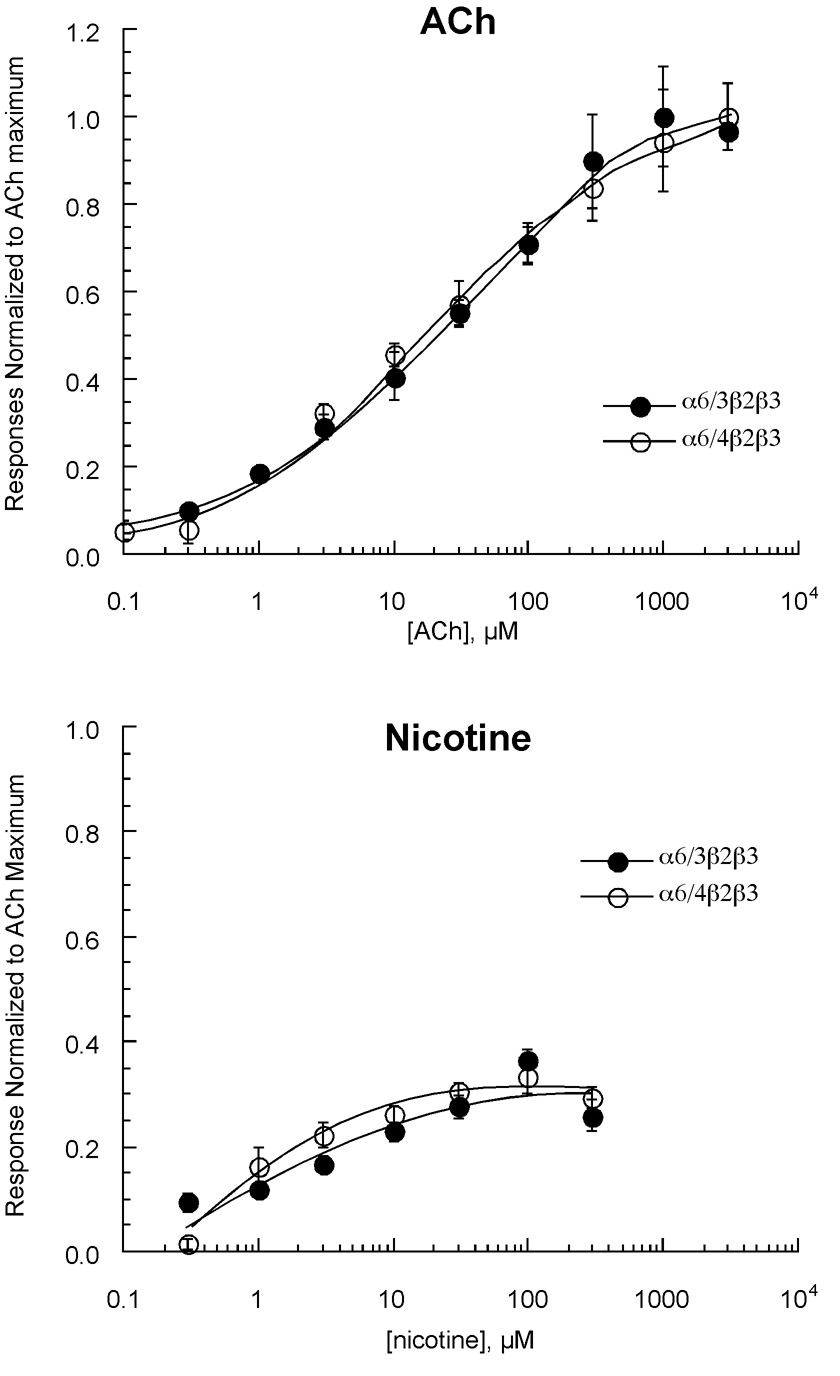
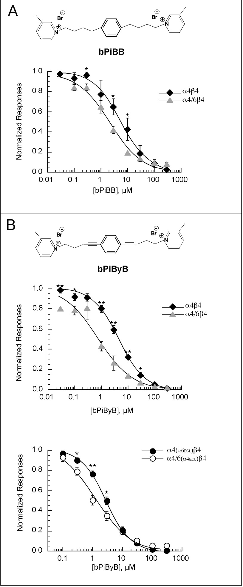
Heterologous expression systems have increased the feasibility of developing selective ligands to target nicotinic acetylcholine receptor (nAChR) subtypes. However, the alpha6 subunit, a component in nAChRs that mediates some of the reinforcing effects of nicotine, is not easily expressed in systems such as the Xenopus oocyte. Certain aspects of alpha6-containing receptor pharmacology have been studied by using chimeric subunits containing the alpha6 ligand-binding domain. However, these chimeras would not be sensitive to an alpha6-selective channel blocker; therefore we developed an alpha6 chimera (alpha4/6) that has the transmembrane and intracellular domains of alpha6 and the extracellular domain of alpha4. We examined the pharmacological properties of alpha4/6-containing receptors and other important nAChR subtypes, including alpha7, alpha4beta2, alpha4beta4, alpha3beta4, alpha3beta2, and alpha3beta2beta3, as well as receptors containing alpha6/3 and alpha6/4 chimeras. Our data show that the absence or presence of the beta4 subunit is an important factor for sensitivity to the ganglionic blocker mecamylamine, and that dihydro-beta-erythroidine is most effective on subtypes containing the alpha4 subunit extracellular domain. Receptors containing the alpha6/4 subunit are sensitive to alpha-conotoxin PIA, while receptors containing the reciprocal alpha4/6 chimera are insensitive. In experiments with novel antagonists of nicotine-evoked dopamine release, the alpha4/6 chimera indicated that structural rigidity was a key element of compounds that could result in selectivity for noncompetitive inhibition of alpha6-containing receptors. Our data extend the information available on prototypical nAChR antagonists, and establish the alpha4/6 chimera as a useful new tool for screening drugs as selective nAChR antagonists.


Figures

Figure 1
Figure 1
Inhibition of responses obtained from oocytes expressing rat neuronal nAChR subunits by mecamylamine (upper panel) or DHβE (lower panel). Each panel shows raw data traces for α4β2 and α3β4 receptors on the left and on the right the averaged normalized responses (± SEM, n ≥ 4) from oocytes expressing α4β2, α3β4, or α7 subunits to the co-application ACh and increasing concentrations of antagonist. Each response to the co-application of ACh and antagonist was normalized based on the response of the same cell to the control application of ACh alone (see Methods for control ACh concentrations used).
Figure 2
Figure 2
Schematic representation of wild-type and chimeric subunits used to study the influence of α6 subdomains of the pharmacological properties of neuronal nAChR. The putative transmembrane domains of α6 are identified in the Kyte-Doolittle plot (11/1 generated by DNA Strider) at the top of the figure. These domains are identified by the wide bars in the schematics below. All the schematics are at the same scale as the Kyte-Doolittle plot but omit the signal sequences. An expansion and overlay of Kyte-Doolittle hydrophobicity plots for the transmembrane domains of α6 (black) and α4 (gray) is shown at the bottom of the figure.
Figure 3
Figure 3
Concentration-response curves obtained from oocytes expressing rat neuronal nAChR α6/3β2β3 or α6/4β2β3 subunits given ACh (upper panel) or nicotine (lower panel). Each panel shows the averaged data (± SEM, n ≥ 4) normalized to the maximum ACh responses obtained from the same oocytes.
Figure 4
Figure 4
Inhibition of responses obtained from oocytes expressing rat neuronal nAChR subunits by mecamylamine (upper panel) or DHβE (lower panel). Each panel shows the averaged normalized data (± SEM, n ≥ 4) from oocytes expressing α3β2, α3β2β3, or α6/3β2β3 subunits to the co-application of ACh and antagonist. Each response to the co-application of ACh and antagonist was normalized based on the response of the same cell to the control application of ACh alone (see Methods for control ACh concentrations used). Significant (p<0.05) differences in inhibition were found between receptor subtypes at the concentrations indicated. ⊗ represents significant difference between α3β2 and α3β2β3 responses, # represents significant difference between α3β2β3 and α3/6β2β3 responses, and * represents significant difference between α3β2 and α3/6β2β3 responses.
Figure 5
Figure 5
Concentration-response curves obtained from oocytes expressing rat neuronal nAChR α4β4 or α4/6β4 subunits given ACh. Shown are the averaged data (± SEM, n ≥ 4) normalized to the maximum ACh responses obtained from the same oocytes.
Figure 6
Figure 6
Inhibition of responses obtained from oocytes expressing rat neuronal α4β4, α6/4β4 or α4/6β4 nAChR subunits to competitive antagonists. The inhibition of responses to DHβE are shown in the upper panel. The lower panel illustrates the effects of 100 nM α-conotoxin PIA on ACh-evoked responses. Initial control responses to 100 µM ACh were obtained, then cells were incubated for 5 min in 100 nM toxin (plus 0.1 mg/ml protease-free BSA), and then ACh was applied in the continued presence of the toxin. Each response to the co-application of ACh and antagonist was normalized based on the response of the same cell to the control application of ACh alone (see Methods for control ACh concentrations used). Also shown are the Ach-evoked responses obtained after a 5 min washout of the toxin. Each panel shows the averaged normalized data (± SEM, n ≥ 4) from oocytes.
Figure 7
Figure 7
Inhibition of peak current responses obtained from oocytes expressing rat neuronal α4β4 or α4/6β4 nAChR subunits by mecamylamine (upper panel) or tetracaine (lower panel). Each panel shows the averaged normalized responses (± SEM, n ≥ 4) from oocytes expressing those subunits to the co-application of ACh and antagonist. Each response to the co-application of ACh and antagonist was normalized based on the response of the same cell to the control application of ACh alone (see Methods for control ACh concentrations used). Significant differences in inhibition were found at the concentrations indicated (* indicates p<0.05, ** indicates p <0.01).
Figure 8
Figure 8
A) The structure of bPiDDB (N,N’-dodecane-1,12-diyl-bis-3-picolinium dibromide), a novel bis-azaaromatic quaternary ammonium compound which functions as a partial antagonist of nicotine-evoked dopamine release (Crooks et al., 2004), is shown at the top of the figure. The upper plot is a competition experiment, comparing responses evoked by ACh or ACh plus 10 µM bPiDDB. The lower plot illustrates the inhibition of peak current responses obtained from oocytes expressing rat neuronal α4β4 or α4/6β4 nAChR subunits by bPiDDB. B) The structure of bPiPyB (1,2-bis-[5-(3-picolinium)-pent-1-ynyl]-benzene dibromide) is shown at the top of the panel. The plot illustrates the inhibition of peak current responses obtained from oocytes expressing rat neuronal α4β4 or α4/6β4 nAChR subunits by bPiPyB. Each panel shows the averaged normalized responses (± SEM, n ≥ 4) from oocytes expressing those subunits to the co-application of ACh and antagonist. Each response to the co-application of ACh and antagonist was normalized based on the response of the same cell to the control application of ACh alone (see Methods for control ACh concentrations used). Significant differences in inhibition were found at the concentrations indicated (* indicates p<0.05, ** indicates p <0.01).
Figure 9
Figure 9
A) The structure of bIQDDB (N,N’-dodecane-1,12-diyl-bis-3-isoquinolinium dibromide) is shown at the top of the panel. The plot illustrates the inhibition of peak current responses obtained from oocytes expressing rat neuronal α4β4 or α4/6β4.nAChR subunits by bIQDDB. B) The structure of bIQPB (1,2-bis-(5-isoquinolinium-pentyl)-benzene dibromide) is shown at the right of the panel. The plot illustrates the inhibition of peak current responses obtained from oocytes expressing rat neuronal α4β4 or α4/6β4 nAChR subunits by bIQPB. C) The structure of bIQPyB (1,2-bis-(5-isoquinolinium-pent-1-ynyl)-benzene dibromide) is shown at the right of the panel. The plot illustrates the inhibition of peak current responses obtained from oocytes expressing rat neuronal α4β4 or α4/6β4 nAChR subunits by bIQPyB. The data in each panel show the averaged normalized responses (± SEM, except where indicated, n ≥ 4) from oocytes expressing those subunits to the co-application of ACh and antagonist. Each response to the co-application of ACh and antagonist was normalized based on the response of the same cell to the control application of ACh alone (see Methods for control ACh concentrations used). Significant differences in inhibition were found at the concentrations indicated (* indicates p<0.05, ** indicates p <0.01).
Figure 10
Figure 10
A) The structure of bPiBB (1,4-bis-[4-(3-picolinium)-butyl]-benzene dibromide) is shown at the top of the panel. The plot illustrates the inhibition of peak current responses obtained from oocytes expressing rat neuronal α4β4 or α4/6β4 nAChR subunits by bPiBB. B) The structure of bPiByB (1,4-bis-[4-(3-picolinium)-but-1-ynyl]-benzene dibromide) is shown at the top of the panel. The upper plot illustrates the inhibition of peak current responses obtained from oocytes expressing rat neuronal α4β4 or α4/6β4 nAChR subunits by bPiByB (n = 4 for the α4β4 data and n = 3 for the α4/6β4 data). The lower plot shows the inhibition produced by bPiByB of α4(α6ECL)β4 or α4/6(α4ECL)β4, double mutants which contain in α4 the two residues from α6 which differ in the extracellular loop between TM2 and TM3 (see Table 3) and in the α4/6 chimera, the two extracellular loop residues from α4, respectively. The data in each panel show the averaged normalized responses (± SEM) from oocytes expressing those subunits to the co-application of ACh and antagonist. Each response to the co-application of ACh and antagonist was normalized based on the response of the same cell to the control application of ACh alone (see Methods for control ACh concentrations used). Significant differences in inhibition were found at the concentrations indicated (* indicates p<0.05, ** indicates p <0.01).

Similar articles

Cited by

References

    1. Ayers JT, Dwoskin LP, Deaciuc AG, Grinevich VP, Zhu J, Crooks PA. bis-Azaaromatic quaternary ammonium analogues: ligands for alpha4beta2* and alpha7* subtypes of neuronal nicotinic receptors. Bioorganic Medicinal Chemistry Letters. 2002;12(21):3067–3071. - PubMed
    1. Azam L, Chen Y, Leslie FM. Developmental regulation of nicotinic acetylcholine receptors within midbrain dopamine neurons. Neuroscience. 2007;144(4):1347–1360. - PMC - PubMed
    1. Azam L, McIntosh JM. Characterization of nicotinic acetylcholine receptors that modulate nicotine-evoked [3H]norepinephrine release from mouse hippocampal synaptosomes. Molecular Pharmacology. 2006;70(3):967–976. - PubMed
    1. Bouzat C, Gumilar F, Spitzmaul G, Wang HL, Rayes D, Hansen SB, Taylor P, Sine SM. Coupling of agonist binding to channel gating in an ACh-binding protein linked to an ion channel. Nature. 2004;430(7002):896–900. - PubMed
    1. Charpantier E, Barneoud P, Moser P, Besnard F, Sgard F. Nicotinic acetylcholine subunit mRNA expression in dopaminergic neurons of the rat substantia nigra and ventral tegmental area. Neuroreport. 1998;9(13):3097–3101. - PubMed

Publication types

MeSH terms

LinkOut - more resources