. 2008 Jun 2;153(4):944-62.
doi: 10.1016/j.neuroscience.2008.02.020. Epub 2008 Feb 21.

Expression of reelin, its receptors and its intracellular signaling protein, Disabled1 in the canary brain: relationships with the song control system

Affiliations

Expression of reelin, its receptors and its intracellular signaling protein, Disabled1 in the canary brain: relationships with the song control system

J Balthazart et al. Neuroscience. .

Abstract

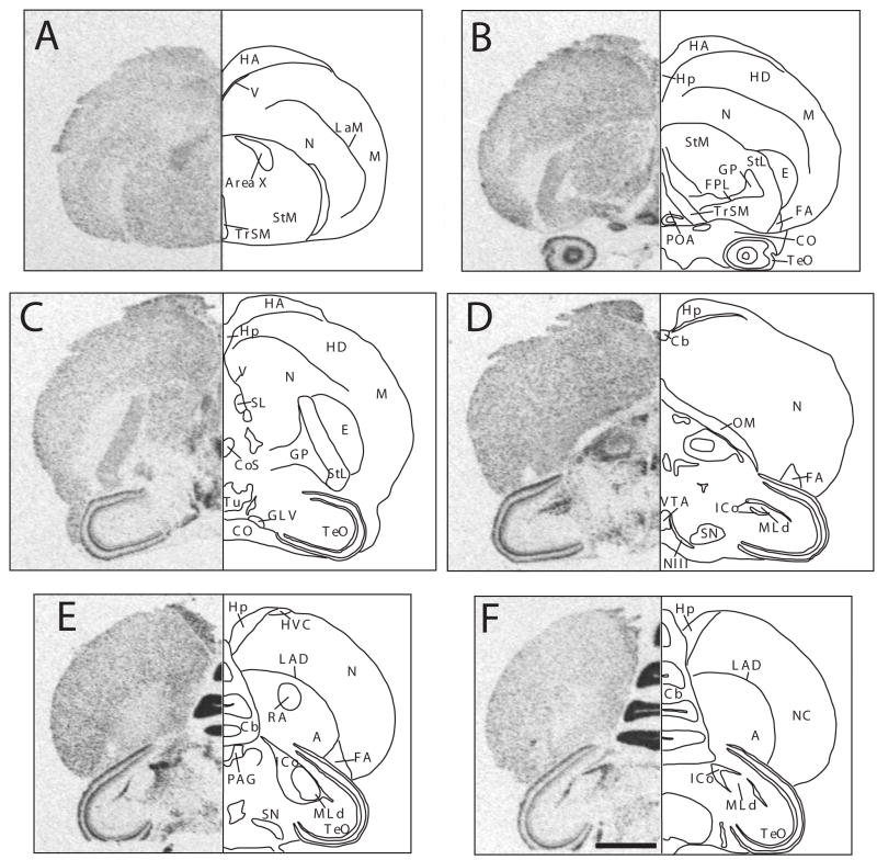
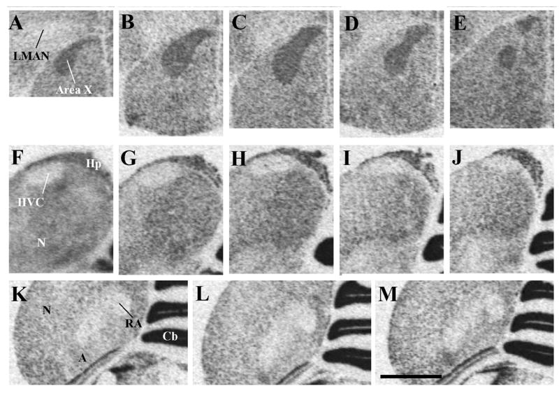
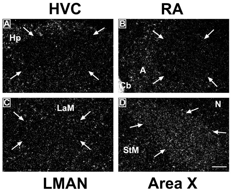
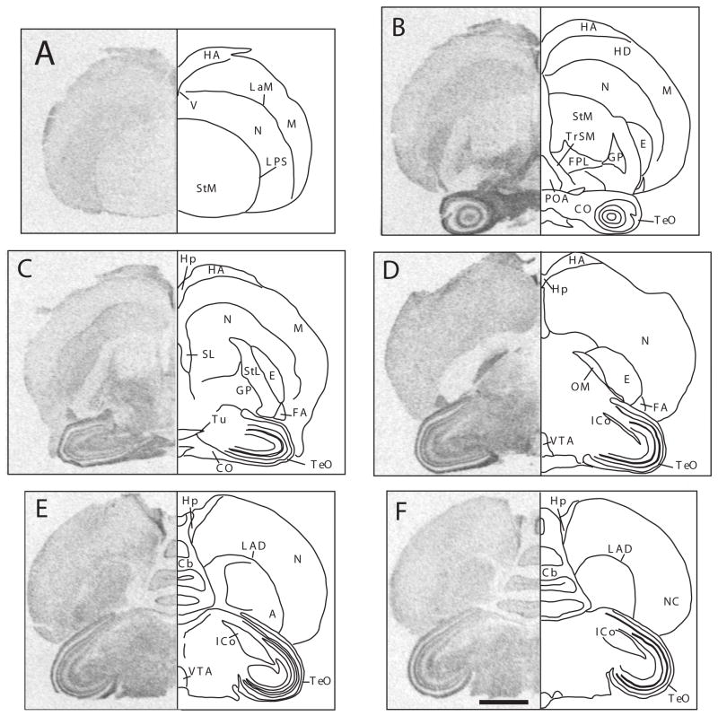
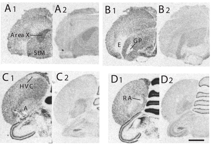
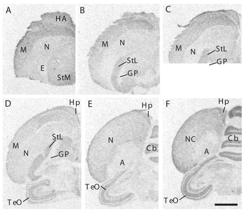
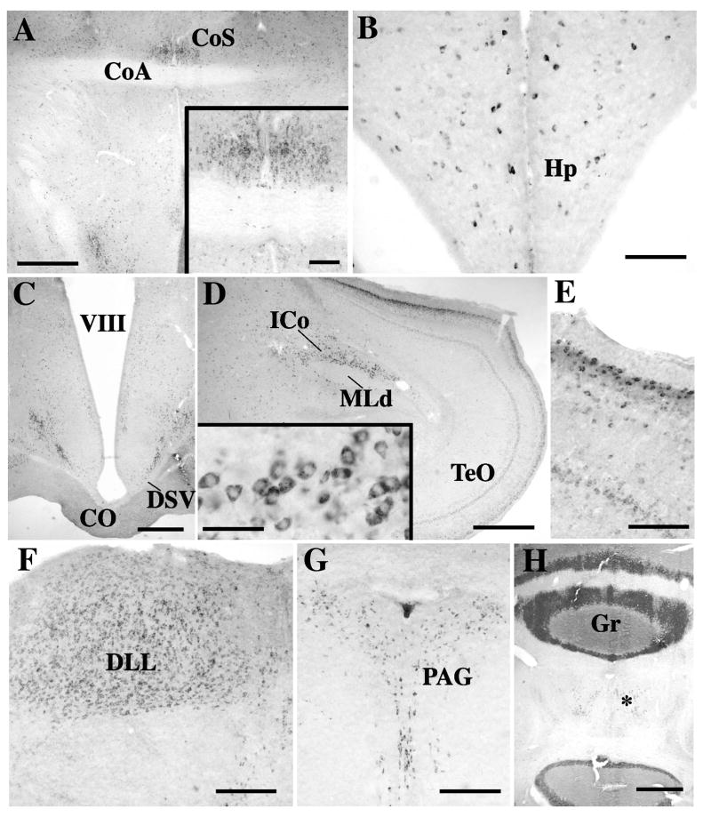
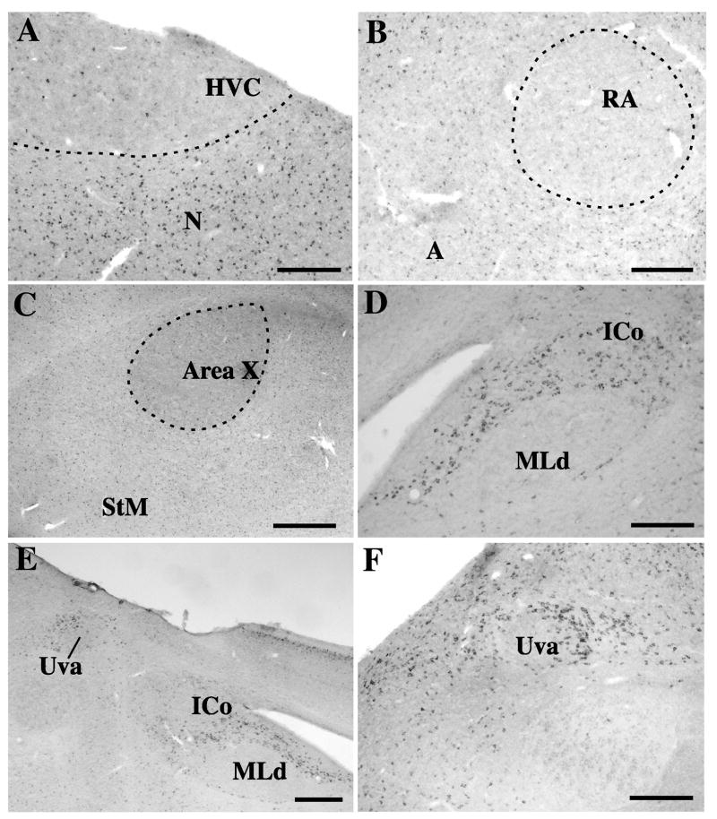
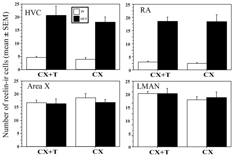
Songbirds produce learned vocalizations that are controlled by a specialized network of neural structures, the song control system. Several nuclei in this song control system demonstrate a marked degree of adult seasonal plasticity. Nucleus volume varies seasonally based on changes in cell size or spacing, and in the case of nucleus HVC and area X on the incorporation of new neurons. Reelin, a large glycoprotein defective in reeler mice, is assumed to determine the final location of migrating neurons in the developing brain. In mammals, reelin is also expressed in the adult brain but its functions are less well characterized. We investigated the relationships between the expression of reelin and/or its receptors and the dramatic seasonal plasticity in the canary (Serinus canaria) brain. We detected a broad distribution of the reelin protein, its mRNA and the mRNAs encoding for the reelin receptors (VLDLR and ApoER2) as well as for its intracellular signaling protein, Disabled1. These different mRNAs and proteins did not display the same neuroanatomical distribution and were not clearly associated, in an exclusive manner, with telencephalic brain areas that incorporate new neurons in adulthood. Song control nuclei were associated with a particular specialized expression of reelin and its mRNA, with the reelin signal being either denser or lighter in the song nucleus than in the surrounding tissue. The density of reelin-immunoreactive structures did not seem to be affected by 4 weeks of treatment with exogenous testosterone. These observations do not provide conclusive evidence that reelin plays a prominent role in the positioning of new neurons in the adult canary brain but call for additional work on this protein analyzing its expression comparatively during development and in adulthood with a better temporal resolution at critical points in the reproductive cycle when brain plasticity is known to occur.


Figures

Figure 1
Figure 1
Autoradiograms of coronal sections through a female canary brain processed by in situ hydridization for the reelin mRNA (left side of each panel) and mirror-image drawing presenting the interpretation of these autoradiograms and names of labeled structures. Panles A through F represent sections in a rostral to caudal order. Magnification bar: 2 mm. Abbreviations: A: arcopallium; Area X: area X of the medial striatum; Cb: cerebellum; CO: chiasma opticum; CoS: nucleus commussuralis septi; E: entopallium; FA: tractus fronto-arcopallialis; FPL: fasciculus prosencephali lateralis; GLV: nucleus geniculatus lateralis, pars ventralis; GP: globus pallidus; HA: hyperpallium apicale; HD: hyperpallium densocellulare; Hp: hippocampus; HVC: song control nucleus HVC, used as a proper name; ICo: nucleus intercollicularis; LAD: lamina arcopallialis dorsalis; LaM: lamina mesopallialis; M: mesopallium; MLd: nucleus mesencephalicus lateralis, pars dorsalis; N: nidopallium; NIII: nervus occulomotorius (third nerve); OM: tractus occipitomesencephalicus; PAG: periaqueductal gray; NC: nidopallium caudale; POA: preoptic area; RA: nucleus robustus arcopallialis; SL: nucleus septalis lateralis; SN: substantia nigra; StL: striatum laterale; StM: striatum mediale; TeO: tectum opticum; TrSM: tractus sepatopallio-mesencephalicus; Tu: tuber; V: ventriculus; VTA: ventral tegmental area.
Figure 2
Figure 2
Autoradiograms of coronal sections through a male canary brain processed by in situ hydridization for the reelin mRNA illustrating the higher density of reelin expression in the song control nucleus area X (panels A–E) compared to the surrounding striatum mediale (StM) but the much lower expression in HVC (panels F–J) and in nucleus robustus arcopallialis (RA, panels K–M) compared to the surrounding nidopallium (N) and arcopallium (A) respectively. Panels located on a same row and illustrating a given nucleus are presented from left to right in a rostral to caudal order. Magnification bar: 2 mm. Abbreviations: A: arcopallium; Cb, cerebellum; Hp, hippocampus; HVC: song control nucleus HVC; LMAN: magnocellular nucleus of the anterior nidopallium; N: nidopallium; StM: striatum mediale;
Figure 3
Figure 3
Darkfield photomicrographs of emulsion-dipped sections illustrating the expression of reelin within the telencephalic song control nuclei HVC (A), robust nucleus of the arcopallium (RA; B), lateral part of the nucleus magnocellularis nidopalli anterioris (LMAN; C) and Area X (D). Arrows indicate the boundaries of the brain regions. Pictures show the right brain hemisphere. Dorsal is at the top and lateral is on the right in these coronal sections. Abbreviations: A, arcopallium; Cb, cerebellum; Hp, hippocampus; LaM, Lamina mesopallialis; StM: striatum mediale. Magnification bar=200 μm.
Figure 4
Figure 4
Autoradiograms of coronal sections through a female canary brain processed by in situ hydridization for the Dab1 mRNA (left side of each panel) and mirror-image drawing presenting the interpretation of these autoradiograms and names of labeled structures. Panels A through F represent sections in a rostral to caudal order. The sections illustrated come from the same subject and are adjacent to sections labeled for reelin that are illustrated in figure 1. Magnification bar: 2 mm. Abbreviations: A: arcopallium; Cb: cerebellum; CO: chiasma opticum; E: entopallium; FA: tractus fronto-arcopallialis; FPL: fasciculus prosencephali lateralis; GP: globus pallidus; HA: hyperpallium apicale; HD: hyperpallium densocellulare; Hp: hippocampus; ICo: nucleus intercollicularis; LAD: lamina arcopallialis dorsalis; LaM: lamina mesopallialis; LPS: lamina pallio-subpalllialis; M: mesopallium; N: nidopallium; OM: tractus occipitomesencephalicus; NC: nidopallium caudale; POA: preoptic area; SL: nucleus septalis lateralis; StM: striatum laterale; StM: striatum mediale; TeO: tectum opticum; TrSM: tractus sepatopallio-mesencephalicus; Tu: tuber; V: ventriculus; VTA: ventral tegmental area.
Figure 5
Figure 5
Autoradiograms of coronal sections through the brain of a male canary processed by in situ hydridization for the reelin (A1, B1, C1, D1) or ApoER2 (A2, B2, C2, D2) mRNA. Panels A through D represent sections in a rostral to caudal order. The sections illustrated in panels designed by a same letter come from adjacent sections from the same subject. Magnification bar: 2 mm. Abbreviations: A: arcopallium; GP: globus pallidus; HVC: song control nucleus HVC; RA: nucleus robustus arcopallialis; StM: striatum mediale.
Figure 6
Figure 6
Autoradiograms of coronal sections through the brain of a male canary processed by in situ hydridization for VLDLR. Magnification bar: 2 mm. Abbreviations: A: arcopallium; Cb: cerebellum; GP: globus pallidus; HA: hyperpallium apicale; Hp: hippocampus; M: mesopallium; N: nidopallium; NC: nidopallium caudale; StL: striatum laterale; StM: striatum mediale; TeO: tectum opticum.
Figure 7
Figure 7
Photomicrographs illustrating the distribution of reelin-ir cells in the canary brain. A. Preoptic area at the level of the anterior commissure. The nucleus of the commisuralis septi (CoS), just dorsal to the commissural anterior (CoA) is clearly highlighted by a dense cluster of positive cells (also shown at higher magnification in the insert). B. Scattered cells are present in the hippocampus (Hp). C. Low densities of positive cells are detected throughout the hypothalamus. In the infundibular hypothalamus two clusters of reelin-ir cells for diagonal bands just dorsal to the chiasma opticum (CO) and the decussatio supraoptica ventralis (DSV). D. In the mesencephalon, two layers of the optic tectum (TeO) contain high density of positive cells. In the tegmentum the nucleus intercollicularis (ICo) is highlighted by a dense cluster of positive cells. These cells are also shown at higher magnification in the insert. E. Higher magnification of a fragment of the optic lobes illustrating the two layers of positive cells surrounding the nucleus mesencephalicus lateralis, pars dorsalis (MLd). F. The nucleus dorsolateralis anterior thalami, pars lateralis (DLL) is entirely filled by reelin-ir cells. G. Positive cells outline the entire periqueductal gray (PAG). H. In the cerebellum a very dense immunoreactive material is present in the granular layer (Gr) and in the Purkinje cells. Reelin-ir cells are also observed in the deeper layers (asterisk). Magnification bars: 500 μm in A, C, D, H; 50 μm in the insert of panel D; 100 μm in insert of panel A, in B, E, 200 μm in F, G.
Figure 8
Figure 8
Photomicrographs illustrating the distribution of reelin-ir cells in and around the song control nuclei of the canary brain. A. HVC and adjacent nidopallium (N). B. nucleus robustus arcopallialis (RA) and adjacent arcopallium (A). C. area X and striatum mediale (StM). D. Nucleus intercollicularis (ICo) surrounding almost completely the nucleus mesencephalicus lateralis, pars dorsalis (MLd). E. low power enlargement of the medial part of the tegmentum containing the ICo, MLd and nucleus uvaeformis (Uva). F. High power magnification illustrating the reelin-ir cells sourounding nucleus Uva. Magnification bars: 200 μm in A, B; D, and F, 500 μm in C and E.
Figure 9
Figure 9
Bar graph illustrating the number of reelin-ir cells inside the 4 song control nuclei HVC, RA, area X and LMAN and in the surrounding tissue just adjacent to the nuclei and effects of the treatment with testosterone on the numbers of these cells in castrated males. Significantly fewer cells are found in HVC and RA than in surrounding tissue but this difference is not observed for area X and MAN. No effect of testosterone could be detected.

Similar articles

Cited by

References

    1. Aamodt SM, Kozlowski MR, Nordeen EJ, Nordeen KW. Distribution and developmental change in [3H]MK-801 binding within zebra finch song nuclei. J Neurobiol. 1992;23:997–1005. - PubMed
    1. Absil P, Pinxten R, Balthazart J, Eens M. Effects of testosterone on Reelin expression in the brain of male European starlings. Cell Tissue Res. 2003;312:81–93. - PubMed
    1. Alcantara S, Ruiz M, D’Arcangelo G, Ezan F, de Lecea L, Curran T, Sotelo C, Soriano E. Regional and cellular patterns of reelin mRNA expression in the forebrain of the developing and adult mouse. J Neurosci. 1998;18:7779–7799. - PMC - PubMed
    1. Alvarez-Buylla A, Kirn JR. Birth, migration, incorporation, and death of vocal control neurons in adult songbirds. J Neurobiol. 1997;33:585–601. - PubMed
    1. Alvarez-Buylla A, Nottebohm F. Migration of young neurons in adult avian brain. Nature. 1988;335:353–354. - PubMed

Publication types

MeSH terms