. 2008 May 30;178(3):167-75.
doi: 10.1016/j.toxlet.2008.03.012. Epub 2008 Mar 27.

Benzo(a)pyrene-caused increased G1-S transition requires the activation of c-Jun through p53-dependent PI-3K/Akt/ERK pathway in human embryo lung fibroblasts

Affiliations

Benzo(a)pyrene-caused increased G1-S transition requires the activation of c-Jun through p53-dependent PI-3K/Akt/ERK pathway in human embryo lung fibroblasts

Shi Jiao et al. Toxicol Lett. .

Abstract

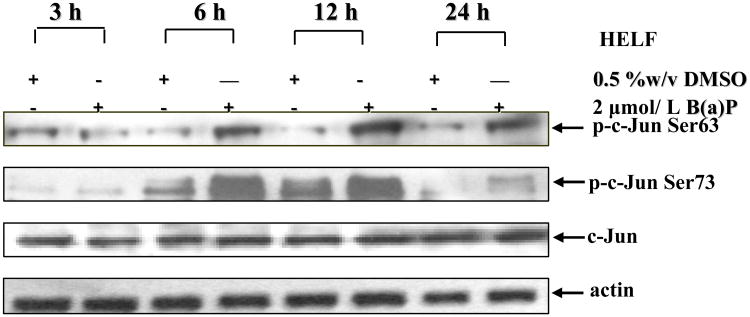
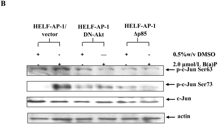
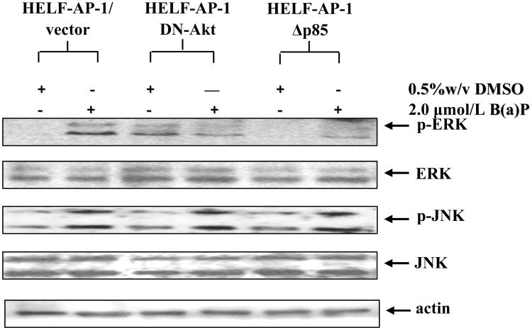
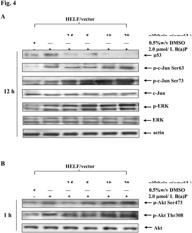
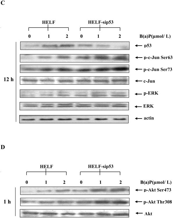
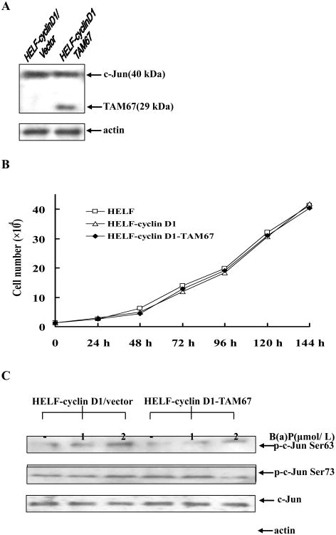
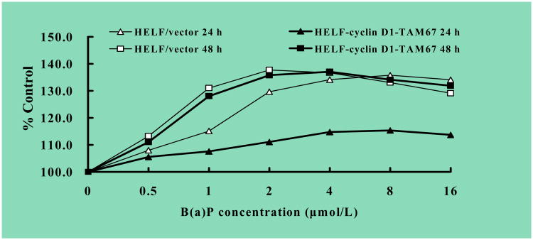
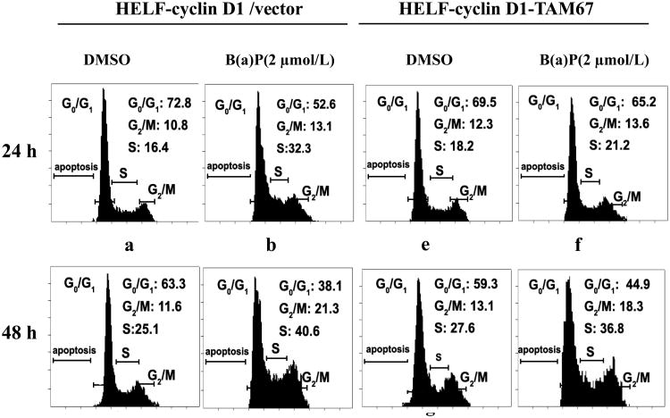
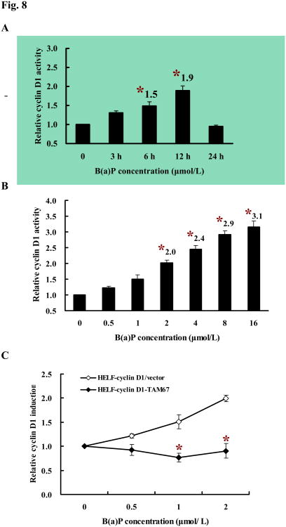
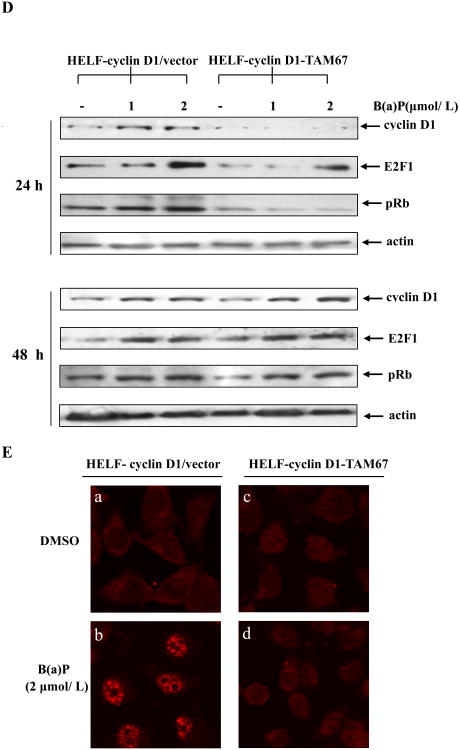
Benzo(a)pyrene (B(a)P) is a potent lung carcinogen mainly derived from tobacco smoking and environmental contamination, however, the molecular mechanisms by which it accelerates the cell cycle progression and induces the abnormal cell proliferation are still far away from understood. Our current analysis of human embryo lung fibroblasts (HELF) showed that B(a)P exposure was able to promote cell cycle G(1)-S phase transition. This effect was correlated with c-Jun activation because inhibition of c-Jun by its dominant negative mutant (TAM67) reversed B(a)P action on cell cycle with the down-regulation of expression of cyclin D1, pRb and E2F1. Further study found that overexpression of dominant negative mutants of, PI-3K or Akt, dramatically reduced B(a)P-induced the activation of c-Jun and extracellular signaling regulated kinase (ERK), but not c-Jun NH2 terminal kinase (JNK). Inhibition of p53 by either its inhibitor pifithrin-alpha or p53 siRNA markedly increased B(a)P-induced the activation of c-Jun, Akt and ERK in this context. Take together, our results indicate that c-Jun activation by p53-dependent PI-3K/Akt/ERK pathway is responsible for B(a)P-induced cell cycle alternations in human embryo lung fibroblasts.


Figures

Fig. 1
Fig. 1. B(a)P enhanced c-Jun phosphorylation in HELF cells
HELF cells were cultured until subconfluent (80-85 %) in 75-cm2 culture flasks with RPMI1640 containing 10% FBS, and then replaced with 0.5% FBS RPMI-1640. After being cultured for 24 h, the cells were exposed to 2 μmol/L B(a)P for various time points as indicated. The cells were then washed once with ice-cold PBS and extracted with SDS sample buffer. The cell extracts were separated on polyacrylamide-SDS gels, transferred, and probed with specific antibodies as indicated, and then detected with secondary antibody as indicated. The results were one representative data from three independent experiments.
Fig. 2
Fig. 2. PI-3K/Akt pathway mediated B(a)P-induced c-Jun activation
A, HELF-AP-1/vector (a and b), HELF-AP-1-DN-Akt (c and d) and HELF-AP-1- DN-Δp85 (e and f) were seeded into six-well plates respectively and cultured in 10% FBS RPMI 1640 medium at 37°C overnight. After being serum-starved for 24 h, cells were untreated (a, c and e) or treated (b, d and f) with 2 μmol/L B(a)P for 6 h, and then fixed. The fixed cells were subsequently blocked with 5% goat serum, incubated with phospho-specific c-Jun at Ser63 antibody, and detected by immunofluorescence assay with FITC-conjugated second antibody (green). B, The aforementioned cells were incubated with 2 μmol/L B(a)P for 12 h, and then extracted, sequentially detected by Western blot with specific antibodies as indicated.
Fig. 3
Fig. 3. ERK activation was essential for PI-3K/Akt pathway mediating B(a)P-induced c-Jun activation
HELF-AP-1/vector, HELF-AP-1-DN-Akt and HELF-AP-1-DN-Δp85 cells were treated with or without 2 μmol/L B(a)P for 12 h. The cells were then washed once with ice-cold PBS and extracted with SDS sample buffer. The cell extracts were separated on polyacrylamide-SDS gels, transferred, and probed with specific antibodies as indicated, and then detected with secondary antibody. The results were one representative data from three independent experiments.
Fig. 4
Fig. 4. p53 inhibition increase B(a)P-induced c-Jun phosphorylation through the activation of PI-3K/Akt/ERK pathway
A, B, After pretreatment with the various concentrations of p53 inhibitor pifithrin-α for 30 min, HELF cells were incubated with or without 2 μmol/L B(a)P for 12 h (A) and 1 h (B). The cells were then extracted with SDS sample buffer and separated on polyacrylamide-SDS gels, and detected with specific antibodies as indicated. C, D, HELF and HELF-sip53 cells were stimulated with several concentrations of B(a)P for 12 h (C) or 1 h (D). The cells were then extracted and analyzed by Western blot with specific antibodies as indicated. The results were one representative data from three independent experiments.
Fig. 5
Fig. 5. Characteristics of TAM67-transfected cells
A, HELF cells were stably transfected with TAM67 as described in Materials and Methods, and extracts were subjected to immunoblotting with an antibody to the C terminus of c-Jun to detect TAM67 at 29 kDa (left). Endogenous c-Jun proteins appeared at the upper region of the gel at about 40 kDa B, TAM67 stable transfectant (HELF-cyclin D1-TAM67 cells), vector transfectant (HELF-cyclin D1 cells) and parent HELF cells were seeded in 6-well plates (1×10⁴/well). Cell viability was measured at daily intervals by counting the number of trypan blue-excluding intact cells. Cell number was determined daily in triplicate for 6 d, and mean values were plotted. C, HELF-cyclin D1/vector or HELF-cyclin D1-TAM67 cells were stimulated with several concentrations of B(a)P for 12 h as indicated. The cells were then extracted and analyzed by Western blot with specific antibodies.
Fig. 6
Fig. 6. B(a)P induced cell proliferation in HELF-cyclin D1/vector but not in TAM67-expressing cells
HELF-cyclin D1/vector (open) and HELF-cyclin D1-TAM67 (filled) cells were seeded in 96-well plates (5 ×10³/ well) at 37°C overnight, and then exposed to B(a)P at various concentrations of 0.5-16 μmol/L. Following treatment for 24 h (triangles) and 48 h (squares), MTT assay was performed as described in materials and methods respectively. All experiments were done in triplicate, and proliferation rate was expressed as a percentage of the control.
Fig. 7
Fig. 7. Overexpression of TAM67 abrogated B(a)P-induced cell cycle alternations
HELF-cyclin D1/vector and TAM67-transfected cells were culture in 75-cm2 culture flasks with RPMI1640 containing 10% FBS at 37°C overnight. After being cultured in 0.5% FBS RMPI1640 for 24 h, cells were unstimulated or stimulated with 2 μmol/L B(a)P, as indicated, for 24 h and 48 h. Cells were sequentially fixed with 70% ice-cold ethanol overnight at 4°C, and then incubated with RNase A (1 mg/ml) for 30 min at room temperature. DNA was stained with propidium iodide (50 μg/mL) at 4°C for 1 h. Cell cycle distribution was determined by flow cytometry. The results were one representative data from three independent experiments.
Fig. 8
Fig. 8. Requirement for c-Jun activation for up-regulation of G1regulators in HELF cells
A, HELF-cyclin D1/vector cells (2×10⁵) were seeded in each well of a 6-well plate. After being serum-starved for 24 h, the cells were exposed to 2 μmol/L B(a)P for the time points as indicated, The luciferase activity was then measured, and the results are presented as relative cyclin D1 induction. Columns, means of triplicate assay wells; bars, standard deviation. The asterisk indicates a significant increase from medium control (P < 0.05). B, HELF-cyclin D1/vector cells were treated with various concentrations of B(a)P as indicated for 12 hours. The luciferase activity was measured as described in Materials and Methods. C, HELF-cyclin D1/vector (open) and HELF-cyclin D1-TAM67 (filled) cells were subjected to reporter gene assay after treatment with the indicated concentrations of B(a)P for 12 h. All treatments were performed in triplicate and the results were expressed as cyclin D1 induction relative to control. The asterisk indicates a significant decrease from HELF-cyclin D1/vector cells (P < 0.05). D, HELF-cyclin D1/vector cells and HELF-cyclin D1-TAM67 cells were untreated or treated with the indicated concentrations of B(a)P for 24 h and 48 h, and then extracted and detected by Western blot with special antibodies as indicated. E, HELF-cyclin D1/vector (a and b) or HELF-cyclin D1-TAM67 cells (c and d) were subjected to incubate with special anti-cyclin D1 antibody and detect by immunofluorescence assay with TRITC-conjugated second antibody (red).

Similar articles

Cited by

References

    1. Andrysík Z, Vondrácek J, Machala M, Krcmár P, Svihálková-Sindlerová L, Kranz A, Weiss C, Faust D, Kozubík A, Dietrich C. The aryl hydrocarbon receptor-dependent deregulation of cell cycle control induced by polycyclic aromatic hydrocarbons in rat liver epithelial cells. Mutat Res. 2007;615:87–97. - PubMed
    1. Bakiri L, Lallemand D, Bossy-Wetzel E, Yaniv M. Cell cycle-dependent variations in c-Jun and Jun B phosphorylation: a role in the control of cyclin D1 expression. EMBO J. 2000;19:2056–5068. - PMC - PubMed
    1. Boström CE, Gerde P, Hanberg A, Jernström B, Johansson C, Kyrklund T, Rannug A, Törnqvis M, Victorin K, Westerholm R. Cancer risk assessment, indicators, and guidelines for polycyclic aromatic hydrocarbons in the ambient air. Environ Health Perspect. 2002;110:451–488. - PMC - PubMed
    1. Behrens A, Sibilia M, David JP, Möhle-Steinlein U, Tronche F, Schetüz G, Wagner EF. Impaired postnatal hepatocyte proliferation and liver regeneration in mice lacking c-jun in the liver. EMBO J. 2002;21:1782–1790. - PMC - PubMed
    1. Chramostova K, Vondracek J, Sindlerova L, Vojtesek B, Kozubik A, Machala M. Polycyclic aromatic hydrocarbons modulate cell proliferation in rat hepatic epithelial stem-like WB-F344 cells. Toxicol Appl Pharmacol. 2004;196:136–148. - PubMed

Publication types

MeSH terms