. 2008 Jun 20;283(25):17250-9.
doi: 10.1074/jbc.M800369200. Epub 2008 Apr 30.

A conserved proliferating cell nuclear antigen-interacting protein sequence in Chk1 is required for checkpoint function

Affiliations

A conserved proliferating cell nuclear antigen-interacting protein sequence in Chk1 is required for checkpoint function

Jennifer Scorah et al. J Biol Chem. .

Abstract

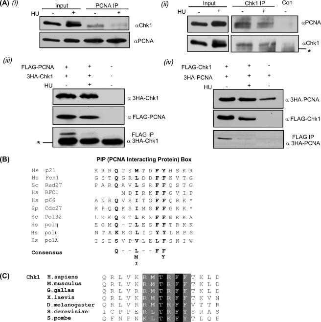
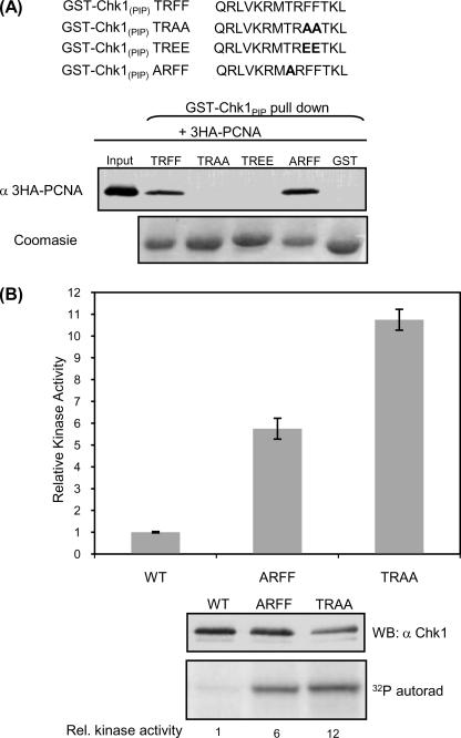
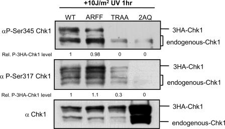
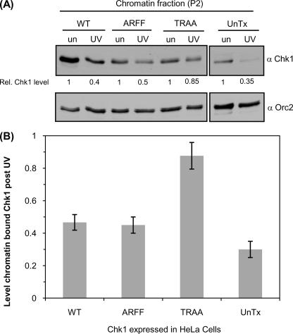
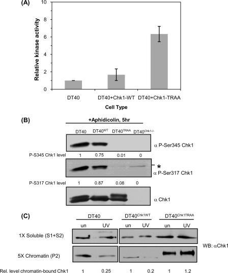
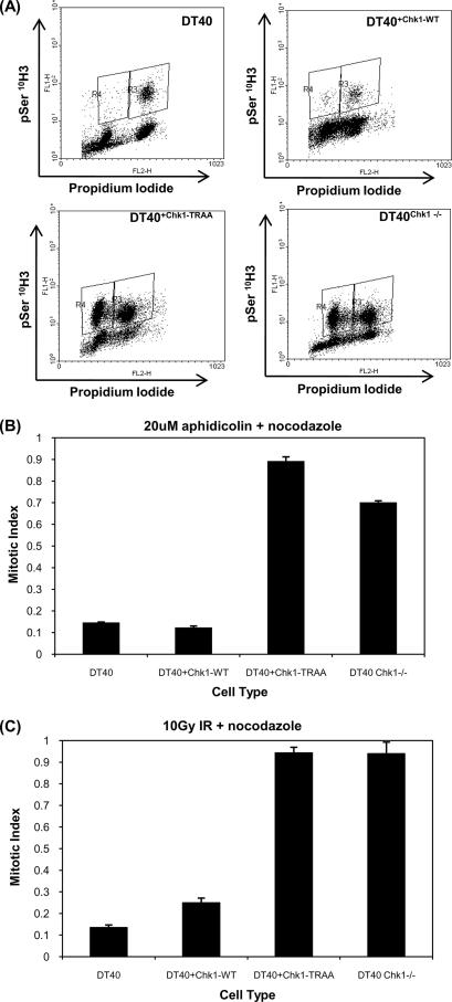
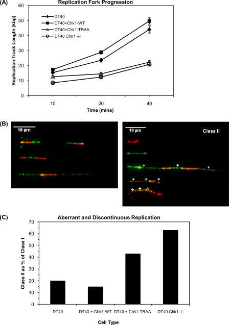
Human checkpoint kinase 1 (Chk1) is an essential kinase required for cell cycle checkpoints and for coordination of DNA synthesis. To gain insight into the mechanisms by which Chk1 carries out these functions, we used mass spectrometry to identify previously uncharacterized interacting partners of Chk1. We describe a novel interaction between Chk1 and proliferating cell nuclear antigen (PCNA), an essential component of the replication machinery. Binding between Chk1 and PCNA was reduced in the presence of hydroxyurea, suggesting that the interaction is regulated by replication stress. A highly conserved PCNA-interacting protein (PIP) box motif was identified in Chk1. The intact PIP box is required for efficient DNA damage-induced phosphorylation and release of activated Chk1 from chromatin. We find that the PIP box of Chk1 is crucial for Chk1-mediated S-M and G(2)-M checkpoint responses. In addition, we show that mutations in the PIP box of Chk1 lead to decreased rates of replication fork progression and increased aberrant replication. These findings suggest an additional mechanism by which essential components of the DNA replication machinery interact with the replication checkpoint apparatus.


Figures

FIGURE 1.
FIGURE 1.
Chk1 and PCNA interact in vivo and interaction is reduced after HU. A, top panels, interaction of endogenous proteins in HeLa cells. i, Chk1 was detected in PCNA immune -precipitates (IP) from HeLa cells. ii, similarly, Chk1 monoclonal antibodies, but not control antibodies (Con), immunoprecipitate PCNA from HeLa cells. The asterisk indicates cross-reaction with IgG heavy chain. Bottom panels, HeLa cells were transfected with PCNA and Chk1 as indicated above each panel. iii, 3HA-Chk1 was detected in FLAG-immune precipitates from cells that express FLAG-PCNA. The asterisk indicates IgG heavy chain. iv, in a reciprocal experiment, 3HA-PCNA was detected in FLAG immune precipitates from cells that express FLAG-Chk1. In all cases association was reduced in samples that had been cultured in 2 mm HU for 16 h before harvesting. B, an alignment of PIP box sequences from known PCNA binding proteins highlights a potential PIP box in the carboxyl terminus of Chk1. C, alignment of Chk1 carboxyl termini from diverse species indicates high conservation of the PIP box. Black indicates identical residues, and gray indicates similar residues.
FIGURE 2.
FIGURE 2.
Mutations within the PIP box activate Chk1 kinase activity. A, wild type or mutant versions of the PIP box (amino acids 371–384) from Chk1 were fused to GST and were probed for the ability to interact with 3HA-PCNA expressed in HeLa cells. GST-Chk1PIP wild type (TRFF) and ARFF sequences bind to PCNA, whereas mutation of conserved hydrophobic residues either to neutral (TRAA) or to acidic residues (TREE) abolishes PCNA binding. B, the kinase activity of FLAG-Chk1ARFF and FLAG-Chk1TRAA is increased compared with FLAG-Chk1WT. Kinase assays were performed on the indicated FLAG-Chk1 proteins immunoprecipitated from HeLa cells. Kinase activity was normalized to protein expression levels. The graph represents the average Chk1 kinase activity measured in four independent experiments. Error bars represent S.E. A representative Western blot (WB) and autoradiograph of indicated FLAG-Chk1 proteins is shown below the graph.
FIGURE 3.
FIGURE 3.
The PIP box of Chk1 is required for efficient damage-induced Chk1 phosphorylation. HeLa cells expressing 3HA-Chk1WT, 3HA-Chk1ARFF, 3HA-Chk1TRAA, and 3HA-Chk12AQ (S317A, S345A) were irradiated with 10 J/m2 UV and harvested 1 h later. The extent of Chk1 phosphorylation was monitored using anti-Ser(P)-345 and anti-Ser(P)-317. Migration of exogenous 3HA-Chk1 and endogenous Chk1 was as indicated. Levels of phosphorylated 3HA-Chk1 were quantified using ImageJ and are shown below the relevant lanes. Excess 3HA-Chk12AQ was used to monitor the extent to which these antibodies react with unphosphorylated Chk1.
FIGURE 4.
FIGURE 4.
The PIP box of Chk1 is required for efficient dissociation of Chk1 from chromatin. A, HeLa cells expressing FLAG-Chk1WT, FLAG-Chk1ARFF, FLAG-Chk1TRAA, or untransfected cells (UnTx) were irradiated with 50 J/m2 UV (UV) or left untreated (un). Two hours after UV treatment subcellular fractions were prepared. The level of Chk1 retained on chromatin (P2) was quantified relative to untreated samples using ImageJ and is shown below the relevant lanes. A longer exposure of the same blot was used to analyze retention of endogenous Chk1 in untransfected cells (right hand panels). Orc2 was used to verify loading of the chromatin fraction. B, quantification showing a defect in release of FLAG-Chk1TRAA from chromatin. The level of Chk1 retained on chromatin after 50J/m2 UV was quantified relative to untreated samples using ImageJ. The graph represents a fraction of chromatin-bound Chk1 measured in three independent experiments. Error bars represent S.E.
FIGURE 5.
FIGURE 5.
PIP-disrupted chicken Chk1 is hyperactive but is not efficiently phosphorylated after DNA damage and is not efficiently released from chromatin after DNA damage. A, the kinase activity of GgChk1TRAA is increased compared with endogenous GgChk1 (DT40) and wild type Chk1 (DT40Chk1WT). The graph represents average Chk1 kinase activity measured in three independent experiments. Error bars represent S.E. B, DT40 cells and DT40Chk1-/- reconstituted with GgChk1WT (DT40Chk1WT) or GgChk1TRAA (DT40Chk1TRAA) were cultured in 20 μm aphidicolin for 5 h. The extent of Chk1 phosphorylation was monitored using anti-Ser(P)-345 and anti-Ser(P)-317. Levels of phospho-Chk1 were quantified using ImageJ and are shown below the relevant lanes. A background/nonspecific band (*) was detected using the P-S317 but not P-S345 antibody. C, DT40, DT40WT, DT40TRAA, and DT40Chk1-/- cells were irradiated with 50 J/m2 UV (UV) or left untreated (un). Two hours later subcellular fractions were prepared. The levels of Chk1 retained on chromatin were quantified relative to untreated samples using ImageJ and are shown below the relevant lanes.
FIGURE 6.
FIGURE 6.
Chk1-TRAA expressing DT40 cells have defective S-M and G2-M checkpoint responses. A, fluorescence due to phosphorylation of histone H3 (pSer 10H3) and PI staining (DNA content) of DT40 cells cultured in 3. 3 nm nocodazole and 20 μm aphidicolin for 16 h. Region R4 includes cells that have high Ser(P)-10 H3 signals and a 2N content of DNA. B, mitotic indices of DT40 cells incubated in the presence of nocodazole and aphidicolin for 16 h. Mitotic index was calculated from the total number of Ser(P)-10 H3-positive cells in aphidicolin plus nocodazole cultures relative to nocodazole-only control cultures. The graph represents average mitotic index measured in three independent experiments. Error bars represent S.E. C, mitotic indices of irradiated (10 gray γ-IdUrd) DT40 cells incubated in the presence of nocodazole. Mitotic index was calculated from the total number of Ser(P)-10 H3-positive cells in irradiated cultures relative to nocodazole only control cultures. The graph represents the average mitotic index measured in three independent experiments. Error bars represent S.E.
FIGURE 7.
FIGURE 7.
The PIP box of Chk1 is required for appropriate progression of replication forks. A, the indicated DT40 cells were labeled with 25 μm BrdUrd for 10, 20, or 40 min before preparing DNA fibers. The mean length of at least 100 replication tracks is plotted for each time point. Error bars indicate S.E. B, examples of dual labeled replication tracks. Class I includes all ongoing replication (red-green patterns). Class II represents discontinuous and aberrant replication tracks (red only, green only, or interspersed patterns). White crosses show active origins. C, DT40 cells were labeled with 25 μm BrdUrd for 10 min followed by 250 μm IdUrd for 20 min before preparing DNA fibers. Replication tracks were assigned as either class I (ongoing) or class II (discontinuous and aberrant) as in B. At least 100 replication tracks were counted, and class II replication structures were quantified as a percentage of ongoing tracks.

Similar articles

Cited by

References

    1. Bartek, J., and Lukas, J. (2007) Curr. Opin. Cell Biol. 19 238-245 - PubMed
    1. Capasso, H., Palermo, C., Wan, S., Rao, H., John, U. P., O'Connell, M. J., and Walworth, N. C. (2002) J. Cell Sci. 115 4555-4564 - PubMed
    1. Chen, Z., Xiao, Z., Chen, J., Ng, S. C., Sowin, T., Sham, H., Rosenberg, S., Fesik, S., and Zhang, H. (2003) Mol. Cancer Ther. 2 543-548 - PubMed
    1. Liu, Q., Guntuku, S., Cui, X. S., Matsuoka, S., Cortez, D., Tamai, K., Luo, G., Carattini-Rivera, S., DeMayo, F., Bradley, A., Donehower, L. A., and Elledge, S. J. (2000) Genes Dev. 14 1448-1459 - PMC - PubMed
    1. Sorensen, C. S., Syljuasen, R. G., Falck, J., Schroeder, T., Ronnstrand, L., Khanna, K. K., Zhou, B. B., Bartek, J., and Lukas, J. (2003) Cancer Cell 3 247-258 - PubMed

Publication types

LinkOut - more resources