. 2008 Jul;82(14):7238-42.
doi: 10.1128/JVI.00425-08. Epub 2008 Apr 30.

Cell adhesion promotes Ebola virus envelope glycoprotein-mediated binding and infection

Affiliations

Cell adhesion promotes Ebola virus envelope glycoprotein-mediated binding and infection

Derek Dube et al. J Virol. 2008 Jul.

Abstract

Ebola virus infects a wide variety of adherent cell types, while nonadherent cells are found to be refractory. To explore this correlation, we compared the ability of pairs of related adherent and nonadherent cells to bind a recombinant Ebola virus receptor binding domain (EboV RBD) and to be infected with Ebola virus glycoprotein (GP)-pseudotyped particles. Both human 293F and THP-1 cells can be propagated as adherent or nonadherent cultures, and in both cases adherent cells were found to be significantly more susceptible to both EboV RBD binding and GP-pseudotyped virus infection than their nonadherent counterparts. Furthermore, with 293F cells the acquisition of EboV RBD binding paralleled cell spreading and did not require new mRNA or protein synthesis.


Figures
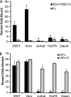
FIG. 1.
FIG. 1.
EboV RBD binding to, and pseudotype infection of, adherent 293T and Vero cells compared to those of B and T lymphocytes. (A) RBD-Fc binding assays were conducted essentially as described by Kuhn et al. (11), using a construct with similar binding properties (D. Dube, S. E. Delos, and J. M. White, unpublished data). Briefly, EboV RBD or control rabbit Fc (200 nM) was added to 5 × 10⁵ cells for 1.5 h at 4°C. The cells were washed three times with 3% bovine serum albumin-phosphate-buffered saline with Ca2+ and Mg2+, and Alexa Fluor 488-conjugated Protein A (Invitrogen) was added at a dilution of 1:250 for 45 min at 4°C. The cells then were washed twice and fixed with 4% paraformaldehyde. The cell surface binding of the EboV RBD was determined by flow cytometry. The percentage of cells that were positive for binding is presented. (B) Infection assays were performed as described by Schornberg et al. (16). Indicated cells were challenged for 18 h with VSV-GPΔ or VSV-G at a multiplicity of infection of 1 and fixed, and the percentage of GFP-positive cells was determined by flow cytometry. Results shown are the averages from three or more experiments, and error bars represent standard deviations. Asterisks indicate statistically significant differences from 293T cell data (P < 0.01).
FIG. 2.
FIG. 2.
EboV RBD binding to, and pseudotype infection of, RA- and PMA-treated THP-1 cells. (A) THP-1 cells were mock treated or treated with 0.1 μM RA or PMA for 24 h. Cells then were assayed for EboV RBD binding as described in the legend to Fig. 1A. (B) THP-1 cells were treated as described for panel A (in a 96-well dish) and then challenged with VSV-GPΔ or VSV-G (multiplicity of infection [MOI] of 2), incubated, and analyzed as described in the legend to Fig. 1B. Results shown in panel A and those for VSV-GPΔ in panel B are the averages from three or more experiments and error bars represent standard deviations, with asterisks indicating statistically significant differences from mock-treated THP-1 cell data (P < 0.02). The data for VSV-G shown in panel B are the averages from duplicate samples from one experiment; similar results were seen at a lower MOI. (C) Mock-, RA-, and PMA-treated THP-1 cells were infected with β-lactamase containing human immunodeficiency virus type 1 virions bearing Ebola virus GPΔ or VSV-G and then loaded with the β-lactamase substrate CCF2/AM. Cells loaded only with CCF2/AM served as a negative control. The extent of CCF2/AM cleavage by the virus-introduced cytoplasmic β-lactamase, which was detected by the change in the dye emission from green to blue, was evaluated using a BD LSR II cell analyzer equipped with a violet laser (407 nm) and appropriate filters for the blue (HQ 450/50; Chroma Technology) and green (HQ 530/30; Chroma Technology) emissions. The averages from duplicate samples from one representative of two experiments are shown. Similar results were seen at a lower MOI.
FIG. 3.
FIG. 3.
EboV RBD binding to, and pseudotype infection of, adherent and suspension 293F cells. (A) 293F cells were maintained in suspension on a rotating platform or allowed to adhere without being shaken for 18 h in the same CO2 incubator. Cells then were assayed for EboV RBD binding as described in the legend to Fig. 1A. (B) 293F cells were treated as described for panel A, challenged with VSV-GPΔ or VSV-G at a multiplicity of infection (MOI) of 2 for 18 h, and scored for infection by flow cytometry. Results shown in panels A and B are the averages from three or more experiments and error bars represent standard deviations, with asterisks indicating statistically significant differences from the suspension 293F cell data (P < 0.015). (C) 293F cells were treated as described for panel A, challenged with VSV-GPΔ or VSV-measles virus F/H at an MOI of 2 for 18 h, and scored for infection by flow cytometry. The averages from duplicate samples from one representative of two experiments are shown. (D) 293F cells were treated as described for panel A and assayed as described in the legend to Fig. 2C, using Ebola virus GPΔ or VSV-G bearing human immunodeficiency virus pseudoparticles harboring β-lactamase. The average results from duplicate samples are shown. (E) 293F cells were left in suspension or allowed to adhere. Cells were photographed at the indicated times using a Spot Insight Color camera attached to a Nikon Diaphot microscope. (F) The cells shown in panel E were examined for cell spreading (▴) and EboV RBD binding (•). Cell spreading was determined by a blind analysis of micrographs such as those shown in panel E by using EZ-C1 Freeviewer 3.0 software from Nikon. The perimeter of 10 cells per field (for 3 or more fields) was outlined to generate cell area data. Cell areas for each time point were averaged and normalized to the area of cells that were plated for 18 h. The binding of the EboV RBD (•) and Fc control (▪) were assayed as described in the legend to Fig. 1B and normalized to the values for EboV RBD binding to cells that were allowed to adhere for 18 h. The data in panels E and F are from one of three experiments that yielded virtually identical results.
FIG. 4.
FIG. 4.
Effects of actinomycin D and cycloheximide on EboV RBD binding to 293F cells. 293F cells maintained on a shaking platform were pretreated with 1 μM actinomycin D (Act D), 10 μM cycloheximide (Cyclo), or vehicle (0.1% dimethylsulfoxide; mock) for 2 h. Cells then were removed from the shaker and allowed to adhere for an additional 2 h in the continued presence of the indicated inhibitor (Adherent Act D or Adherent Cyclo). Mock-treated cells either were allowed to adhere (Adherent Mock) or were kept on the shaker (Suspension Mock) for an additional 2 h. The cells then were examined for EboV RBD binding as described in the legend to Fig. 1A, and the data were normalized to those for EboV RBD binding to mock-treated adherent cells. Results shown are the averages from five determinations from two experiments, and error bars represent standard deviations. The asterisk indicates a statistically significant difference from mock-treated adherent 293F cell data (P < 0.015). Parallel cells were transfected with a GFP expression plasmid and treated with 1 μM Act D, 10 μM cycloheximide, or 0.1% dimethylsulfoxide (mock). Whereas GFP was robustly expressed in the mock-treated samples, no GFP was detected in samples treated with Act D or cycloheximide.

Similar articles

Cited by

References

    1. Balasubramanian, N., D. W. Scott, J. D. Castle, J. E. Casanova, and M. A. Schwartz. 2007. Arf6 and microtubules in adhesion-dependent trafficking of lipid rafts. Nat. Cell Biol. 91381-1391. - PMC - PubMed
    1. Bavari, S., C. M. Bosio, E. Wiegand, G. Ruthel, A. B. Will, T. W. Geisbert, M. Hevey, C. Schmaljohn, A. Schmaljohn, and M. J. Aman. 2002. Lipid raft microdomains: a gateway for compartmentalized trafficking of Ebola and Marburg viruses. J. Exp. Med. 195593-602. - PMC - PubMed
    1. Brindley, M. A., L. Hughes, A. Ruiz, P. B. McCray, Jr., A. Sanchez, D. A. Sanders, and W. Maury. 2007. Ebola virus glycoprotein 1: identification of residues important for binding and postbinding events. J. Virol. 817702-7709. - PMC - PubMed
    1. Chesnokov, V. N., and N. P. Mertvetsov. 1990. The effect of translation inhibitor cycloheximide on expression of mammalian genes. Biokhimiia 551276-1278. - PubMed
    1. Graham, F. L., J. Smiley, W. C. Russell, and R. Nairn. 1977. Characteristics of a human cell line transformed by DNA from human adenovirus type 5. J. Gen. Virol. 3659-74. - PubMed

Publication types

Substances