Comparative Study
. 2008 Jul;82(13):6721-33.
doi: 10.1128/JVI.02250-07. Epub 2008 Apr 30.

Molecular basis of cytotoxicity of Epstein-Barr virus (EBV) latent membrane protein 1 (LMP1) in EBV latency III B cells: LMP1 induces type II ligand-independent autoactivation of CD95/Fas with caspase 8-mediated apoptosis

Affiliations
Comparative Study

Molecular basis of cytotoxicity of Epstein-Barr virus (EBV) latent membrane protein 1 (LMP1) in EBV latency III B cells: LMP1 induces type II ligand-independent autoactivation of CD95/Fas with caspase 8-mediated apoptosis

Christophe Le Clorennec et al. J Virol. 2008 Jul.

Abstract

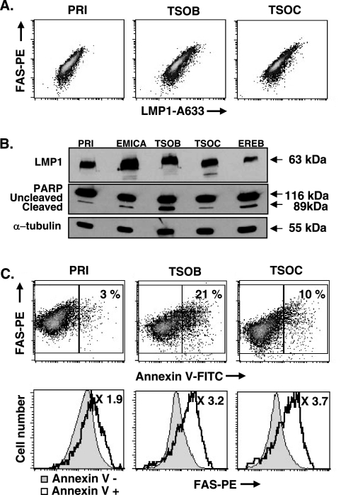
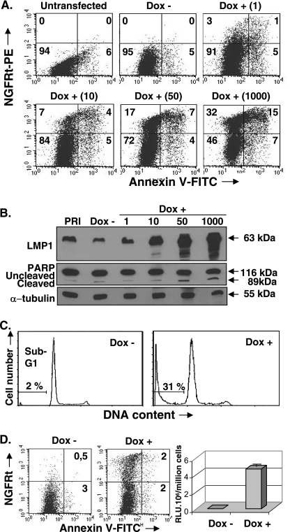
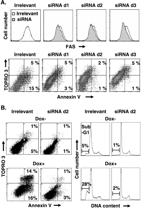
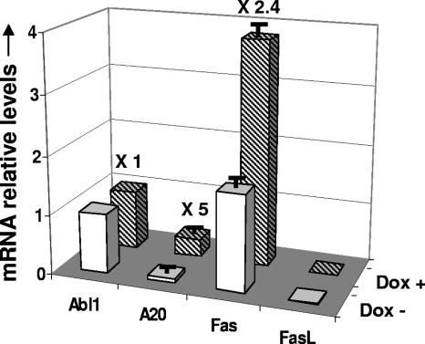
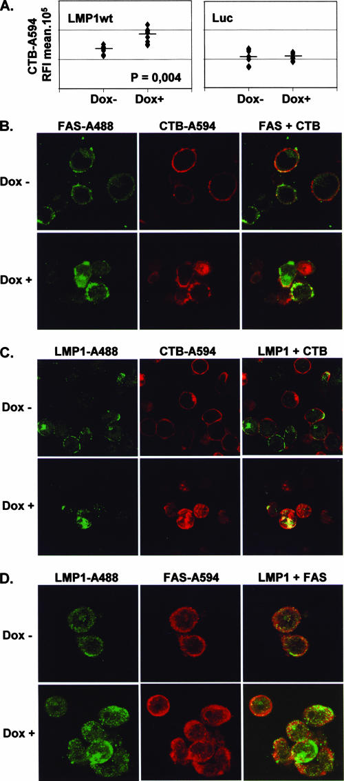
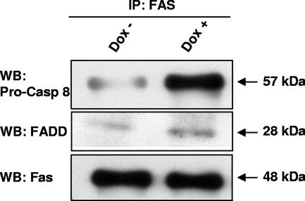
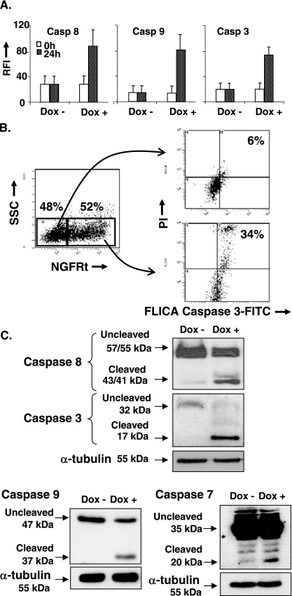
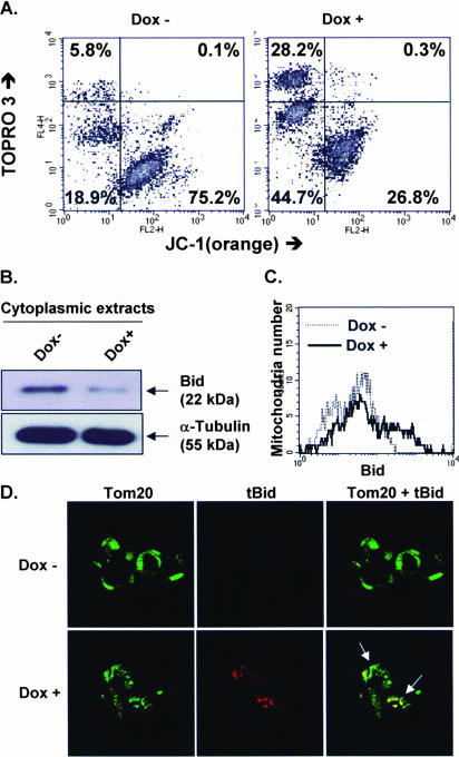
The Epstein-Barr virus (EBV) oncoprotein latent membrane protein 1 (LMP1) is thought to act as the major transforming protein in various cell types, by rerouting the tumor necrosis factor receptor family signaling pathway. Despite this implication in EBV-associated transformation of cells, LMP1 toxicity is a well-known but poorly studied feature, perhaps because it contradicts its role in transformation. We show that LMP1 physiological levels are very heterogeneous and that the highest levels of LMP1 correlate with Fas overexpression and spontaneous apoptosis in lymphoblastoid cell lines (LCLs). To understand the cytotoxic effect of LMP1 in LCLs, we cloned wild-type LMP1 into a doxycycline double-inducible episomal vector pRT-1, with a truncated version of NGFR as a surrogate marker of inducibility. We found that LMP1 overexpression induced apoptosis in LCL B cells, as shown by annexin V labeling, sub-G(1) peak, and poly(ADP ribose) polymerase cleavage. Knocking down Fas expression by small interfering RNA abolished LMP1-induced apoptosis. The absence of detectable levels of Fas ligand mRNA suggested a ligand-independent activation of Fas. LMP1 induced Fas overexpression with its relocalization in lipid raft microdomains of the membrane. Fas immunoprecipitation detected FADD (Fas-associated death domain protein) and caspase 8, suggesting a Fas-dependent formation of the death-inducing signaling complex. Caspases 8, 9, 3, and 7 were activated by LMP1. Caspase 8 activation was associated with BID cleavage and truncated-BID mitochondrial relocalization, consistent with type II apoptosis. Therefore, our results are in agreement with a model where LMP1-dependent NF-kappaB activation induces Fas overexpression and autoactivation that could overwhelm the antiapoptotic effect of NF-kappaB, revealing an ambivalent function of LMP1 in cell survival and programmed cell death.


Figures

FIG. 1.
FIG. 1.
In LCLs, cells with high LMP1wt and Fas expression levels are more sensitive to induction of apoptosis, (A) LMP1 and total Fas (intracellular and cytoplasmic membrane) expression levels of three LCLs (PRI, TSOB, and TSOC) were analyzed by flow cytometry after immunostaining, as described in Materials and Methods. The use of two lasers for excitation allowed independent fluorescence analysis for Fas and LMP1. (B) Five LCLs (PRI, EMICA, TSOB, TSOC, and EREB) were analyzed by Western blotting with anti-LMP1, anti-PARP, and anti-α-tubulin antibodies, as described in Materials and Methods. (C) PRI, TSOB, and TSOC cells were stained with annexin V-FITC and PE-conjugated Fas MAb and analyzed by flow cytometry. For each cell line, histograms of cytoplasmic membrane Fas expression levels were reported for annexin V-negative against annexin V-positive cells.
FIG. 2.
FIG. 2.
LMP1wt overexpression induces apoptosis in PRI-LCLs. (A) Latency III LMP1-positive PRI-LCLs were stably transfected with the inducible vector pRT-1, which contains cDNA coding for LMP1wt and NGFRt as a reporter gene. After 24 h doxycycline induction, untreated cells (Dox −) and doxycycline (1 to 1,000 ng/ml)-treated cells (Dox +) were stained with annexin V-FITC and PE-conjugated NGFR MAb and analyzed by flow cytometry. (B) Whole-cell extracts were analyzed in parallel for LMP1 and PARP expression by Western blotting. (C) LMP1-transfected PRI-LCLs were stained with propidium iodide to analyze cell DNA content before (Dox −) or after (Dox +) doxycycline induction at 1 μg/ml by flow cytometry. (D) PRI-LCLs were stably transfected with the inducible vector pRT-1, which contains cDNA coding for luciferase instead of LMP1. Annexin V-FITC and PE-conjugated NGFR MAb staining (left) were analyzed by flow cytometry, and luciferase activity (right) was assessed by luciferase assay, as described in Materials and Methods.
FIG. 3.
FIG. 3.
Downregulation of Fas expression decreases apoptosis in LCLs and LMP1wt-induced cells. Downregulation of Fas expression was optimized using siRNA against Fas gene, as described in Materials and Methods. (A) Downregulation of Fas expression in the LCL TSOB, after transient transfection with an irrelevant siRNA or with Fas-specific siRNA duplexes 1 to 3 (d1, d2, and d3), were analyzed by flow cytometry at 24 h by staining Fas with a PE-conjugated Fas MAb for 15 min (top). siRNA-transfected TSOB cells were stained with annexin V-FITC and TOPRO-3 as an impermeant DNA dye (bottom). (B) LMP1wt-transfected LCL PRI cells, 24 h after transient transfection with irrelevant siRNA or Fas-specific siRNA d2, were induced with doxycycline for 24 h. Then, untreated cells (Dox−) and doxycycline-treated cells (Dox+) were stained with annexin V-FITC and with TOPRO-3 as an impermeant DNA dye. Induced cells were previously identified with PE-conjugated NGFR MAb and analysis performed by flow cytometry (left). LMP1wt-transfected PRI cells, 24 h after transient transfection with irrelevant siRNA or Fas-specific siRNA d2, were stained with propidium iodide to analyze cell DNA content by flow cytometry, before (Dox−) or after (Dox+) doxycycline induction at 1 μg/ml (right).
FIG. 4.
FIG. 4.
Absence of FasL induction in LMP1wt-induced PRI-LCLs cells. After 24 h doxycycline induction, cells were harvested and mRNA from untreated cells (Dox −) and doxycycline cells (Dox +) was extracted. A20 mRNA, Fas mRNA, and FasL mRNA were analyzed by real-time RT-PCR. The Abl1 gene was used as a reference gene. Error bars correspond to the standard deviations of three experiments.
FIG. 5.
FIG. 5.
LMP1 and Fas are recruited in dissimilar GM1 lipid rafts. (A) GM1 levels in untreated cells (Dox−) or in doxycycline-treated cells (Dox+) were analyzed after 24 h doxycycline induction, by staining with 2.5 μg/ml AlexaFluor 594 CTB, fixation with 4% PFA, and permeabilization with PBS-0.05% Triton X-100. LMP1wt/NGFRt or luciferase/NGFRt vector-transfected cells were analyzed by confocal microscopy, and mean cell fluorescence was extracted with ImageJ software (A). Colocalization of Fas and CTB (B) or of LMP1 and CTB (C) was detected as described in Materials and Methods. After 24 h doxycycline induction, cells were incubated with 2.5 μg/ml AlexaFluor 594 CTB (red) at 4°C for 30 min. Cells were washed twice with cold PBS and fixed with 4% PFA. Then, cells were permeabilized with PBS-0.05% Triton X-100 and incubated with primary antibodies (anti-LMP1 antibody CS1-4 or anti-Fas antibody B-10) for 30 min, and proteins were visualized with AlexaFluor 488-conjugated goat anti-mouse immunoglobulin secondary antibodies (green) for 30 min. Colocalization between LMP1 and Fas (D) was detected by incubation with primary antibodies (CS1-4 [anti-LMP1] and C-20 [anti-Fas]) for 30 min, revealed with AlexaFluor 488-conjugated goat anti-mouse immunoglobulin and AlexaFluor 594-conjugated goat anti-rabbit immunoglobulin secondary antibodies, respectively, for 30 min. Colocalization experiments were performed by confocal microscopy.
FIG. 6.
FIG. 6.
Apoptosis involves Fas-dependent DISC formation. After 24 h doxycycline induction, untreated cells (Dox −) and doxycycline-treated cells (Dox +) were harvested, and whole-cell extracts were immunoprecipitated with an anti-Fas (C-20) MAb. Immunoprecipitates were subjected to sodium dodecyl sulfate-polyacrylamide gel electrophoresis analysis using anti-caspase 8, anti-FADD, and anti-Fas antibodies.
FIG. 7.
FIG. 7.
LMP1wt-induced apoptosis involves caspase activation. (A) After doxycycline induction, cells were subjected to FLICA labeling by adding labeled-caspase inhibitor FAM-LETD-FMK, FAM-LEHD-FMK, FAM-DEVD-FMK, respectively, to detect the presence of active caspases 8, 9 and 3. Induced cells were previously identified with PE-conjugated NGFR MAb and analyses were performed by flow cytometry. Propidium iodide was used as a DNA impermeant dye. Fluorescence unit was reported as RFI (Relative Fluorescence Intensity). (B) Example of graphs for caspase 3 analysis after FLICA labeling. After 24 h of doxycycline induction, NGFRt positive subpopulation selected on the cytogram NGFRt versus SSC (side scatter) showed an increase in the percentage of cells with positive fluorescence (caspase active form), compared to NGFRt negative cells (from 6 to 34%). For the NGFR positive subpopulation, caspase 3 activated cells were not, partially or highly stained by the permeability marker propidium iodide (PI), corresponding to the different stages seen during apoptosis (intact, plasma membrane disturbed and late apoptotic or necrotic cells, respectively). (C) After 24 h doxycycline induction, untreated cells (Dox −) and doxycycline-treated cells (Dox +) were harvested, and whole-cell extracts were analyzed by Western blotting with anti-caspase 8, anti-caspase 9, anti-caspase 3, anti-caspase 7, and anti-α-tubulin antibodies, as described in Materials and Methods.
FIG. 8.
FIG. 8.
LMP1wt induction induces mitochondrial transmembrane potential loss and tBID relocalization at the organelle level. (A) After 24 h doxycycline induction, untreated cells (Dox−) and doxycycline-treated cells (Dox+, NGFRt-expressing sorted cells) were stained by JC-1 (1 μg/ml, 20 min) and the impermeant DNA dye TOPRO-3 (1 μM) before analysis by flow cytometry. Cell physiological status was defined according to transmembrane Δψ and cytoplasmic membrane permeability. Three stages were identified: intact (JC-1+/TOPRO-3), apoptotic (JC-1/TOPRO-3), and late apoptotic or necrotic (JC-1/TOPRO-3+) cells. (B) Mitochondrion-depleted cytoplasmic extracts were subjected to Western blot analysis with anti-Bid and anti-α-tubulin (cytoplasm-specific) antibodies. (C) Isolated mitochondria were incubated with anti-Bid antibodies, visualized with AlexaFluor 594, and analyzed by flow cytometry. (D). tBID expression and its colocalization with Tom20 were assessed by incubation with primary antibodies (anti-Tom20 and anti-tBID) for 30 min and visualization with AlexaFluor 488-conjugated (green) and AlexaFluor 594-conjugated (red) antibodies, respectively, for 30 min. Analyses were carried out by confocal microscopy. Arrows indicate cells with colocalized tBID and Tom20.

Similar articles

Cited by

References

    1. Baran-Marszak, F., J. Feuillard, I. Najjar, C. Le Clorennec, J. Béchet, I. Dusanter-Fourt, G. W. Bornkamm, M. Raphaël, and R. Fagard. 2004. Differential roles of STAT1α and STAT1β in fludarabine-induced cell cycle arrest and apoptosis in human B cells. Blood 1042475-2483. - PubMed
    1. Bornkamm, G. W., C. Berens, C. Kuklik-Roos, J. Bechet, G. Laux, J. Bachl, M. Korndoerfer, M. Schlee, M. Hölzel, A. Malamoussi, R. D. Chapman, F. Nimmerjahn, J. Mautner, W. Hillen, H. Bujard, and J. Feuillard. 2005. Stringent doxycycline-dependent control of gene activities using an episomal one-vector system. Nucleic Acids Res. 33e137. - PMC - PubMed
    1. Bornkamm, G. W., and W. Hammerschmidt. 2001. Molecular virology of Epstein-Barr virus. Philos. Trans. R. Soc. Lond. B 356437-459. - PMC - PubMed
    1. Busch, L. K., and G. A. Bishop. 1999. The EBV transforming protein, latent membrane protein 1, mimics and cooperates with CD40 signaling in B lymphocytes. J. Immunol. 1622555-2561. - PubMed
    1. Canman, C. E., and M. B. Kastan. 1995. Induction of apoptosis by tumor suppressor genes and oncogenes. Semin. Cancer Biol. 617-25. - PubMed

Publication types