Structural analysis of major species barriers between humans and palm civets for severe acute respiratory syndrome coronavirus infections
- PMID: 18448527
- PMCID: PMC2446986
- DOI: 10.1128/JVI.00442-08
Structural analysis of major species barriers between humans and palm civets for severe acute respiratory syndrome coronavirus infections
Abstract
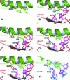
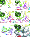
It is believed that a novel coronavirus, severe acute respiratory syndrome coronavirus (SARS-CoV), was passed from palm civets to humans and caused the epidemic of SARS in 2002 to 2003. The major species barriers between humans and civets for SARS-CoV infections are the specific interactions between a defined receptor-binding domain (RBD) on a viral spike protein and its host receptor, angiotensin-converting enzyme 2 (ACE2). In this study a chimeric ACE2 bearing the critical N-terminal helix from civet and the remaining peptidase domain from human was constructed, and it was shown that this construct has the same receptor activity as civet ACE2. In addition, crystal structures of the chimeric ACE2 complexed with RBDs from various human and civet SARS-CoV strains were determined. These structures, combined with a previously determined structure of human ACE2 complexed with the RBD from a human SARS-CoV strain, have revealed a structural basis for understanding the major species barriers between humans and civets for SARS-CoV infections. They show that the major species barriers are determined by interactions between four ACE2 residues (residues 31, 35, 38, and 353) and two RBD residues (residues 479 and 487), that early civet SARS-CoV isolates were prevented from infecting human cells due to imbalanced salt bridges at the hydrophobic virus/receptor interface, and that SARS-CoV has evolved to gain sustained infectivity for human cells by eliminating unfavorable free charges at the interface through stepwise mutations at positions 479 and 487. These results enhance our understanding of host adaptations and cross-species infections of SARS-CoV and other emerging animal viruses.
Figures


References
-
- Brunger, A. T., P. D. Adams, G. M. Clore, W. L. DeLano, P. Gros, R. W. Grosse-Kunstleve, J. S. Jiang, J. Kuszewski, M. Nilges, N. S. Pannu, R. J. Read, L. M. Rice, T. Simonson, and G. L. Warren. 1998. Crystallography & NMR system: a new software suite for macromolecular structure determination. Acta Crystallogr. D 54905-921. - PubMed
-
- Cowtan, K. 1994. Joint CCP4 and ESF-EACBM Newsl. Protein Crystallogr. 3134-38.
-
- Fenn, T. D., D. Ringe, and G. A. Petsko. 2003. POVScript+: a program for model and data visualization using persistence of vision ray-tracing. J. Appl. Crystallogr. 36944-947.
-
- Guan, Y., B. J. Zheng, Y. Q. He, X. L. Liu, Z. X. Zhuang, C. L. Cheung, S. W. Luo, P. H. Li, L. J. Zhang, Y. J. Guan, K. M. Butt, K. L. Wong, K. W. Chan, W. Lim, K. F. Shortridge, K. Y. Yuen, J. S. M. Peiris, and L. L. M. Poon. 2003. Isolation and characterization of viruses related to the SARS coronavirus from animals in Southern China. Science 302276-278. - PubMed
Publication types
MeSH terms
Substances
Grants and funding
LinkOut - more resources
Full Text Sources
Other Literature Sources
Miscellaneous

