. 2008 Jul;82(14):6798-811.
doi: 10.1128/JVI.00154-08. Epub 2008 Apr 30.

Hepatitis B virus HBx protein localizes to mitochondria in primary rat hepatocytes and modulates mitochondrial membrane potential

Affiliations

Hepatitis B virus HBx protein localizes to mitochondria in primary rat hepatocytes and modulates mitochondrial membrane potential

Amy J Clippinger et al. J Virol. 2008 Jul.

Abstract

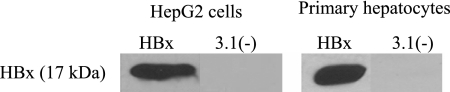
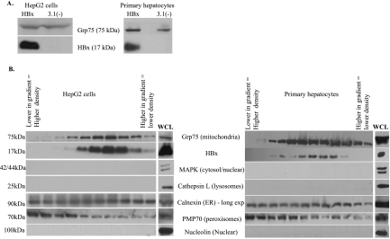
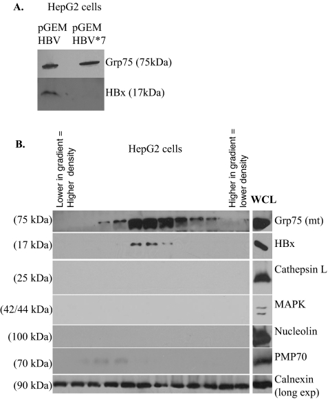
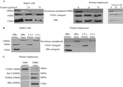
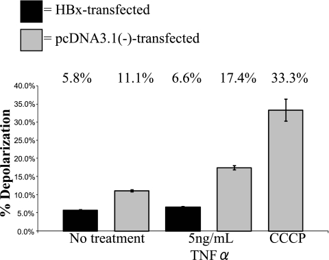
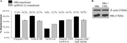
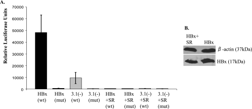
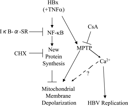
Over 350 million people are chronically infected with hepatitis B virus (HBV), and a significant number of chronically infected individuals develop primary liver cancer. HBV encodes seven viral proteins, including the nonstructural X (HBx) protein. The results of studies with immortalized or transformed cells and with HBx-transgenic mice demonstrated that HBx can interact with mitochondria. However, no studies with normal hepatocytes have characterized the precise mitochondrial localization of HBx or the effect of HBx on mitochondrial physiology. We have used cultured primary rat hepatocytes as a model system to characterize the mitochondrial localization of HBx and the effect of HBx expression on mitochondrial physiology. We now show that a fraction of HBx colocalizes with density-gradient-purified mitochondria and associates with the outer mitochondrial membrane. We also demonstrate that HBx regulates mitochondrial membrane potential in hepatocytes and that this function of HBx varies depending on the status of NF-kappaB activity. In primary rat hepatocytes, HBx activation of NF-kappaB prevented mitochondrial membrane depolarization; however, when NF-kappaB activity was inhibited, HBx induced membrane depolarization through modulation of the mitochondrial permeability transition pore. Collectively, these results define potential pathways through which HBx may act in order to modulate mitochondrial physiology, thereby altering many cellular activities and ultimately contributing to the development of HBV-associated liver cancer.


Figures

FIG. 1.
FIG. 1.
HBx expression in HepG2 cells and primary rat hepatocytes. HepG2 cells or primary rat hepatocytes were transfected 24 h after plating with FL1-154 HBx or pcDNA3.1(−). At 48 h posttransfection, cells were collected and proteins were resolved by SDS-PAGE. Western blot analysis was performed using an anti-Flag antibody.
FIG. 2.
FIG. 2.
Confirmation of hepatocyte differentiation in culture. RNA was extracted either from primary rat hepatocytes freshly isolated from a rat liver or from cultured primary rat hepatocytes maintained in Williams E medium as described in Materials and Methods. RT-PCR analysis was performed to analyze expression of albumin (A), transferrin (T), and HNF4 (H) as markers of differentiated hepatocytes. Expected band sizes are 914 bp for albumin, 530 bp for transferrin, and 530 bp for HNF4. Note that the primers for albumin and transferrin were combined in the same sample due to the substantial difference in PCR product size. Lanes AT (-RT) and H(-RT) represent samples in which isolated RNA was analyzed directly by PCR to confirm the absence of contaminating DNA.
FIG. 3.
FIG. 3.
Copurification of HBx with a mitochondrially enriched fraction isolated from HepG2 cells or primary rat hepatocytes. (A) Hepatocytes were transfected at 24 h after plating with FL1-154 HBx or pcDNA3.1(−), and a mitochondrially enriched fraction was isolated at 48 h posttransfection as described in Materials and Methods. Proteins were resolved by SDS-PAGE, and Western blot analysis was performed using an anti-Grp75 antibody and an anti-Flag antibody. (B) HepG2 cells or primary rat hepatocytes were transfected with FL1-154 HBx, followed by separation over a Percoll density gradient. Western blot analysis was performed using anti-Grp75, anti-Flag, anti-MAPK, anti-cathepsin L, anti-calnexin, anti-PMP70, and anti-nucleolin antibodies. WCL, whole-cell lysate.
FIG. 4.
FIG. 4.
Copurification of mitochondria with HBx expressed in the context of HBV replication in HepG2 cells. (A) HepG2 cells were transfected with pGEMHBV or pGEMHBV*7, and at 48 h posttransfection a mitochondrially enriched fraction was isolated. Proteins were resolved by SDS-PAGE and Western blot analysis was performed using an anti-Grp75 antibody and an anti-HBx antibody. (B) HepG2 cells were transfected with pGEMHBV, and at 48 h posttransfection isolated mitochondria were separated over a Percoll density gradient. Western blot analysis was performed using anti-Grp75, anti-Flag, anti-cathepsin L, anti-calnexin, anti-MAPK, anti-nucleolin, and anti-PMP70 antibodies. WCL, whole-cell lysate.
FIG. 5.
FIG. 5.
Localization of HBx to the outer mitochondrial membrane. (A) HepG2 cells or primary rat hepatocytes were transfected with FL1-154 HBx, and at 48 h posttransfection a mitochondrially enriched fraction was isolated. Trypsin treatment of isolated mitochondria was performed using increasing concentrations of trypsin. Western blot analysis was performed using anti-hexokinase, anti-VDAC, and anti-Flag antibodies. Treatment of mitochondria isolated from HBx-transfected primary hepatocytes with trypsin and Triton X-100 rendered HBx and VDAC susceptible to trypsin treatment. (B) HepG2 cells and primary rat hepatocytes were transfected with FL1-154 HBx, and at 48 h posttransfection a mitochondrially enriched fraction was isolated. Alkaline treatment of isolated mitochondria was performed, and proteins were resolved by SDS-PAGE. Western blot analysis was performed using anti-hexokinase, anti-VDAC, and anti-Flag antibodies. (C) Primary rat hepatocytes were transfected with FL1-154, followed by mitochondrial isolation and KCl treatment. Following differential centrifugation, proteins were resolved by SDS-PAGE, and Western blot analysis was performed using COXIV as an inner mitochondrial membrane (IMM) marker and Bcl-2 and TOM20 as markers of the outer mitochondrial membrane (OMM).
FIG. 6.
FIG. 6.
HBx prevents mitochondrial membrane depolarization in primary rat hepatocytes. Primary rat hepatocytes were transfected with FL1-154 HBx. At 24 h posttransfection, hepatocytes were treated with TNF-α, as shown. Twenty-four hours later, cells were treated with CCCP for 5 min, where shown, and cells were then collected and stained with JC-1, followed by flow cytometry analysis. Statistical analysis, conducted using Student's t test, verified that these differences are statistically significant (P ≤ 0.01). Error bars represent the standard deviation.
FIG. 7.
FIG. 7.
Mitochondrial membrane potential in primary rat hepatocytes. (A) At 24 h after plating, primary rat hepatocytes were transfected with FL1-154 HBx, pcDNA3.1(−), or IκB-α mutant, as shown. At 24 h posttransfection, hepatocytes were treated with CHX, CsA, or FK506, where applicable. Cells were collected 24 h later, stained with JC-1, and analyzed by flow cytometry. Statistical analysis, conducted using Student's t test, verified that the differences between the HBx-transfected and negative control samples are statistically significant for CHX, IκB-SR, CsA, and CHX-FK506 treatment (P ≤ 0.05). Error bars represent the standard deviation. (B) Primary rat hepatocytes were collected at 48 h posttransfection as for panel A, and Western blot analysis was conducted to confirm that treatment with CHX did not substantially change the level of HBx expression.
FIG. 8.
FIG. 8.
NF-κB activation in primary rat hepatocytes. At 24 h after plating, primary rat hepatocytes were transfected with pNF-κB-Luc (wild type [wt]), pNF-κB-Luc-MUT (mut), FL1-154, pcDNA3.1(−), or IκB-SR, where applicable, and then collected at 48 h posttransfection. (A) Luciferase assays were performed as described in the Materials and Methods. This is a representative graph from three separate experiments, each performed in triplicate. Statistical analysis, conducted using Student's t test, verified that these differences are statistically significant (P ≤ 0.01). Error bars represent the standard deviation. (B) Hepatocytes were collected at 48 h posttransfection, and Western blot analysis was conducted to confirm that cotransfection of IκB-SR did not substantially change the level of HBx expression.
FIG. 9.
FIG. 9.
HBx regulation of mitochondrial membrane potential in primary rat hepatocytes. See Discussion for explanation.

Similar articles

Cited by

References

    1. Andrisani, O., and S. Barnabas. 1999. The transcriptional function of the hepatitis B virus X protein and its role in hepatocarcinogenesis. Int. J. Oncol. 151-8. - PubMed
    1. Anflous, K., O. Blondel, A. Bernart, M. Khrestchatisky, and R. Ventura-Clapier. 1998. Characterization of rat porin isoforms: cloning of a cardiac type-3 variant encoding an additional methionine at its putative N-terminal region. Biochim. Biophys. Acta 139947-50. - PubMed
    1. Beasley, R., C.-C. Lin, L.-Y. Hwang, and C.-S. Chien. 1981. Hepatocellular carcinoma and hepatitis B virus: a prospective study of 22 707 men in Taiwan. Lancet ii1129-1133. - PubMed
    1. Bergametti, F., D. Sitterlin, and C. Transy. 2002. Turnover of hepatitis B virus X protein is regulated by damaged DNA-binding complex. J. Virol. 766495-6501. - PMC - PubMed
    1. Bernardi, P. 1999. Mitochondrial transport and cations: channels, exchangers, and permeability transition. Physiol. Rev. 791127-1155. - PubMed

Publication types

LinkOut - more resources