Comparative Study
. 2008 Jul;82(13):6654-66.
doi: 10.1128/JVI.00257-08. Epub 2008 Apr 30.

The UL25 gene product of herpes simplex virus type 1 is involved in uncoating of the viral genome

Affiliations
Comparative Study

The UL25 gene product of herpes simplex virus type 1 is involved in uncoating of the viral genome

Valerie G Preston et al. J Virol. 2008 Jul.

Abstract

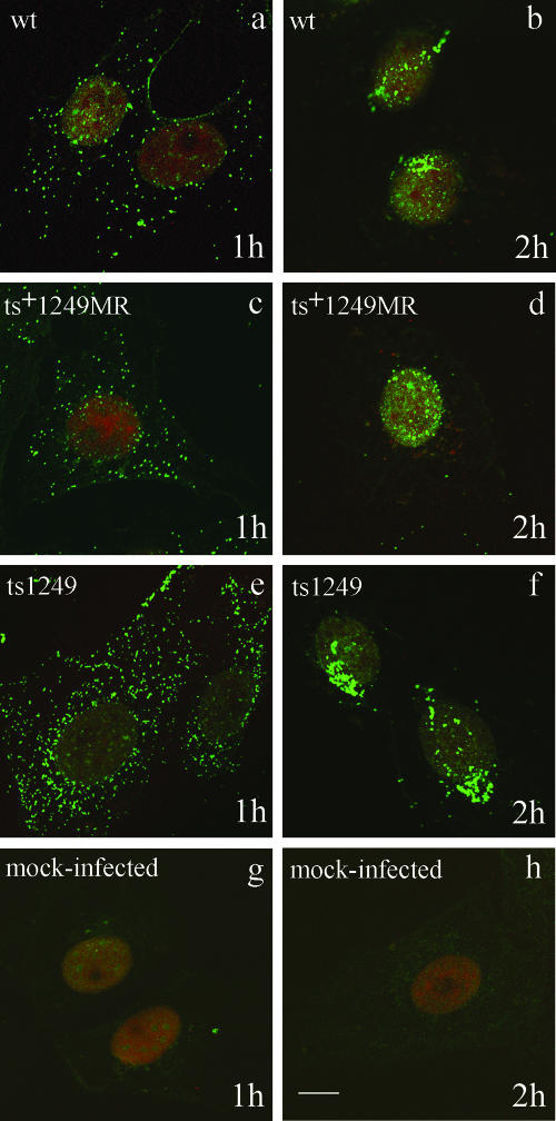
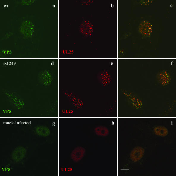
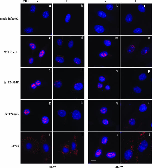
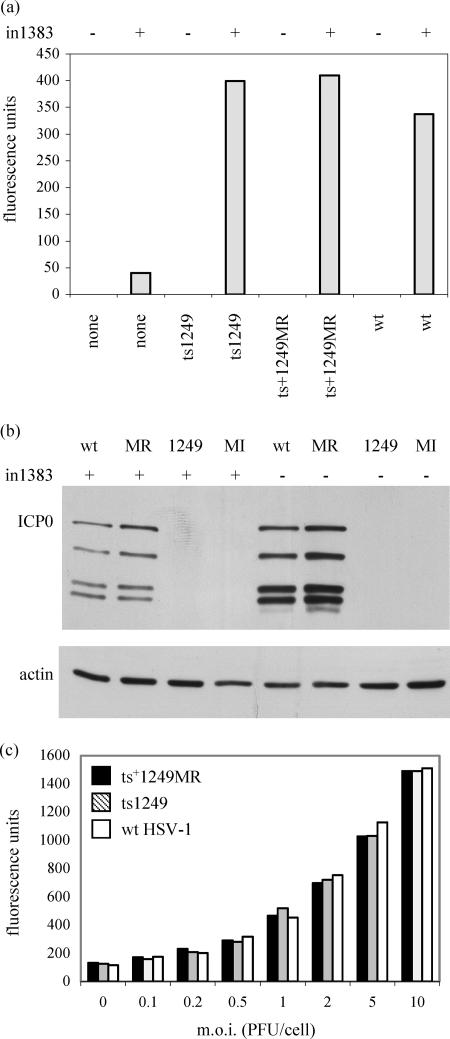
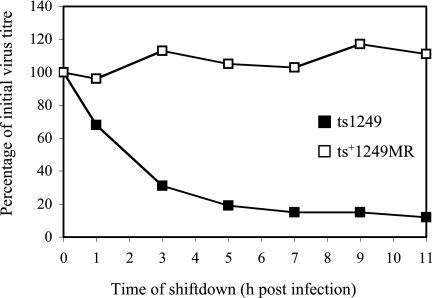
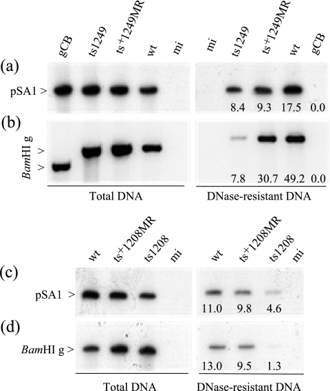
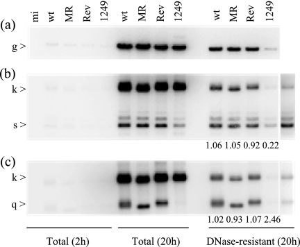
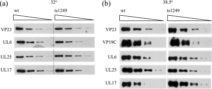
Studies on the herpes simplex virus type 1 UL25-null mutant KUL25NS have shown that the capsid-associated UL25 protein is required at a late stage in the encapsidation of viral DNA. Our previous work on UL25 with the UL25 temperature-sensitive (ts) mutant ts1204 also implicated UL25 in a role at very early times in the virus growth cycle, possibly at the stage of penetration of the host cell. We have reexamined this mutant and discovered that it had an additional ts mutation elsewhere in the genome. The ts1204 UL25 mutation was transferred into wild-type (wt) virus DNA, and the UL25 mutant ts1249 was isolated and characterized to clarify the function of UL25 at the initial stages of virus infection. Indirect immunofluorescence assays and in situ hybridization analysis of virus-infected cells revealed that the mutant ts1249 was not impaired in penetration of the host cell but had an uncoating defect at the nonpermissive temperature. When ts1249-infected cells were incubated initially at the permissive temperature to allow uncoating of the viral genome and subsequently transferred to the restrictive temperature, a DNA-packaging defect was evident. The results suggested that ts1249, like KUL25NS, had a block at a late stage of DNA packaging and that the packaged genome was shorter than the full-length genome. Examination of ts1249 capsids produced at the nonpermissive temperature revealed that, in comparison with wt capsids, they contained reduced amounts of UL25 protein, thereby providing a possible explanation for the failure of ts1249 to package full-length viral DNA.


Figures

FIG. 1.
FIG. 1.
(a) Structure of the HSV-1 genome showing the unique regions (UL and US), the repeated regions (TRL, IRL, TRS, and IRS), and the a sequence, which contains the cis-acting packaging signals (shaded box). The positions of the BamHI fragments k, q, s, and g are shown. (b) Expanded section of BamHI fragments s, k, and q showing the HSV-1 regions present in the plasmids pBE1 and pST17.
FIG. 2.
FIG. 2.
Identification of ts1204 and ts1208 UL25 mutations. The solid black box indicates the location of the ts mutation within the BamHI u genomic fragment obtained previously by marker rescue experiments. The numbers refer to the positions of the restriction endonuclease sites in the HSV-1 strain 17 genome. The precise base pair change determined by DNA sequence analysis and the effect of the mutation on the UL25 amino acid sequence are shown below the marker rescue data. ORF, open reading frame.
FIG. 3.
FIG. 3.
Comparison of [35S]methionine-labeled mutant virus-infected cell polypeptides synthesized at the PT and NPT. Vero cells were mock infected (mi) or infected with wt HSV-1, ts1204, ts1249, ts+1249MR, ts1208, or ts+1208MR, and the virus-infected cells were incubated at 32°C, 38.5°C, or 39.5°C. At 5 h p.i., [35S]methionine was added to the cells and incubation continued for a further 15 h. Samples were analyzed on an SDS-10% polyacrylamide gel and the proteins detected by autoradiography. Selected virus polypeptides are labeled on the left-hand side, and on the right-hand side the positions of molecular mass markers (kDa) are indicated. TK, thymidine kinase.
FIG. 4.
FIG. 4.
Localization of incoming capsids in the cell. Vero cells were either mock infected or infected with wt HSV-1, ts+1249 MR, or ts1249 in the presence of cycloheximide and incubated for 1 (a, c, e, g) or 2 (b, d, f, h) h at 38.5°C. Capsids were detected by indirect immunofluorescence with MAb DM165, specific for VP5, and the secondary IgG antibody FITC-GAM. The nuclei were stained with propidium iodide. The cells were examined by confocal microscopy and digital images taken. Bar, 10 μm.
FIG. 5.
FIG. 5.
Colocalization of UL25 with VP5 in incoming ts1249 capsids at the NPT. Vero cells were either mock infected or infected with wt HSV-1 or ts1249 in the presence of cycloheximide and incubated for 4 h at 38.5°C. Capsids were detected in the cell by indirect immunofluorescence with MAb DM165, specific for VP5, and the secondary IgG antibody FITC-GAM (a, d, e), and UL25 was identified with rabbit antibody 335 and the secondary IgG antibody Cy5-GAR (b, e, h). Each set of three images shows FITC staining of VP5 (left), Cy5 staining of UL25 (middle), and a merged image of the two for the same field of infected cells (right). The cells were examined by confocal microscopy and digital images taken. Bar, 10 μm.
FIG. 6.
FIG. 6.
Localization of incoming virion DNA in the cell by in situ hybridization. Vero cells were either mock infected or infected with wt HSV-1, ts+1249MR, ts+1249rev, or ts1249 in the presence or absence of cycloheximide (CHI) and incubated for 5 h at 38.5°C or 36.5°C as indicated. HSV-1 DNA was detected by in situ hybridization and confocal microscopy. Bar, 10 μm.
FIG. 7.
FIG. 7.
(a) β-Galactosidase activity in cell extracts. Cells were mock infected (−) or infected with in1383 (+), followed by superinfection with ts1249, ts+1249MR, or wt HSV-1 virions as indicated. The activities shown are expressed in arbitrary fluorescence units and represent the means of readings obtained for duplicate samples. (b) Western blot analysis of protein extracts from cells infected with ts1249 (1249), ts+1249MR (MR), or wt HSV-1 (wt) or mock infected (MI) cells in the presence or absence of in1383 infecting virus, screened with ICP0 or actin antibodies. (c) β-Galactosidase activity in extracts from cells infected with in1383 and different MOI of ts1249, ts+1249MR, or wt HSV-1 virions.
FIG. 8.
FIG. 8.
Reversibility of the ts1249 uncoating defect. BHK cells were infected with either ts1249 or ts+1249MR (MR) at the NPT and transferred to the PT at the times indicated. After 3 days at 32°C, the virus plaques were counted. The values shown are the means of values obtained for duplicate samples.
FIG. 9.
FIG. 9.
Amplification and packaging of the plasmid pSA1 in Vero cells by ts1249 and ts1208. One set of cell monolayers was transfected with pSA1, and cells subsequently were infected with wt HSV-1, gCB, ts+1249MR, or ts1249 or were mock infected (mi) in the presence of cycloheximide at 36.5°C. After 2 h, the cycloheximide block was removed and the samples were transferred to 38.5°C. The second set of cell monolayers was transfected with pSA1, and cells were subsequently infected with ts1208, ts+1208MR, or wt HSV-1 or were mock infected. These samples were incubated only at 39.5°C in the absence of cycloheximide. At 20 h p.i., the cells were harvested and total cellular DNA and DNase-resistant DNA were prepared and digested with DpnI and EcoRI (a, c) or BamHI (b, d). Southern blot analysis was carried out, using 32P-labeled plasmid vector pAT153 (a, c) or the 32P-labeled, cloned HSV-1 genomic fragment BamHI g (b, d) as a probe. The numbers at the bottom of the lanes indicate the percentage of radioactivity present in the band in the DNase-treated sample relative to that in the band in the total DNA sample. Note that gCB BamHI g is smaller than that of wt HSV-1 strain 17.
FIG. 10.
FIG. 10.
Analysis of ts1249 total and DNase-resistant viral DNAs at the NPT. Duplicate samples of cells were infected with wt HSV-1, ts+1249MR (MR), ts+1249rev (Rev), or ts1249 or were mock infected (mi) in the presence of cycloheximide at 36.5°C. After 2 h, the cycloheximide block was removed and the samples were transferred to 38.5°C. One set of samples was harvested at 4 h p.i. and the other set at 20 h p.i. Total cellular DNA and DNase-resistant DNA were prepared and digested with BamHI. Southern blot analysis was carried out, using 32P-labeled, cloned HSV-1 genomic fragment BamHI g (a), pBE1 (b), or pST17 (c) as a probe. The far right-hand lanes in panels b and c are the ts1249 lanes after longer exposure. The numbers at the bottom of the lanes indicate the ratio of radioactivity present in the joint fragment k to that in the terminal s or q fragment in the DNase-treated sample.
FIG. 11.
FIG. 11.
(a) Association of UL25 with ts1249 B capsids produced at the PT. Cells were infected with wt HSV-1 and ts1249 at 36.5°C for 2 h and transferred to 32°C for 18 h. Serial twofold dilutions of purified B capsids were prepared and equalized on the basis of their VP23 content by Western blotting. The polypeptides from the equalized capsids were resolved by SDS-PAGE, transferred onto nitrocellulose, and probed sequentially with UL6, UL17, and UL25 MAbs as indicated on the left side. (b) Association of UL25 with ts1249 B capsids produced at the NPT. Capsids were treated in the same way as those produced at the PT and the equalized capsids screened sequentially with UL6, UL17, UL25, and VP19C MAbs.

Similar articles

Cited by

References

    1. Addison, C., F. J. Rixon, J. W. Palfreyman, M. O'Hara, and V. G. Preston. 1984. Characterization of a herpes simplex virus type-1 mutant which has a temperature-sensitive defect in penetration of cells and assembly of capsids. Virology 138246-259. - PubMed
    1. Baines, J. D., and S. K. Weller. 2005. Cleavage and packaging of herpes simplex virus 1 DNA, p. 135-150. In C. Catalano (ed.), Viral genome packaging machines. Kluwer Academic/Plenum Publishers, New York, NY.
    1. Batterson, W., D. Furlong, and B. Roizman. 1983. Molecular genetics of herpes simplex virus. VIII. Further characterization of a temperature-sensitive mutant defective in the release of viral DNA and in other stages of the viral reproductive cycle. J. Virol. 45397-407. - PMC - PubMed
    1. Booy, F. P., W. W. Newcomb, B. L. Trus, J. C. Brown, T. S. Baker, and A. C. Steven. 1991. Liquid-crystalline, phage-like packing of encapsidated DNA in herpes simplex virus. Cell 641007-1015. - PMC - PubMed
    1. Bowman, B. R., R. L. Welschhans, H. Jayaram, N. D. Stow, V. G. Preston, and F. A. Quiocho. 2006. Structural characterization of the UL25 DNA-packaging protein from herpes simplex virus type 1. J. Virol. 802309-2317. - PMC - PubMed

Publication types

LinkOut - more resources