. 2008 Jul;82(13):6618-30.
doi: 10.1128/JVI.00140-08. Epub 2008 Apr 30.

The temperate marine phage PhiHAP-1 of Halomonas aquamarina possesses a linear plasmid-like prophage genome

Affiliations

The temperate marine phage PhiHAP-1 of Halomonas aquamarina possesses a linear plasmid-like prophage genome

Jennifer M Mobberley et al. J Virol. 2008 Jul.

Abstract

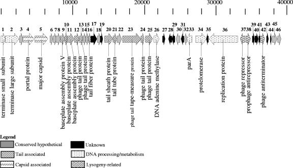
A myovirus-like temperate phage, PhiHAP-1, was induced with mitomycin C from a Halomonas aquamarina strain isolated from surface waters in the Gulf of Mexico. The induced cultures produced significantly more virus-like particles (VLPs) (3.73 x 10(10) VLP ml(-1)) than control cultures (3.83 x 10(7) VLP ml(-1)) when observed with epifluorescence microscopy. The induced phage was sequenced by using linker-amplified shotgun libraries and contained a genome 39,245 nucleotides in length with a G+C content of 59%. The PhiHAP-1 genome contained 46 putative open reading frames (ORFs), with 76% sharing significant similarity (E value of <10(-3)) at the protein level with other sequences in GenBank. Putative functional gene assignments included small and large terminase subunits, capsid and tail genes, an N6-DNA adenine methyltransferase, and lysogeny-related genes. Although no integrase was found, the PhiHAP-1 genome contained ORFs similar to protelomerase and parA genes found in linear plasmid-like phages with telomeric ends. Southern probing and PCR analysis of host genomic, plasmid, and PhiHAP-1 DNA indicated a lack of integration of the prophage with the host chromosome and a difference in genome arrangement between the prophage and virion forms. The linear plasmid prophage form of PhiHAP-1 begins with the protelomerase gene, presumably due to the activity of the protelomerase, while the induced phage particle has a circularly permuted genome that begins with the terminase genes. The PhiHAP-1 genome shares synteny and gene similarity with coliphage N15 and vibriophages VP882 and VHML, suggesting an evolutionary heritage from an N15-like linear plasmid prophage ancestor.


Figures

FIG. 1.
FIG. 1.
Growth of host (A) and prophage production (B) during the 24-h mitomycin C induction experiment. Asterisks indicate when mitomycin C was added. Mean BDC for each time point was used to chart bacterial growth. Error bars indicate the standard deviations of the results from triplicate slides. VDC, enumeration of viruses by the direct-count method.
FIG. 2.
FIG. 2.
Transmission electron micrographs of ΦHAP-1 particles. Black scale bars represent 50 nm.
FIG. 3.
FIG. 3.
Genomic map of ΦHAP-1. KODON was used to construct the gene map. ORFs were numbered based on the arbitrary start of the genome at the terminase small subunit. Patterns were assigned based on functional assignments of the ORFs as indicated in the key.
FIG. 4.
FIG. 4.
SDS-PAGE of proteins associated with ΦHAP-1 particles. Molecular-mass standard is in lane 1, with sizes in kilodaltons to the left. ΦHAP-1 proteins are in lane 2, with ORF designations based on peptide sequence to the right. The contrast of the image was digitally enhanced to increase the visibility of the bands.
FIG. 5.
FIG. 5.
Cartoon of the nucleotide sequence of the ΦHAP-1 92-base-pair inverted repeat found in the DNA region between the partitioning and protelomerase genes. The predicted cut site of the protelomerase is indicated by the dashed line. The sequences below the repeat are the predicted right and left telomeric ends of ΦHAP-1.
FIG. 6.
FIG. 6.
Genomic comparisons of six completed phage genomes that contained a protelomerase gene. KODON was used to construct the gene maps. The genomes of N15, ΦKO2, PY54, VHML, and VP882 were collected from GenBank. The location of the protelomerase gene in each genome is denoted by a “P.” Patterns were assigned based on functional assignment of the ORFs as indicated in the key.
FIG. 7.
FIG. 7.
Schematic representation of the two conformations of the ΦHAP-1 genome. The cut sites of restriction enzymes are indicated by vertical lines. The predicated sizes of fragments are noted to the right of each map. The numbered arrows below each map represent the primers used for PCR analysis. Figures are not drawn to scale. Pt, protelomerase; Pa, ParA; Ts, terminase small subunit; 46, ORF 46 hypothetical protein; cI, ORF 36 prophage repressor, which was used as the probe.
FIG. 8.
FIG. 8.
Southern gel transfer (right) and PFGE (left) of ΦHAP-1 DNA fractions. Lanes: 1, 8- to 48-kb standard; 2 to 6, host chromosomal DNA (2, undigested; 3, XbaI and Nar I digested; 4, Nar I digested; 5, XbaI digested; 6, BamHI digested); 7 to 11, H. aquamarina plasmid DNA (7, undigested; 8, XbaI and Nar I digested; 9, Nar I digested; 10, XbaI digested; 11, BamHI digested); 12 to 16, ΦHAP-1 DNA (12, undigested; 13, XbaI and Nar I digested; 14, Nar I digested; 15, XbaI digested; 16, BamHI digested). Numbers at left are sizes in kilobases.
FIG. 9.
FIG. 9.
Gel electrophoresis of H. aquamarina DNA PCR amplicons. Lanes: 1, MidRange Plus DNA ladder, with sizes in base pairs on the left; 2, host chromosomal DNA amplified with primer set 1 and 2; 3, host chromosomal DNA amplified with primer set 3 and 4; 4, H. aquamarina plasmid DNA amplified with primer set 1 and 2; 5, H. aquamarina plasmid DNA amplified with primer set 3 and 4; 6, ΦHAP-1 DNA amplified with primer set 1 and 2; 7, ΦHAP-1 DNA amplified with primer set 3 and 4.

References

    1. Ackermann, H. W., and M. S. DuBow. 1987. Viruses of prokaryotes. CRC Press, Boca Raton, FL.
    1. Reference deleted.
    1. Altschul, S. F., T. L. Madden, A. A. Schaffer, J. Zhang, Z. Zhang, W. Miller, and D. J. Lipman. 1997. Gapped BLAST and PSI-BLAST: a new generation of protein database search programs. Nucleic Acids Res. 253389-3402. - PMC - PubMed
    1. Bazinet, C., and J. King. 1985. The DNA translocating vertex of dsDNA bacteriophage. Annu. Rev. Microbiol. 39109-129. - PubMed
    1. Bergh, O., K. Y. Borsheim, G. Bratbak, and M. Heldal. 1989. High abundance of viruses found in aquatic environments. Nature 340467-468. - PubMed

Publication types

MeSH terms

Associated data

LinkOut - more resources