Comparative Study
. 2008 Jul;82(13):6600-9.
doi: 10.1128/JVI.00137-08. Epub 2008 Apr 30.

Small interfering RNAs that deplete the cellular translation factor eIF4H impede mRNA degradation by the virion host shutoff protein of herpes simplex virus

Affiliations
Comparative Study

Small interfering RNAs that deplete the cellular translation factor eIF4H impede mRNA degradation by the virion host shutoff protein of herpes simplex virus

Nandini Sarma et al. J Virol. 2008 Jul.

Abstract

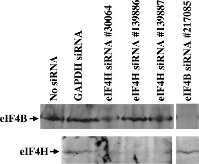
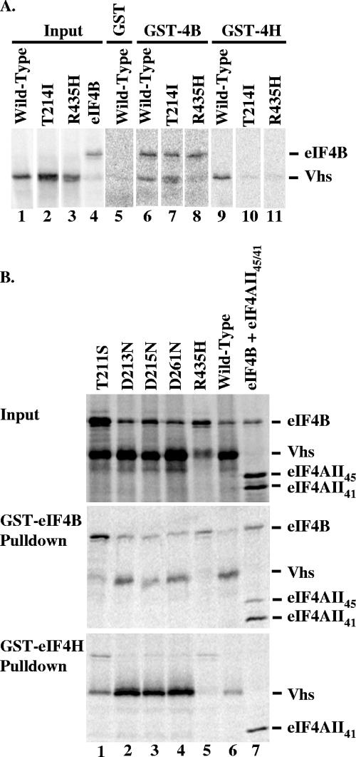
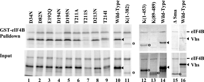
The herpes simplex virus (HSV) virion host shutoff (Vhs) protein is an endoribonuclease that accelerates decay of many host and viral mRNAs. Purified Vhs does not distinguish mRNAs from nonmessenger RNAs and cuts target RNAs at many sites, yet within infected cells it is targeted to mRNAs and cleaves those mRNAs at preferred sites including, for some, regions of translation initiation. This targeting may result in part from Vhs binding to the translation initiation factor eIF4H; in particular, several mutations in Vhs that abrogate its binding to eIF4H also abolish its mRNA-degradative activity, even though the mutant proteins retain endonuclease activity. To further investigate the role of eIF4H in Vhs activity, HeLa cells were depleted of eIF4H or other proteins by transfection with small interfering RNAs (siRNAs) 48 h prior to infection or mock infection in the presence of actinomycin D. Cellular mRNA levels were then assayed 5 h after infection. In cells transfected with an siRNA for the housekeeping enzyme glyceraldehyde-3-phosphate dehydrogenase, wild-type HSV infection reduced beta-actin mRNA levels to between 20 and 30% of those in mock-infected cells, indicative of a normal Vhs activity. In contrast, in cells transfected with any of three eIF4H siRNAs, beta-actin mRNA levels were indistinguishable in infected and mock-infected cells, suggesting that eIF4H depletion impeded Vhs-mediated degradation. Depletion of the related factor eIF4B did not affect Vhs activity. The data suggest that eIF4H binding is required for Vhs-induced degradation of many mRNAs, perhaps by targeting Vhs to mRNAs and to preferred sites within mRNAs.


Figures

FIG. 1.
FIG. 1.
Structures of eIF4H and eIF4B siRNAs. The structures of eIF4B and eIF4H are depicted at the top of the figure, with the regions of sequence homology shared by eIF4B (amino acids 90 to 172) and eIF4H (amino acids 36 to 117) represented by the black rectangles. The region of eIF4H shown by deletion analysis to be required for Vhs binding (amino acids 90 to 136) (17) is depicted by the solid line below the rectangle for eIF4H. Alteration of any of three amino acids (E97, D102, or D114) to alanine significantly reduces the binding of eIF4H to Vhs (18). The locations of these amino acids are depicted by the arrows below the rectangle for eIF4H. The sequences of the three eIF4H siRNAs and one eIF4B siRNA used in these experiments are shown at the bottom of the figure, along with their positions within eIF4H or eIF4B mRNAs. The identification number assigned by the manufacturer (Ambion) to each siRNA is shown at the left. UTR, untranslated region.
FIG. 2.
FIG. 2.
Transfection with siRNAs depletes the levels of eIF4H or eIF4B. HeLa cells were transfected with no siRNA, with siRNAs for GAPDH or eIF4B, or with three different siRNAs for eIF4H as indicated above the gel lanes. Cell lysates were prepared 48 h after transfection and assayed by Western blotting for eIF4B or eIF4H as indicated to the left of the gel. Each eIF4H-specific siRNA significantly reduced the level of eIF4H, but not eIF4B. The eIF4B siRNA depleted the level of eIF4B, but not eIF4H.
FIG. 3.
FIG. 3.
siRNA-mediated depletion of eIF4H impedes Vhs-induced degradation of β-actin mRNA. HeLa cells were transfected with no siRNA, with an siRNA for GAPDH, or with three different siRNAs for eIF4H as indicated. The cells were infected 48 h after transfection with 20 PFU/cell of wild-type (WT) HSV-1 or mock infected, both in the presence of actinomycin D. Total cytoplasmic RNAs were prepared 5 h after infection or mock infection and analyzed by real-time reverse transcription-PCR for the relative amounts of the housekeeping β-actin mRNA, using 18S rRNA as an endogenous control. For each type of transfection, the amount of β-actin mRNA in infected cells (gray bar) was normalized to the amount in mock-infected cells (black bar). Error bars represent the standard deviations of replicate samples and multiple experiments.
FIG. 4.
FIG. 4.
Binding of wild-type and mutant Vhs polypeptides to GST-eIF4B and GST-eIF4H. (A) [35S]methionine-labeled wild-type polypeptide and the mutant Vhs polypeptides T214I and R435H were produced by in vitro transcription and translation, as was [35S]methionine-labeled eIF4B. Aliquots of the in vitro-translated material are shown in lanes 1 through 4. Each of the Vhs polypeptides was mixed with an approximately equal amount (as judged by [35S]methionine incorporation) of eIF4B and analyzed for the ability to bind GST-eIF4B and GST-eIF4H as described previously (14, 17, 18). Equal aliquots of the same mixture of wild-type Vhs and eIF4B were assayed for binding GST (lane 5), GST-eIF4B (lane 6), and GST-eIF4H (lane 9) (14, 17, 18). Similarly, equal aliquots of the same mixture of the T214I polypeptide and eIF4B were assayed for binding GST-eIF4B (lane 7) and GST-eIF4H (lane 10), while equal aliquots of the same mixture of the R435H polypeptide and eIF4B were assayed for binding GST-eIF4B (lane 8) and GST-eIF4H (lane 11). Bound proteins were eluted from the beads and analyzed by SDS-PAGE and autoradiography. (B) [35S]methionine-labeled HSV-1(KOS) and mutant Vhs polypeptides were produced by in vitro transcription and translation and mixed with [35S]methionine-labeled, in vitro-translated eIF4B (lanes 1 to 6). The Vhs polypeptides and eIF4B were analyzed for the ability to bind GST-eIF4B and GST-eIF4H as described previously (14, 17, 18). Proteins that bound to GST-eIF4B (middle panel) and GST-eIF4H (bottom panel) were analyzed by SDS-PAGE and autoradiography. Aliquots of the input in vitro-translated material are shown in the top panel. The wild-type HSV-1(KOS) and mutant Vhs polypeptides are indicated at the top of each lane. Their structures are diagrammed in Fig. 6. In a parallel reaction, [35S]methionine-labeled eIF4B was mixed with full-length in vitro-translated eIF4AII (eIF4AII45) and a truncated form of eIF4AII containing amino acids 38 through 407 of the full-length protein (lane 7) and analyzed for binding to GST-eIF4B (middle panel) and GST-eIF4H (bottom panel).
FIG. 5.
FIG. 5.
Binding of wild-type HSV-1(KOS) and mutant Vhs polypeptides to GST-eIF4B. [35S]methionine-labeled HSV-1(KOS) and mutant Vhs polypeptides were produced by in vitro transcription and translation, mixed with [35S]methionine-labeled in vitro-translated eIF4B, and analyzed for the ability to bind GST-eIF4B (14, 17, 18). Proteins that bound to GST-eIF4B were analyzed by SDS-PAGE and autoradiography in the upper panel (GST-eIF4B Pulldown), while aliquots of the input in vitro-translated material are shown in the lower panel (Input). The wild-type HSV-1(KOS) and mutant Vhs polypeptides are indicated at the top of each lane. Their structures are diagrammed in Fig. 6. All binding reactions were performed in the same experiment, but lanes 1 through 9 were run on one gel, lanes 10 and 11 on another, lanes 12 through 14 on a third gel, and lanes 15 and 16 on a fourth. The gels were not run for precisely the same length of time, which is why the electrophoretic mobility of wild-type Vhs, relative to eIF4B, was not the same for the different gels. The location of the wild-type Vhs protein is indicated by filled arrowheads to the right of lanes 10, 14, and 16. The locations of mutant Vhs polypeptides that are not full length are shown by open circles to the right of lanes 11 to 13 and 15.
FIG. 6.
FIG. 6.
Summary of the structures and in vivo mRNA-degradative activities of wild-type and mutant Vhs polypeptides and their abilities to bind eIF4H and eIF4B. The Vhs polypeptide encoded by wild-type HSV-1(KOS) is represented by the solid rectangle in line 1, and the structures of deletion and point mutants of the HSV-1(KOS) polypeptide are in lines 2 through 17. For deletion mutants, the Vhs residues included in the mutant proteins are indicated. For each point mutant, the location of the altered residue is indicated by the vertical line above the bar representing the protein. The in vivo mRNA-degradative activity of each Vhs protein is shown in the third column from the right and summarizes, in a qualitative fashion, quantitative measurements that were reported previously (14-16, 49, 57). Activity was assayed for all Vhs alleles using a cotransfection assay of Vhs activity and during virus infections for those alleles marked by an asterisk. The double plus sign indicates activity similar to that of wild-type HSV-1(KOS), and the minus sign indicates no detectable mRNA-degradative activity. The second column from the right indicates whether a Vhs protein binds (++) or does not bind (−) eIF4H and summarizes data that have been reported previously (14, 17, 18). The binding of various mutant Vhs polypeptides to GST-eIF4B was expressed relative to the binding of wild-type Vhs to GST-eIF4B, as explained in Materials and Methods, and is shown in the rightmost column.
FIG. 7.
FIG. 7.
siRNA-mediated depletion of eIF4B does not affect Vhs-induced degradation of β-actin mRNA. HeLa cells were transfected with no siRNA or with siRNAs for GAPDH or eIF4B as indicated. The cells were infected 48 h after transfection with 20 PFU/cell of wild-type (WT) HSV-1 or mock infected, both in the presence of actinomycin D. Total cytoplasmic RNAs were prepared 5 h after infection or mock infection and analyzed by real-time reverse transcription-PCR for the relative amounts of β-actin mRNA, using 18S rRNA as an endogenous control. For each type of transfection, the amount of β-actin mRNA in infected cells (gray bar) was normalized to the amount in mock-infected cells (black bar). Error bars represent the standard deviations of replicate samples and multiple experiments.

Similar articles

Cited by

References

    1. Barzilai, A., I. Zivony-Elbom, R. Sarid, E. Noah, and N. Frenkel. 2006. The herpes simplex virus type 1 vhs-UL41 gene secures viral replication by temporarily evading apoptotic cellular response to infection: Vhs-UL41 activity might require interactions with elements of cellular mRNA degradation machinery. J. Virol. 80505-513. - PMC - PubMed
    1. Becker, Y., E. Tavor, Y. Asher, C. Berkowiltz, and M. Moyal. 1993. Effect of herpes simplex virus type-1 UL41 gene on the stability of mRNA from the cellular genes: beta-actin, fibronectin, glucose transporter-1, and docking protein, and on virus intraperitoneal pathogenicity of newborn mice. Virus Genes 7133-143. - PubMed
    1. Bevington, P. R. 1969. Data reduction and error analysis for the physical sciences. McGraw-Hill Book Company, New York, NY.
    1. Bookout, A. L., C. L. Cummins, D. J. Mangelsdorf, J. M. Pesola, and M. F. Kramer. 2006. High-throughput real-time quantitative reverse transcription PCR, p. 15.8.1-15.8.28. In F. M. Ausubel, R. Brent, R. E. Kingston, D. D. Moore, J. G. Seidman, J. A. Smith, and K. Struhl (ed.), Current protocols in molecular biology. John Wiley & Sons, Inc., New York, NY. - PubMed
    1. Chee, A. V., and B. Roizman. 2004. Herpes simplex virus 1 gene products occlude the interferon signaling pathway at multiple sites. J. Virol. 784185-4196. - PMC - PubMed

Publication types

MeSH terms