Comparative Study
. 2008 Jul;82(13):6631-43.
doi: 10.1128/JVI.00316-08. Epub 2008 Apr 30.

Antibodies to envelope glycoprotein of dengue virus during the natural course of infection are predominantly cross-reactive and recognize epitopes containing highly conserved residues at the fusion loop of domain II

Affiliations
Comparative Study

Antibodies to envelope glycoprotein of dengue virus during the natural course of infection are predominantly cross-reactive and recognize epitopes containing highly conserved residues at the fusion loop of domain II

Chih-Yun Lai et al. J Virol. 2008 Jul.

Abstract

The antibody response to the envelope (E) glycoprotein of dengue virus (DENV) is known to play a critical role in both protection from and enhancement of disease, especially after primary infection. However, the relative amounts of homologous and heterologous anti-E antibodies and their epitopes remain unclear. In this study, we examined the antibody responses to E protein as well as to precursor membrane (PrM), capsid, and nonstructural protein 1 (NS1) of four serotypes of DENV by Western blot analysis of DENV serotype 2-infected patients with different disease severity and immune status during an outbreak in southern Taiwan in 2002. Based on the early-convalescent-phase sera tested, the rates of antibody responses to PrM and NS1 proteins were significantly higher in patients with secondary infection than in those with primary infection. A blocking experiment and neutralization assay showed that more than 90% of anti-E antibodies after primary infection were cross-reactive and nonneutralizing against heterologous serotypes and that only a minor proportion were type specific, which may account for the type-specific neutralization activity. Moreover, the E-binding activity in sera of 10 patients with primary infection was greatly reduced by amino acid replacements of three fusion loop residues, tryptophan at position 101, leucine at position 107, and phenylalanine at position 108, but not by replacements of those outside the fusion loop of domain II, suggesting that the predominantly cross-reactive anti-E antibodies recognized epitopes involving the highly conserved residues at the fusion loop of domain II. These findings have implications for our understanding of the pathogenesis of dengue and for the future design of subunit vaccine against DENV as well.


Figures

FIG. 1.
FIG. 1.
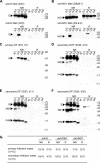
Antibody responses to different DENV proteins of four serotypes in patients with primary and secondary DENV2 (D2) infections. Convalescent-phase sera from two patients with primary infection (A and B) and two with secondary infection (C and D) as well as anti-E (4G2), anti-PrM (70-21), anti-C (DB32-40-30), and anti-NS1 (DB29-1) MAbs were subjected to Western blot analysis using lysates derived from mock (M)-, DENV1 (Hawaii strain)-, DENV2 (NGC strain)-, DENV3 (H87 strain)-, or DENV4 (H241 strain)-infected C6/36 cells. DF and DHF were classified according to the WHO case definition (60). Primary or secondary infection was determined as described previously (53, 59). Day 1 was defined as the day of onset of fever. Arrowheads indicate the E, PrM, C, and NS1 proteins recognized. Molecular size marker units are kDa. d14, day 14.
FIG. 2.
FIG. 2.
Antibody responses to different DENV proteins of four serotypes in sequential sera from patients with primary (A and B) and secondary (C to E) DENV2 (D2) infection. Sera were subjected to Western blot analysis, and the data are presented as described for Fig. 1. Arrowheads indicate the E, PrM, and NS1 proteins recognized. Molecular size marker units are kDa. d5, day 5.
FIG. 3.
FIG. 3.
Antibody responses to different DENV proteins of four serotypes under nonreducing and reducing conditions in patients with primary and secondary DENV2 (D2) infections. Anti-E MAbs (4G2 and 3H5 in panel A) and anti-NS1 MAbs (DB29-1 and D2-8-2 in panel B), as well as convalescent-phase sera from patients with primary (C) or secondary infection (D to F) were subjected to Western blot analysis under nonreducing (NR) and reducing (R) conditions as described in Materials and Methods. DF and DHF were classified according to the WHO case definition (60). Primary or secondary infection was determined as described previously (53, 59). Day 1 was defined as the day of onset of fever. Arrowheads indicate E, PrM, and NS1 proteins. Molecular size marker units are kDa. d7, day 7. (G) Summary of the results for 25 patients, including 15 with primary infection and 10 with secondary infection. One representative experiment of more than two is shown.
FIG. 4.
FIG. 4.
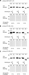
Antibody blocking experiment to determine the DENV2 (D2)-specific E-binding activity relative to DENV1, DENV3, and DENV4 in sera of three patients with primary DENV2 infection, including (A) ID17, (B) ID4, and (C) ID5. The antibody blocking experiment was as described in Materials and Methods. The ratio of the intensity of the anti-DENV2 E protein band from postblocking serum to that of the band from nonblocked serum (in percent) was determined as the DENV2-specific E-binding activity for that serotype. Primary infection was determined as described previously (53, 59). Day 1 was defined as the day of onset of fever. Arrowheads indicate E proteins. Molecular size marker units are kDa. One representative experiment of more than three was shown. d12, day 12.
FIG. 5.
FIG. 5.
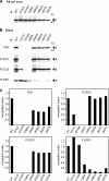
MAb binding to DENV1 E proteins with mutations within (101WA, 107LD, and 108FA) and outside (209HA, 258GA, 262TA, and 265TA) the fusion loop of domain II. Cell lysates derived from 293T cells transfected with mock (M), wild-type (WT; pCB-D1), and mutant E constructs were subjected to Western blot analysis by using mixed sera (A) and four anti-E MAbs (B) (26). Mixed sera consisted of a pool of nine sera of DENV2 patients with secondary infection (59). Arrowheads indicate E proteins. Molecular size marker units are kDa. One representative experiment of more than two was shown. (C) The intensities of E protein bands of wild type and mutants were analyzed as described in Materials and Methods. The recognition index of a MAb to a mutant E protein is given by the following formula: (intensity of the mutant E band/intensity of wild-type E band [by MAb]) × (intensity of wild-type E band/intensity of mutant E band [by mixed sera]).
FIG. 6.
FIG. 6.
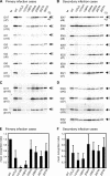
Binding of convalescent-phase sera to DENV1 E proteins with mutations within (101WA, 107LD, and 108FA) and outside (209HA, 258GA, 262TA, and 265TA) the fusion loop of domain II. Cell lysates derived from 293T cells transfected with mock (M), wild-type (WT; pCB-D1), and mutant E constructs were subjected to Western blot analysis by using mixed sera (shown in Fig. 5A): sera from 10 patients with primary DENV2 infection, 7 at early (A) and 3 at later time points (C), and from 11 patients with secondary DENV2 infection, 7 at early (B) and 4 at later time points (D). Primary or secondary infection was determined as described previously (53, 59). Day 1 was defined as the day of onset of fever. The data are presented as described for Fig. 5. One representative experiment of more than two was shown. d7, day 7. (E and F) The intensities of E protein bands of the wild type and mutants as well as recognition indices were analyzed as described for Fig. 5. Data are mean recognition indices ± standard errors for patients with primary (E) and secondary (F) infections to each mutant E protein.

Similar articles

Cited by

References

    1. Alvarez, M., R. Rodriguez-Roche, L. Bernardo, S. Vazquez, L. Morier, D. Gonzalez, O. Castro, G. Kouri, S. B. Halstead, and M. G. Guzman. 2006. Dengue hemorrhagic Fever caused by sequential dengue 1-3 virus infections over a long time interval: Havana epidemic, 2001-2002. Am. J. Trop. Med. Hyg. 751113-1117. - PubMed
    1. Beasley, D. W. C., and A. D. T. Barrett. 2002. Identification of neutralizing epitopes within structural domain III of the West Nile virus envelope protein. J. Virol. 7613097-13100. - PMC - PubMed
    1. Bray, M., and C. J. Lai. 1991. Dengue virus premembrane and membrane proteins elicit a protective immune response. Virology 185505-508. - PubMed
    1. Burke, D. S., A. Nisalak, D. E. Johnson, and R. M. Scott. 1988. A prospective study of dengue infections in Bangkok. Am. J. Trop. Med. Hyg. 38172-180. - PubMed
    1. Churdboonchart, V., N. Bhamarapravati, S. Peampramprecha, and S. Sirinavin. 1991. Antibodies against dengue viral proteins in primary and secondary dengue hemorrhagic fever. Am. J. Trop. Med. Hyg. 44481-493. - PubMed

Publication types