Comparative Study
. 2008 Jul;82(13):6470-80.
doi: 10.1128/JVI.00117-08. Epub 2008 Apr 30.

Cholesterol effectively blocks entry of flavivirus

Affiliations
Comparative Study

Cholesterol effectively blocks entry of flavivirus

Chyan-Jang Lee et al. J Virol. 2008 Jul.

Erratum in

Abstract

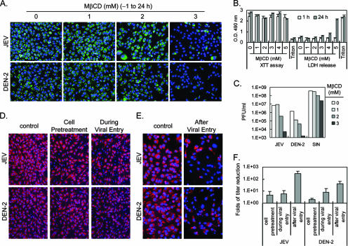
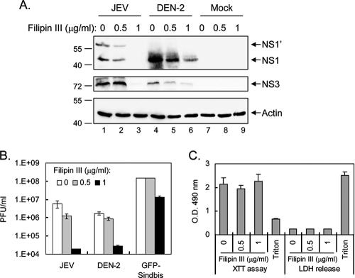
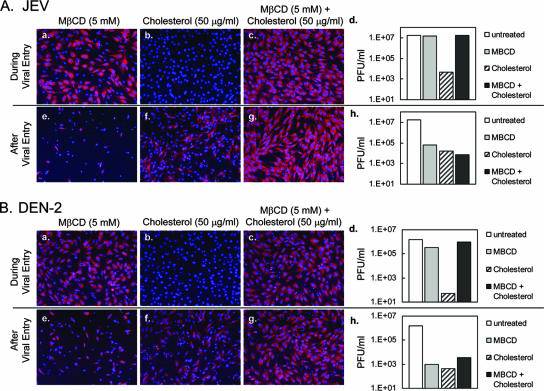
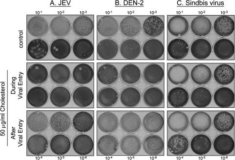
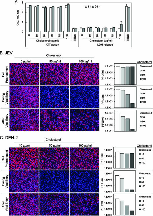
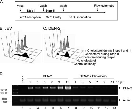
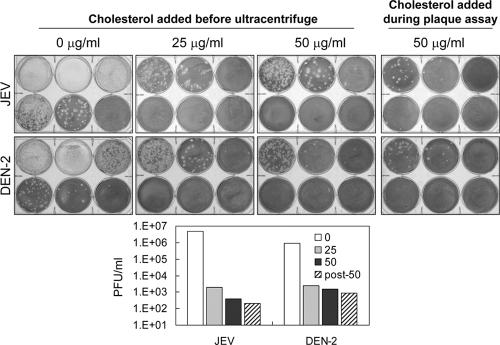
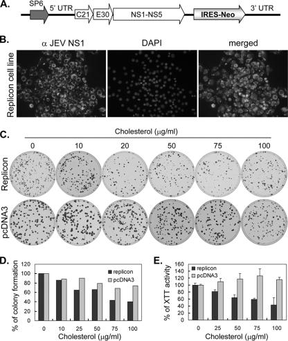
Japanese encephalitis virus (JEV) and dengue virus serotype 2 (DEN-2) are enveloped flaviviruses that enter cells through receptor-mediated endocytosis and low pH-triggered membrane fusion and then replicate in intracellular membrane structures. Lipid rafts, cholesterol-enriched lipid-ordered membrane domains, are platforms for a variety of cellular functions. In this study, we found that disruption of lipid raft formation by cholesterol depletion with methyl-beta-cyclodextrin or cholesterol chelation with filipin III reduces JEV and DEN-2 infection, mainly at the intracellular replication steps and, to a lesser extent, at viral entry. Using a membrane flotation assay, we found that several flaviviral nonstructural proteins are associated with detergent-resistant membrane structures, indicating that the replication complex of JEV and DEN-2 localizes to the membranes that possess the lipid raft property. Interestingly, we also found that addition of cholesterol readily blocks flaviviral infection, a result that contrasts with previous reports of other viruses, such as Sindbis virus, whose infectivity is enhanced by cholesterol. Cholesterol mainly affected the early step of the flavivirus life cycle, because the presence of cholesterol during viral adsorption greatly blocked JEV and DEN-2 infectivity. Flavirial entry, probably at fusion and RNA uncoating steps, was hindered by cholesterol. Our results thus suggest a stringent requirement for membrane components, especially with respect to the amount of cholesterol, in various steps of the flavivirus life cycle.


Figures

FIG. 1.
FIG. 1.
Depletion of cholesterol by MβCD affects JEV, DEN-2, and SIN infection. (A and C) N18 cells were pretreated with MβCD (0, 1, 2, or 3 mM) for 1 h, and then the cells were adsorbed with JEV, DEN-2, or SIN (MOI = 10) in the presence of various concentrations of MβCD at 37°C. At 90 min after adsorption, the inoculated virus was washed away and the cells were cultured in fresh medium containing MβCD. At 24 h after infection, the cells were stained for IFA using anti-NS1 antibody to visualize viral protein expression (green) (A), and the culture supernatants were harvested for viral titration as determined by a plaque-forming assay (C). The cell nuclei were stained with DAPI (4′,6′-diamidino-2-phenylindole) (blue). (B) The drug effect of MβCD was determined in the mock-infected N18 cells by culturing with MβCD (0 to 5 mM) for 1 or 24 h, and cell proliferation and cytotoxicity were detected by XTT and LDH assays, respectively. O.D., optical density. (D) In the “Cell Pretreatment” group, N18 cells were pretreated with MβCD (5 mM) for 1 h, washed, and infected with JEV or DEN-2 (MOI = 10). In the “During Viral Entry” group, N18 cells were adsorbed with JEV or DEN-2 (MOI = 10) in the presence of MβCD (5 mM) at 37°C. After 90 min of viral adsorption, the cells were washed and then cultured in fresh medium without MβCD. The IFA and viral titration assay were performed at 24 h after infection. (E) In the “After Viral Entry” group, N18 cells were adsorbed with JEV or DEN-2 (MOI = 10) for 90 min at 37°C and then cultured in medium supplemented with MβCD (3 mM). The IFA and viral titration assay were performed at 24 h postinfection. (F) Severalfold decreases in titers of JEV and DEN-2 after treatment with MβCD. The viral production determined as shown in panels D and E is presented as severalfold reductions in viral levels compared with the results seen with cells not treated with MβCD. The data represent the averages and standard deviations of the results of two to three independent experiments.
FIG. 2.
FIG. 2.
Filipin III blocks JEV and DEN-2 infection. (A and B) N18 cells were adsorbed with JEV or DEN-2 (MOI = 10) for 90 min at 37°C and then cultured in the medium supplemented with various doses of filipin III, as indicated. At 20 h postinfection, the cell lysates were immunoblotted with antibodies against NS1 or NS3 of JEV and DEN-2 or an actin control (A), and the culture supernatants were harvested and subjected to a plaque formation assay (B). (C) The drug effect of filipin in the mock-infected N18 cells was determined by culturing with filipin (0, 0.5, and 1 μg/ml) for 24 h, and then cell proliferation and cytotoxicity were detected by XTT and LDH assays, respectively. O.D., optical density.
FIG. 3.
FIG. 3.
Association of flaviviral nonstructural proteins with DRM fractions. (A) N18 cells were infected with JEV or DEN-2 (MOI = 10) for 18 h. The cells were lysed using 1% Triton X-100 in TNE buffer and then subjected to sucrose gradient ultracentrifugation. The floating bands were collected for Western blotting analysis as described in Materials and Methods. (B) N18 cells were infected with JEV (MOI = 10) for 16 h, and the cell lysates collected by use of 1% NP-40-LSB were subjected to sucrose gradient ultracentrifugation as described in Materials and Methods. The samples in each fraction were analyzed by Western blotting using the antibodies indicated on the right side of the panel.
FIG. 4.
FIG. 4.
The effects of MβCD are partially compensated by coincubation with cholesterol. In the “During Viral Entry” group, MβCD (5 mM) (a), cholesterol (50 μg/ml) (b), or MβCD (5 mM) plus cholesterol (50 μg/ml) (c) was added to the medium during JEV (A) or DEN-2 (B) (MOI = 5) adsorption at 37°C for 90 min. The cells were washed and cultured in fresh medium without any treatment. In the “After Viral Entry” group, after 90 min of JEV (A) or DEN-2 (B) (MOI = 5) adsorption at 37°C, the cells were cultured in medium with MβCD (5 mM) (e), cholesterol (50 μg/ml) (f), or MβCD (5 mM) plus cholesterol (50 μg/ml) (g). The viral protein IFA results (a, b, c, e, f, and g) and viral titer results (d and h) were determined at 24 h postinfection.
FIG. 5.
FIG. 5.
Cholesterol decreases plaque formation of JEV and DEN-2 in BHK-21 cells. Viral plaque formation assays were performed using BHK-21 cells for JEV (A), DEN-2 (B), and SIN (C). The effect of cholesterol on viral entry was assessed by adding 50 μg/ml of cholesterol into the culture medium during viral adsorption at 37°C (middle panels). The cells were then washed and overlaid with agarose for plaque formation. The effect of cholesterol on viral replication was assessed by including cholesterol (50 μg/ml) in the agarose overlay (bottom panels). The plaques were stained with crystal violet as described in Materials and Methods.
FIG. 6.
FIG. 6.
Cholesterol blocks flaviviral infection at the viral entry and replication steps in N18 cells. (A) The drug effect of cholesterol was determined in the mock-infected N18 cells by culturing with cholesterol (0 to 100 μg/ml) for 1 or 24 h, and then cell proliferation and cytotoxicity were detected by XTT and LDH assays, respectively. O.D., optical density. (B and C) In the “Cell Pretreatment” group, N18 cells were pretreated with cholesterol (10 to 100 μg/ml) for 60 min, washed, and infected with JEV (B) or DEN-2 (C) (MOI = 10). In the “During Viral Entry” group, cholesterol (10 to 100 μg/ml) was added in the culture medium during viral adsorption at 37°C. In the “After Viral Entry” group, media with various doses of cholesterol (10 to 100 μg/ml) were used after viral entry. IFA with anti-NS1 antibody (red) and viral titration assays were performed at 18 h postinfection. The cell nuclei were stained with DAPI (blue).
FIG. 7.
FIG. 7.
Cholesterol may block flaviviral entry and viral RNA uncoating. As outlined in panel A, BHK-21 cells were adsorbed with JEV (B) or DEN-2 (C) (MOI = 5) for 90 min on ice with or without cholesterol (100 μg/ml). The cells were washed twice with cold medium and incubated at 37°C in the presence or absence of cholesterol for 15 min. The cells were washed and incubated for 20 h before harvest for intracellular staining with anti-NS3 and flow cytometric analysis. (D) N18 cells were infected with DEN-2 (MOI = 5) for 60 min at 37°C with or without cholesterol treatment (100 μg/ml). The cells were then washed and incubated further for the times indicated (h p.i., hours postinfection). Total RNA was isolated and analyzed by reverse transcription-PCR for DEN-2 positive-sense viral RNA and actin as described in Materials and Methods.
FIG. 8.
FIG. 8.
Cholesterol may block flaviviral entry through intercalating into the lipid bilayer of virions. JEV and DEN-2 were incubated with cholesterol (0, 25, or 50 μg/ml) for 30 min and then ultracentrifuged through a 35% sucrose cushion at 35,000 rpm in a Beckman SW41 rotor for 3.5 h at 4°C. The pellets were then resuspended, and plaque levels were assayed using BHK-21 cells as described in Materials and Methods. The same batch of virus stock without ultracentrifugation was also treated with cholesterol (50 μg/ml) during the adsorption step of the plaque assay to serve as the positive control in the investigation of cholesterol's antiviral effect.
FIG. 9.
FIG. 9.
JEV replicon cell growth is slightly reduced by cholesterol treatment. (A) Schematic representation of the JEV replicon construct. SP6, SP6 promoter. “5′ UTR” and “3′ UTR” represent the 5′ and 3′ UTRs, respectively. C21 corresponds to the first 21 amino acids of JEV core protein, and E30 corresponds to the last 30 amino acids of JEV E protein. NS1-NS5 corresponds to the sequence coding for the JEV nonstructural proteins. IRES-Neo represents a sequence of an IRES of encephalomyocarditis virus followed by a neomycin resistance gene (Neo). (B) Establishment of a JEV replicon cell line. BHK-21 cells were transfected with in vitro-transcribed replicon RNA and selected with G418. The G418-resistant cells were stained with anti-JEV NS1 plus fluorescein isothiocyanate-conjugated secondary antibody and DAPI. (C) Colony formation of JEV replicon cells in the presence of cholesterol. The replicon cells and the pcDNA3-transfected BHK-21 cells were cultured with a G418-containing agarose overlay plus different concentrations of cholesterol as listed at the top of the panel. Colony formation was assessed by crystal violet staining after 1 week of incubation. (D) Replicon cell colony formation was slightly reduced by cholesterol treatment. The crystal violet-stained cells were lysed, and the level of optical density absorbance at an optical density of 570 nm was determined. The data are derived from the average results obtained with four plates for each experimental group. (E) JEV replicon cell growth was reduced by cholesterol treatment. The indicated cells were cultured with G418 plus cholesterol at different concentrations for 24 h before the cell growth was monitored by XTT assays. The averages and standard deviations of the results obtained with triplicate samples are shown.

Similar articles

Cited by

References

    1. Aizaki, H., K. J. Lee, V. M. Sung, H. Ishiko, and M. M. Lai. 2004. Characterization of the hepatitis C virus RNA replication complex associated with lipid rafts. Virology 324450-461. - PubMed
    1. Amano, T., E. Richelson, and M. Nirenberg. 1972. Neurotransmitter synthesis by neuroblastoma clones (neuroblast differentiation-cell culture-choline acetyltransferase-acetylcholinesterase-tyrosine hydroxylase-axons-dendrites). Proc. Natl. Acad. Sci. USA 69258-263. - PMC - PubMed
    1. Anderson, R. 2003. Manipulation of cell surface macromolecules by flaviviruses. Adv. Virus Res. 59229-274. - PMC - PubMed
    1. Bender, F. C., J. C. Whitbeck, M. Ponce de Leon, H. Lou, R. J. Eisenberg, and G. H. Cohen. 2003. Specific association of glycoprotein B with lipid rafts during herpes simplex virus entry. J. Virol. 779542-9552. - PMC - PubMed
    1. Briggs, J. A., T. Wilk, and S. D. Fuller. 2003. Do lipid rafts mediate virus assembly and pseudotyping? J. Gen. Virol. 84757-768. - PubMed

Publication types