. 2008 Jun;135(12):2083-91.
doi: 10.1242/dev.020099. Epub 2008 Apr 30.

Early patterning of the chorion leads to the trilaminar trophoblast cell structure in the placental labyrinth

Affiliations

Early patterning of the chorion leads to the trilaminar trophoblast cell structure in the placental labyrinth

David G Simmons et al. Development. 2008 Jun.

Abstract

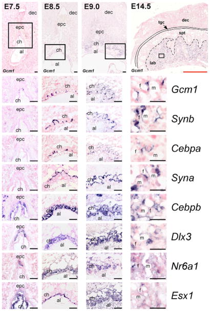
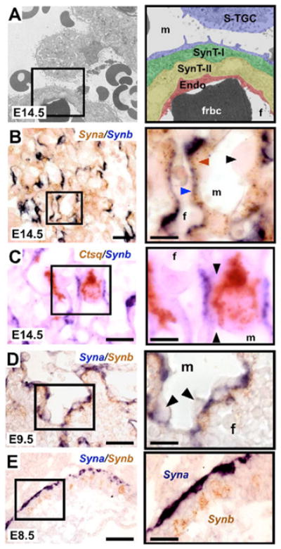
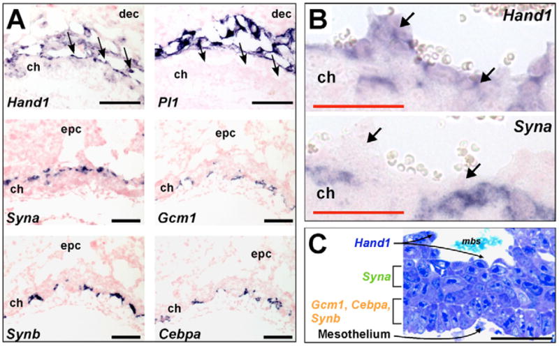
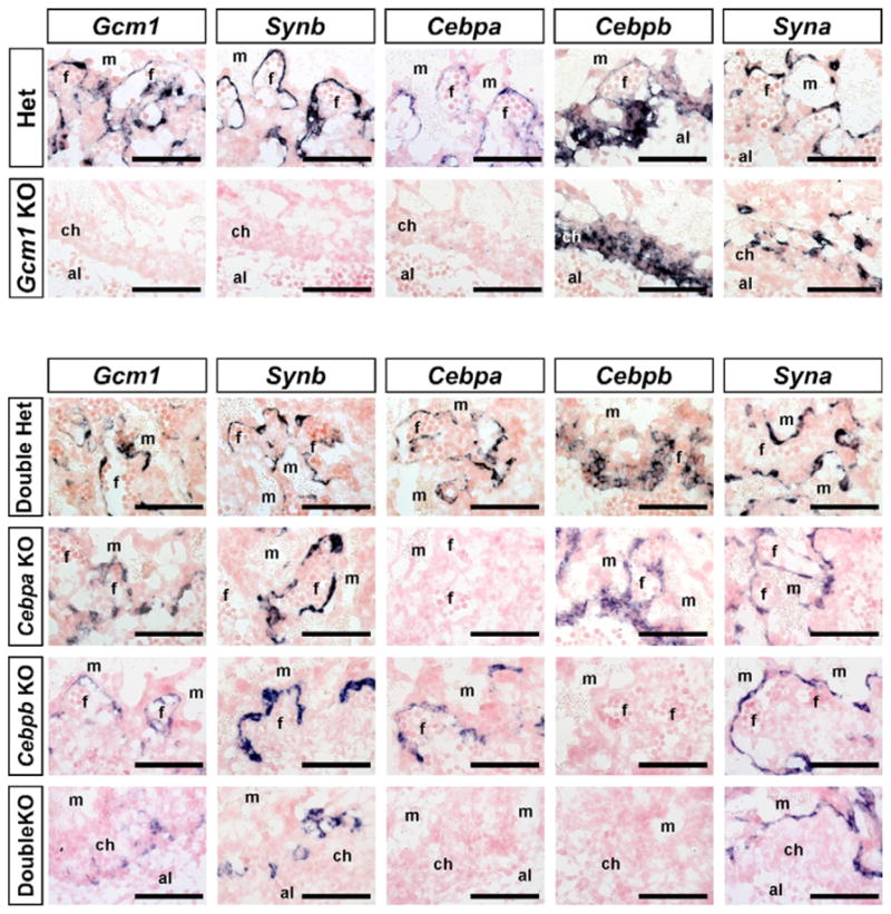
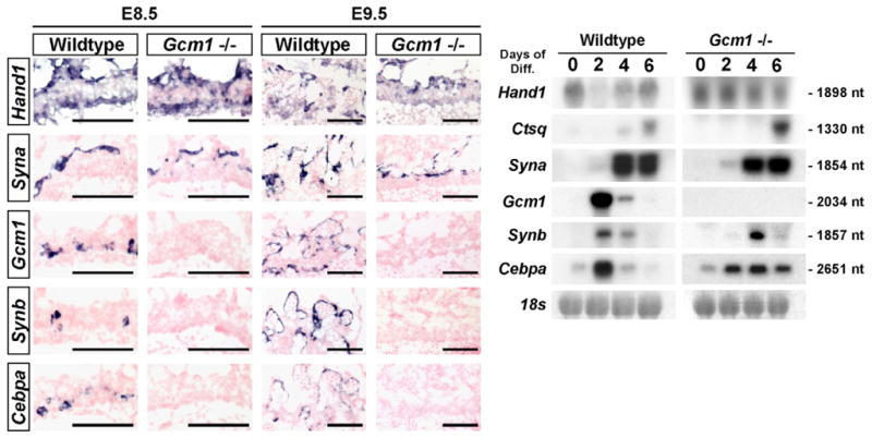
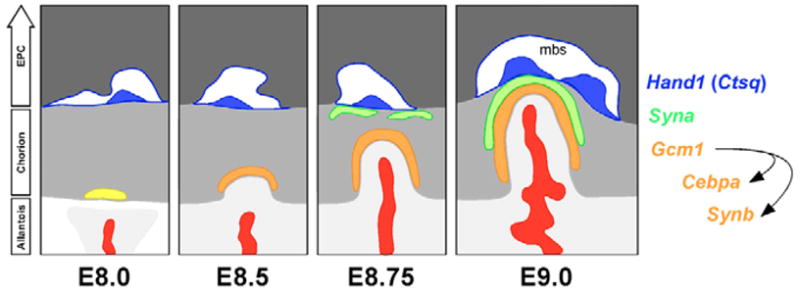
The labyrinth of the rodent placenta contains villi that are the site of nutrient exchange between mother and fetus. They are covered by three trophoblast cell types that separate the maternal blood sinusoids from fetal capillaries--a single mononuclear cell that is a subtype of trophoblast giant cell (sinusoidal or S-TGC) with endocrine function and two multinucleated syncytiotrophoblast layers, each resulting from cell-cell fusion, that function in nutrient transport. The developmental origins of these cell types have not previously been elucidated. We report here the discovery of cell-layer-restricted genes in the mid-gestation labyrinth (E12.5-14.5) including Ctsq in S-TGCs (also Hand1-positive), Syna in syncytiotrophoblast layer I (SynT-I), and Gcm1, Cebpa and Synb in syncytiotrophoblast layer II (SynT-II). These genes were also expressed in distinct layers in the chorion as early as E8.5, prior to villous formation. Specifically, Hand1 was expressed in apical cells lining maternal blood spaces (Ctsq is not expressed until E12.5), Syna in a layer immediately below, and Gcm1, Cebpa and Synb in basal cells in contact with the allantois. Cebpa and Synb were co-expressed with Gcm1 and were reduced in Gcm1 mutants. By contrast, Hand1 and Syna expression was unaltered in Gcm1 mutants, suggesting that Gcm1-positive cells are not required for the induction of the other chorion layers. These data indicate that the three differentiated trophoblast cell types in the labyrinth arise from distinct and autonomous precursors in the chorion that are patterned before morphogenesis begins.


Figures

Fig. 1
Fig. 1. Expression of mouse labyrinth trophoblast markers from E7.5-14.5
In situ hybridizations for the labyrinth trophoblast markers Gcm1, Synb, Cebpa, Syna, Cebpb, Dlx3, Nr6a1 and Esx1 at E7.5, E8.5, E9.0 and E14.5. Low-magnification images (top row) are of Gcm1 expression. Black boxes indicate the areas shown at high-magnification in the panels beneath. al, allantois; ch, chorion; dec, decidua; epc, ectoplacental cone; f, fetal blood space; lab, labyrinth; m, maternal blood space; spt, spongiotrophoblast; tgc, trophoblast giant cell. Scale bars: black, 100 μm; red, 1.4 mm.
Fig. 2
Fig. 2. Gcm1, Synb and Cebpa are expressed in SynT-II cells and Syna is expressed in SynT-I cells
(A) Electron micrograph of the trilaminar trophoblast structure that separates the maternal and fetal blood compartments. The black box indicates the area shown at high-magnification on the right. S-TGC, sinusoidal trophoblast giant cell; SynT-I, syncytiotrophoblast layer I; SynT-II, syncytiotrophoblast layer II; Endo, fetal endothelial cell; m, maternal blood space; f, fetal blood space; frbc, fetal red blood cell. (B) Double in situ hybridization for Syna (brown) and Synb (purple) in the labyrinth at E14.5 indicating that Syna expression is restricted to SynT-I cells and Synb expression is restricted to SynT-II cells. Note that Syna/Gcm1 and Syna/Cebpa double in situ hybridizations produced similar results (data not shown). Black arrowhead indicates unstained S-TGC which lines maternal blood spaces. Brown arrowhead indicates Syna-positive SynT-I cells located closer to maternal blood spaces. Blue arrowhead indicates Synb-positive SynT-II cells located closest to fetal blood spaces. (C) Double in situ hybridization for Ctsq (brown) and Synb (purple) in the labyrinth at E14.5. Black arrows indicate unstained SynT-I cell between Ctsq-positive S-TGC and Synb-positive SynT-II cell. (D) Double in situ hybridization for Syna (purple) and Synb (brown) in the labyrinth at E9.5. Syna and Synb expression do not co-localize. Note that Syna/Gcm1 and Syna/Cebpa double in situ hybridizations showed similar results (data not shown). (E) Double in situ hybridization for Syna (purple) and Synb (brown) in chorion at E8.5. Syna and Synb expression do not co-localize at E8.5 and demarcate distinct trophoblast populations within the chorion. Note that Syna/Gcm1 and Syna/Cebpa double in situ hybridizations showed similar results (data not shown). Scale bars: 100 μm (left); 50 μm (right).
Fig. 3
Fig. 3. Early patterning of the mouse chorion
(A) In situ hybridizations for Hand1 and Pl1 at E9.0 indicate that Hand1+/Pl1 cells line the maternal blood sinuses located on the apical side of the chorion. At E8.5, Syna expression is restricted to cells near the top of the chorion, whereas Gcm1, Synb and Cebpa staining (on serial sections) indicates co-localization within a subset of trophoblast located on the basal side of the chorion (confirmed by double in situ hybridization, data not shown). The black arrows indicate Hand1+/Pl1 cells lining maternal sinusoids. (B) Higher magnification images of Hand1 and Syna in situ hybridizations on E8.5 chorion. Note that Syna-positive cells are separated from the maternal blood sinuses by a single layer of cells, typically Hand1-positive. Black arrows indicate the cell layer lining the maternal sinusoids. (C) Ultrathin epoxy section of E8.5 chorion (2 μm) stained with Tissue Epoxy Stain (EMS). Note the unique morphology of the different layers of trophoblast cells in the chorion. Cells that line the maternal sinusoids (Hand1+/Pl1 at E8.5) are positive for Ctsq expression by E12.5 (data not shown). Scale bars: black, 100 μm; red, 50 μm.
Fig. 4
Fig. 4. Synb and Cebpa are downstream of Gcm1, but Synb is not downstream of Cebpa
In situ hybridizations for Gcm1, Synb, Cebpa, Cebpb and Syna on 10 μm sections of E9.5 Gcm1+/− (Het) and Gcm1−/− (Gcm1 KO) mouse placentas and Cebpa+/−;Cebpb+/− (Double Het), Cebpa−/− (Cebpa KO), Cebpb−/− (Cebpb KO) and Cebpa−/−;Cebpb−/− (DoubleKO) placentas. al, allantois; ch, chorion; f, fetal blood space; m, maternal blood space. Scale bars: 100 μm.
Fig. 5
Fig. 5. Gcm1/Synb/Cebpa-expressing cells are not required to induce or maintain Syna or Hand1 expression
(Left) In situ hybridizations for Hand1, Syna, Gcm1, Cebpa and Synb were performed on E8.5 and E9.5 wild-type and Gcm1−/− mouse placentas. (Right) Northern blot analysis of Hand1, Ctsq, Syna, Gcm1, Cebpa and Synb expression in differentiating TS cultures following the removal of human recombinant FGF4, heparin and embryonic fibroblast-conditioned media. 18s rRNA was used as a loading control.
Fig. 6
Fig. 6. Model for mouse labyrinth formation from a pre-patterned chorion
Note that Ctsq expression within S-TGCs is not fully evident until E12.5. No exclusive marker has been identified for S-TGC precursors, although cells lining the early maternal sinusoids are Hand1+/Pl1. However, Hand1 expression is not restricted to cells lining the maternal sinusoids in the way that Ctsq is restricted to S-TGCs in mid-gestation; Hand1 is expressed in all TGC subtypes and within the ectoplacental cone and chorion (not shown). It is the combined Hand1+/Pl1 nature of the cells lining the maternal sinusoids, together with their location, that implies they are S-TGC progenitors. For simplicity, cell bodies of Hand1+/Pl1 cells are shown only along the top of the chorion. Light gray, allantois; gray, chorion; dark gray, ectoplacental cone (EPC); red, extraembryonic mesoderm-derivatives; yellow, Gcm1+ chorionic trophoblast cells; orange, Gcm1/Synb/Cebpa+ chorionic trophoblast cells; green, Syna+ trophoblast cells; blue, developing sinusoidal giant cells (S-TGCs) which will subsequently express Ctsq; mbs, maternal blood sinus.

Similar articles

Cited by

References

    1. Akiyama Y, Hosoya T, Poole AM, Hotta Y. The gcm-motif: a novel DNA-binding motif conserved in Drosophila and mammals. Proc Natl Acad Sci USA. 1996;93:14912–14916. - PMC - PubMed
    1. Anson-Cartwright L, Dawson K, Holmyard D, Fisher SJ, Lazzarini RA, Cross JC. The glial cells missing-1 protein is essential for branching morphogenesis in the chorioallantoic placenta. Nat Genet. 2000;25:311–314. - PubMed
    1. Basyuk E, Cross JC, Corbin J, Nakayama H, Hunter P, Nait-Oumesmar B, Lazzarini RA. Murine Gcm1 gene is expressed in a subset of placental trophoblast cells. Dev Dyn. 1999;214:303–311. - PubMed
    1. Beck F, Erler T, Russell A, James R. Expression of Cdx-2 in the mouse embryo and placenta: possible role in patterning of the extra-embryonic membranes. Dev Dyn. 1995;204:219–227. - PubMed
    1. Begay V, Smink J, Leutz A. Essential requirement of CCAAT/enhancer binding proteins in embryogenesis. Mol Cell Biol. 2004;24:9744–9751. - PMC - PubMed

Publication types

MeSH terms

Grants and funding