. 2008 Jul;295(1):F18-26.
doi: 10.1152/ajprenal.00573.2007. Epub 2008 Apr 30.

Regulation of epithelial sodium transport by promyelocytic leukemia zinc finger protein

Affiliations

Regulation of epithelial sodium transport by promyelocytic leukemia zinc finger protein

Anikó Náray-Fejes-Tóth et al. Am J Physiol Renal Physiol. 2008 Jul.

Abstract

Aldosterone is the principal regulator of Na homeostasis, and thereby blood pressure. One of the main targets of aldosterone is the epithelial Na channel (ENaC) located in the apical membrane of target cells. Previous studies identified several genes involved in the regulation of ENaC such as SGK1; however, SGK1 knockout mice have only a mild salt-losing phenotype, indicating that further genes must be involved in the action of aldosterone. In our search for further aldosterone-regulated genes, we discovered that aldosterone, at physiological concentrations, induces the expression of the promyelocytic leukemia zinc finger protein (PLZF) in renal cortical collecting duct (CCD) cell lines that stably express mineralocorticoid receptors (MRs). This effect is rapid and does not require de novo protein synthesis, suggesting a direct action. Surprisingly, stable overexpression of human or mouse PLZF isoforms significantly decreased transepithelial Na transport in CCD cells while having no effect on the integrity of the monolayers. In parallel with the decline in Na transport, PLZF suppressed the mRNA levels of beta- and gamma-ENaC subunits. These observations suggest that PLZF is a negative regulator of ENaC in renal epithelial cells and might be part of a negative feedback loop that limits aldosterone's stimulatory effects on sodium reabsorption.


Figures
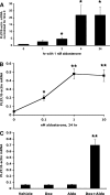
Fig. 1.
Fig. 1.
A: Aldosterone induces promyelocytic leukemia zinc finger protein (PLZF) expression in M1-MR+ cells. Cells were grown for 48 hrs in steroid-free medium, and then medium was changed to the same (control) or supplemented with 1 nM aldosterone, for 1–24 h. PLZF and β-actin mRNA levels were determined by RT-PCR from the same samples. Values are RE (relative expression) of PLZF/β-actin mRNA, and are expressed as fold change. *, P < 0.05 vs. time 0, using 1-tailed unpaired t-test; n = 5 for time 0 and 2 h; n = 3 for other time points. B: Dose-response effect of aldosterone on PLZF mRNA levels in M1/TetON-MR cells. Cells were treated with 500 ng/ml dox for 2 days to induce MR expression, then with 0–10 nM aldosterone for an additional 24 h, and PLZF RNA levels determined as above. Values shown are RE *, P < 0.05 vs. untreated cells using 1-tailed unpaired t-test, n = 3. C: effect of aldosterone is mediated via the MR. Cells were incubated with or without 500 ng/ml dox for 2 days, then with 1 nM aldosterone for an additional 24 h. Aldosterone increased PLZF expression only when MR expression was induced by dox. n = 3 for each treatment. **, P < 0.01 vs. untreated cells using 2-tailed unpaired t-test.
Fig. 2.
Fig. 2.
The effect of protein synthesis inhibition on the induction of PLZF by aldosterone. M1/TetON-MR cells were grown in steroid-free medium for 2 days, then expression of MR was induced with 500 ng/ml dox for 2 days. Cells were then preincubated for 30 min with vehicle (no inhibitor), or the protein synthesis inhibitors cycloheximide (CHX; 5 μg/ml) or anisomycin (aniso; 10 μM). Aldosterone (1 nM) was given (or not) at 30 min and cells were incubated for an additional 2 h before RNA was prepared. PLZF and β-actin mRNA levels were determined by RT-PCR from the same samples. Values are RE (relative expression) of PLZF/β-actin mRNA, and are expressed as fold change vs. untreated cells (no inhibitor, no aldosterone) *, P < 0.01, vs. control cells, using one-tailed unpaired t-test. The values of PLZF mRNA levels in the presence aldosterone alone were not significantly different from those in cells incubated with aldosterone and CHX or anisomycin (using 2-tailed unpaired t-tests); n = 3 for each cell type.
Fig. 3.
Fig. 3.
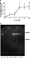
Induction of PLZF expression by doxycycline (dox) in cells stably expressing hPLZF. Expression of hPLZF in M1/TetON-hPLZF cells was induced by 500 ng/ml dox treatment for different periods, and the levels of hPLZF transcript (A) or protein (B) were determined as described in the Experimental Procedures. A: Values shown are RE, and are expressed as fold increase in PLZF expression vs. time 0. *, P < 0.01; **, P < 0.005. B: Western blot analysis of expression of hPLZF. Cells were induced by dox at different concentrations (0–1,000 ng/ml) for 24 h, lysed and PLZF protein expression determined by immunoblotting using 1:1,000 dilution of a PLZF-specific mouse monoclonal antibody from Calbiochem. Forty micrograms of total protein was loaded in each lane; Lane 1: + control (HL-60 cell extract); Lanes 2–4, no dox treated cell lysate; lane 5–8: cells were treated with 1,000, 500, 250, 125 ng/ml dox, respectively.
Fig. 4.
Fig. 4.
Stable expression of hPLZF decreases transepithelial Na+ current in M1 cells. A: Time course of PLZF effect on transepithelial Na current (Isc). M1/TETOn-hPLZF cells were grown in complete growth medium (PC1) to confluence on Millicell membranes. Cells were then maintained in steroid-free medium for 2 days, then 500 ng/ml dox (dashed line) or vehicle (solid line) was added to the cells, and transepithelial voltage (Vte) and resistance (Rte) determined at different times and Isc calculated as described in materials and methods. Values are expressed as % of time 0 value of the same culture. Note spontaneous decrease in Isc values of control cells, which is due to maintenance on steroid-free medium. Isc in cells in which PLZF expression was induced by dox is significantly reduced at 24 and 48. **, P < 0.01; ***, P < 0.001, vs. time 0 using 2-tail unpaired t-test. n = 16 for times 0, 24 and 48 h, n = 8 for times 4 and 8 h. B: Effects of PLZF on Vte, Rte and Isc. PLZF decreases Vte (left) and Isc (right) but has no significant effect on Rte (middle). PLZF expression was induced for 24 h with 500 ng/ml dox. ***, P < 0.001, n = 16.
Fig. 5.
Fig. 5.
A: Expression of the full-length (FL) and C-terminally truncated (mPLZF2) PLZF isoforms in mouse cortical collecting duct cells. RNA was prepared from parent M1 cells, reverse transcribed, and splice variant primers pairs were used in RT-PCR as described in the materials and methods. The expected size of the FL-PLZF-specific amplicon is 315 bp, while that of the mPLZF2-specific amplicon is 399 bp. B: Effect of corticosteroids on the expression of PLZF isoforms in M1 cells. M1/TetON-MR cells were treated or not (filled bars) with 1 nM aldosterone (aldo, hatched bars) or 10 nM dexamethasone (grey bars) for 24 h and expression of PLZF isoforms determined with RT-PCR as above. n = 3 in each group. *P < 0.05 vs. no steroid group.
Fig. 6.
Fig. 6.
Stable expression of mouse PLZF2 decreases transepithelial Na+ current in M1. Individual clonal lines (clones 1 -3 and 5) of M1/TetOn-mPLZF2 cells were grown in complete growth medium (PC1) to confluence on Millicell membranes, then maintained in steroid-free medium for 2 days, and then mPLZF2 expression was induced by 500 ng/ml dox (striped bars) or not (vehicle; filled bars) added to the cells. In clones 2 and 3 separate cultures received 100 nM dexamethasone (dex, open bars) or dox plus dex (hatched bars). Isc values shown are derived from Vte and Rte determined 48 h after treatment. For each treatment group n = 2 for clones 1 and 5, n = 3 for clones 2 and 3. *, P < 0.05 vs. no steroid group; #, P < 0.05 vs. dox-treated cells.
Fig. 7.
Fig. 7.
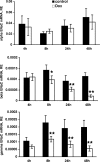
Effect of PLZF on the expression of epithelial Na channel (ENaC) subunit mRNAs. M1/TETOn-hPLZF cells were grown as above, and hPLZF expression induced with 500 ng/ml dox for 4–48 hrs. Cells were then lysed and the levels of alpha (upper panel), beta (middle panel) and gamma (lower panel). ENaC mRNA determined as described in the materials and methods. *, P < 0.05; **, P < 0.01; n = 5 for each time point.
Fig. 8.
Fig. 8.
Inhibition of histone deacetylase and p300 does not prevent repression of β- and γ-ENaC by PLZF. M1-PLZF cells were grown in complete medium until reaching confluence. Cells were then steroid-starved for 48 h prior to an 24-h treatment with 500 ng/ml dox (open bars or vehicle (filled bars). Where indicated, 100 ng/ml trichostatin A (TSA) or 100 μM curcumin was added and cells incubated for 24 h. ENaC and β-actin mRNA levels were determined as above. Values are RE of ENaC/β-actin mRNA, normalized for control (no dox) in each group. *, P < 0.05 control vs. dox treated cells; n = 3 in each group.

Similar articles

Cited by

References

    1. Alvarez de la Rosa D, Canessa CM. Role of SGK in hormonal regulation of epithelial sodium channel in A6 cells. Am J Physiol Cell Physiol 284: C404–C414, 2003. - PubMed
    1. Barna M, Hawe N, Niswander L, Pandolfi PP. Plzf regulates limb and axial skeletal patterning. Nat Genet 25: 166–172, 2000. - PubMed
    1. Berger S, Bleich M, Schmid W, Cole TJ, Peters J, Watanabe H, Kriz W, Warth R, Greger R, Schutz G. Mineralocorticoid receptor knockout mice: pathophysiology of Na+ metabolism. Proc Natl Acad Sci USA 95: 9424–9429, 1998. - PMC - PubMed
    1. Boyd C, Naray-Fejes-Tóth A. Gene regulation of ENaC subunits by serum- and glucocorticoid-inducible kinase-1. Am J Physiol Renal Physiol 288: F505–F512, 2005. - PubMed
    1. Chen SY, Bhargava A, Mastroberardino L, Meijer OC, Wang J, Buse P, Firestone GL, Verrey F, Pearce D. Epithelial sodium channel regulated by aldosterone-induced protein sgk. Proc Natl Acad Sci USA 96: 2514–2519, 1999. - PMC - PubMed

Publication types

MeSH terms

LinkOut - more resources