. 2008 Apr 30;28(18):4604-12.
doi: 10.1523/JNEUROSCI.5074-07.2008.

The neurogenesis-controlling factor, Pax6, inhibits proliferation and promotes maturation in murine astrocytes

Affiliations

The neurogenesis-controlling factor, Pax6, inhibits proliferation and promotes maturation in murine astrocytes

Katsuyasu Sakurai et al. J Neurosci. .

Abstract

Astrocytes serve various important functions in the CNS, but the molecular mechanisms of their generation and maturation are still enigmatic. Here, we show that Pax6, a key transcription factor that controls neurogenesis, also regulates proliferation, differentiation, and migration of astrocytes in the CNS. We first reveal that Pax6 is expressed in astrocytes during development as well as postnatally in the wild-type mouse. Astrocytes derived from Pax6 homozygous mutants (Sey/Sey) mice exhibited aberrant proliferation together with immature differentiation, both in vivo and in vitro, with higher migration potential in scratch-wound assays in vitro. Furthermore, a larger population of Sey/Sey astrocytes expresses neural stem cell markers such as nestin, Sox2, and prominin-1. These phenotypes of Pax6-deficient astrocytes putatively occur via higher Akt activity. Thus, the breakdown of Pax6 function induces the retention of neural stem-like characteristics and inhibits astrocyte maturation.


Figures

Figure 1.
Figure 1.
Pax6 is expressed in mature astrocytes in vivo. Double staining of Pax6 (green) together with cell-type-specific markers (GFAP, S100β, and NeuN; magenta) in the wild-type (WT) mouse at 8 weeks. A, A′, In the hippocampus, 98% GFAP+ astrocytes express with Pax6 (911 of 925 cells; n = 3). B, B′, In the cerebral cortex, 85% S100β+ astrocytes express Pax6 (980 of 1150 cells; n = 3). C, C′, Pax6+ cells are totally negative for NeuN in the cerebral cortex. Scale bars, 20 μm.
Figure 2.
Figure 2.
Pax6-deficient astrocytes exhibit aberrant proliferation in vitro. A, Experimental design of cultured astrocytes for different assays shown in Figures 2–4. Primary cultures of astrocytes were prepared from WT, Sey/+, and Sey/Sey mice cerebral cortex at E18.5. After cells became confluent, cells were replated on proper dishes or plates and used for various experiments. B, Immunostaining of Pax6 (magenta) and S100β (green) in astrocytes derived from the WT, Sey/+, and Sey/Sey mice at 4 d after replating. In astrocytes derived from WT mice, 99% of S100β+ astrocytes are colabeled with Pax6 (1639 of 1643 cells; n = 3). Astrocytes are generated in a Pax6-deficient condition (Sey/Sey). C, D, Quantification of BrdU+ cells in cultured astrocytes. Astrocytes were cultured for 4 d with the treatment of BrdU for the final 24 h. The number of BrdU+ cells is expressed as a percentage of the total number of DAPI-stained cells. The ratio of BrdU+ (magenta) cells is almost similar in astrocytes taken from both WT (24%) and Sey/+ (27%) mice. The number of BrdU+ cells in Sey/Sey astrocytes (37%) is quite larger than that in WT (p = 0.000006) and Sey/+ (p = 0.0006) astrocytes. The results are presented as the mean ± SD of the samples (n = 4; **p < 0.001, ***p < 0.00001). E, To evaluate growth rates of WT, Sey/+, and Sey/Sey astrocytes, the number of DAPI-stained cells is calculated in five fields per well. In WT and Sey/+ astrocytes, the cell number reaches a plateau at 6 d after plating, and there is no significant difference in the cell numbers between WT and Sey/+ cultures except for those at 9 d (p = 0.001). The cell number of Sey/Sey astrocytes is significantly higher than that of WT and Sey/+ at 3 and 6 d, without reaching a plateau even at 9 d (p = 0.0009, 0.001, and 0.0003 for 3, 6, and 9 d, respectively). The results are presented as the mean ± SD of the samples (n = 5; *p < 0.01, **p < 0.001). F, To block the function of Pax6 in WT astrocytes, we introduced pCAX-Pax6-EnR plus pCAX-GFP or pCAX plus pCAX-GFP and performed BrdU incorporation assay. Four days after transfection by electroporation, the percentage of BrdU+ cells/GFP+ in pCAX-Pax6-EnR-expressing astrocytes (33.7%) is larger than that in control astrocytes (13.7%) (p = 0.0002) (n = 6). Scale bars, 100 μm.
Figure 3.
Figure 3.
Cultured, Pax6-deficient astrocytes are immature and express neural stem cell markers. A, Immunostaining of GAFP, nestin, and prominin-1 (CD133) in WT and Sey/Sey astrocytes. Most WT astrocytes strongly express GFAP, whereas only a small number express nestin or prominin-1. In contrast, a smaller number of Sey/Sey astrocytes express GFAP, and a larger number strongly express nestin and prominin-1 compared with the WT astrocytes. Scale bar, 100 μm. The bottom two panels show the high-power images of prominin-1 expression in WT and Sey/Sey astrocytes. Scale bar, 25 μm. B, Cell lysates of cultured WT and Sey/Sey astrocytes were subjected to Western blot analysis with antibodies against GFAP, nestin, prominin-1, and Hsp90. The expression level of GFAP protein is slightly lower in Sey/Sey astrocytes than in WT astrocytes. In contrast, expression levels of nestin and prominin-1, markers for neural stem cells, are higher in Sey/Sey astrocytes than in WT astrocytes. C, Relative expression levels of GFAP, nestin, and prominin-1 protein normalized by Hsp90. The expression level of GFAP protein in Sey/Sey astrocytes is 0.73-fold lower than that in Sey/Sey astrocytes. The expression level of nestin and prominin-1 protein in Sey/Sey astrocytes is 1.61- and 6.2-fold higher than that in Sey/Sey astrocytes, respectively. The results are presented as the mean ± SD of samples (n = 3; p < 0.05).
Figure 4.
Figure 4.
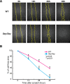
Cultured astrocytes exhibit higher migration potential in the Pax6-deficient condition. Scratch-wound assay was performed as shown in Figure 2A. A, B, At 14 (p = 0.03) and 28 (p = 0.04) hours after scratch, Sey/Sey astrocytes robustly migrate and cover larger cell-free regions compared with the WT astrocytes. At 49 h after scratch, almost all of the cell-free regions are covered with Sey/Sey astrocytes (p = 0.007), whereas astrocytes derived from WT cover ∼62% of the cell-free regions. The yellow dotted lines represent the edge of the cell-free regions. The results are presented as the mean values ± SD of the samples (n = 3; *p < 0.05, **p < 0.01). Scale bar, 500 μm.
Figure 5.
Figure 5.
Sey/Sey astrocytes exhibit transiently increased proliferation in the spinal cord. A, A′, Immunostaining of Pax6 and GLAST in coronal sections of E14.5 mouse spinal cord. Pax6+ (green) cells are located in the ventricular zone and gray matter. Almost all Pax6+ cells are colabeled with GLAST (magenta). Scale bars, 20 μm. B, Immunostaining of BrdU and FABP7 (BLBP) in coronal sections of E16.5 mouse spinal cord. BrdU+ cells (magenta) are observed all over the spinal cord, and almost all of these cells coexpress FABP7 (green), a marker for astrocytic progenitors and mature astrocytes, in both WT and Sey/Sey (seen as white cells). Scale bar, 40 μm. C, Quantification of BrdU+ cells in WT and Sey/Sey spinal cord at E14.5, E16.5, and E18.5. The number of BrdU+ cells in the Sey/Sey spinal cord is significantly higher (p = 0.006) than that in WT at E16.5, whereas there is no statistical difference in the number of BrdU+ cells between WT and Sey/Sey spinal cords at E14.5 and E18.5. The results are presented as the mean values ± SD of the samples (n = 3; **p < 0.01). D, Immunostaining of Sox2 and FABP7 in coronal sections of E18.5 WT and Sey/Sey spinal cord. The number of Sox2 (magenta) and FABP7 (green) double-positive cells (seen as white cells) are dramatically larger in Sey/Sey spinal cord (1374 cells/section) than that in WT (1149 cells/section) (p = 0.0003; n = 3). Scale bar, 20 μm. E, Immunostaining of GFAP in coronal sections of E16.5 WT and Sey/Sey spinal cord. Expression of GFAP in the spinal cord of Sey/Sey mouse is lower than that in WT mouse. Scale bar, 100 μm.
Figure 6.
Figure 6.
Akt activity is increased in cultured astrocytes in Pax6-deficient condition. A, The equal amount of total protein (10 μg) from cell lysates of cultured WT and Sey/Sey astrocytes (Fig. 2A) are subjected to Western blot analysis with antibodies against Akt, pAkt, or Hsp90. The level of expression of pAkt is higher in Sey/Sey astrocytes than in the WT astrocytes. B, Relative expression level of Akt and pAkt protein normalized by Hsp90. The expression level of Akt protein in WT astrocytes is similar that in Sey/Sey astrocytes. In contrast, the expression level of pAkt protein in Sey/Sey astrocytes is 1.8-fold higher than that in the WT astrocytes (p = 0.00003). The results are presented as the mean values ± SD of the samples (n = 5; ***p < 0.0001).
Figure 7.
Figure 7.
Pax6 expression persists from neuroepithelial/radial glial cells to astrocytes and adult neural stem cell. Schematic illustration of Pax6 expression in the course of cortical neurogenesis and gliogenesis. Neuroepithelial/radial glial cells (Pax6+++) initially undergo highly regulated self-renewing divisions and eventually generate lineage-committed progenitor cells, which subsequently differentiate into the three main neural lineages [i.e., neurons (Pax6−), astrocytes (Pax6++), and oligodendrocytes (data not shown)]. The expression of Pax6 (++) continues in postnatal adult neural stem/progenitor cells. Pax6 expression also persists in astrocytic progenitors (Pax6++) and in mature astrocytes (Pax6++). The same expression pattern of Pax6 is also observed in the hippocampus.

Similar articles

Cited by

References

    1. Abe K, Saito H. Developmental changes in cyclic AMP-stimulated stellation of cultured rat cortical astrocytes. Jpn J Pharmacol. 1997;75:433–438. - PubMed
    1. Allen NJ, Barres BA. Signaling between glia and neurons: focus on synaptic plasticity. Curr Opin Neurobiol. 2005;15:542–548. - PubMed
    1. Alvarez-Buylla A, Seri B, Doetsch F. Identification of neural stem cells in the adult vertebrate brain. Brain Res Bull. 2002;57:751–758. - PubMed
    1. Arai Y, Funatsu N, Numayama-Tsuruta K, Nomura T, Nakamura S, Osumi N. Role of Fabp7, a downstream gene of Pax6, in the maintenance of neuroepithelial cells during early embryonic development of the rat cortex. J Neurosci. 2005;25:9752–9761. - PMC - PubMed
    1. Bader AG, Kang S, Zhao L, Vogt PK. Oncogenic PI3K deregulates transcription and translation. Nat Rev Cancer. 2005;5:921–929. - PubMed

Publication types

MeSH terms

LinkOut - more resources