. 2008 Apr 30;28(18):4712-25.
doi: 10.1523/JNEUROSCI.5735-07.2008.

Zic deficiency in the cortical marginal zone and meninges results in cortical lamination defects resembling those in type II lissencephaly

Affiliations

Zic deficiency in the cortical marginal zone and meninges results in cortical lamination defects resembling those in type II lissencephaly

Takashi Inoue et al. J Neurosci. .

Abstract

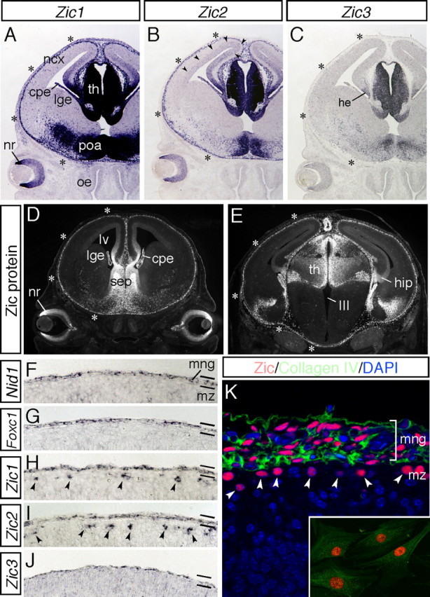
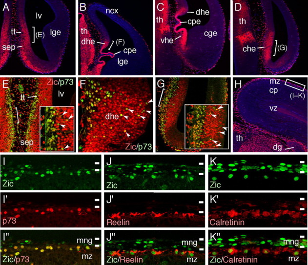
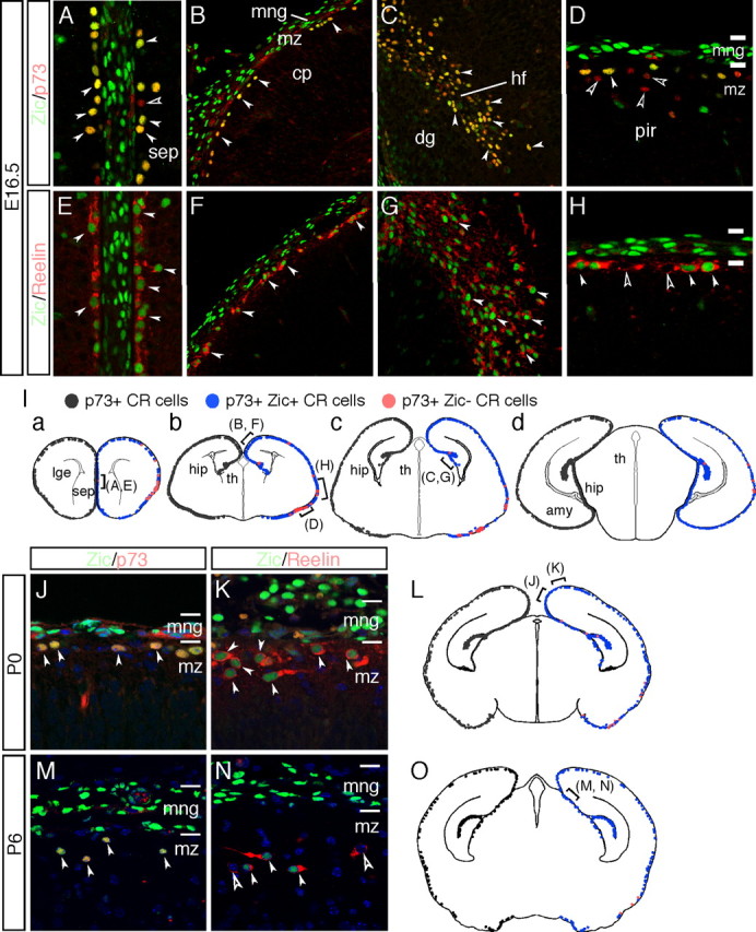
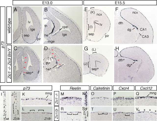
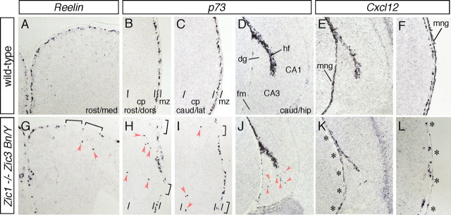
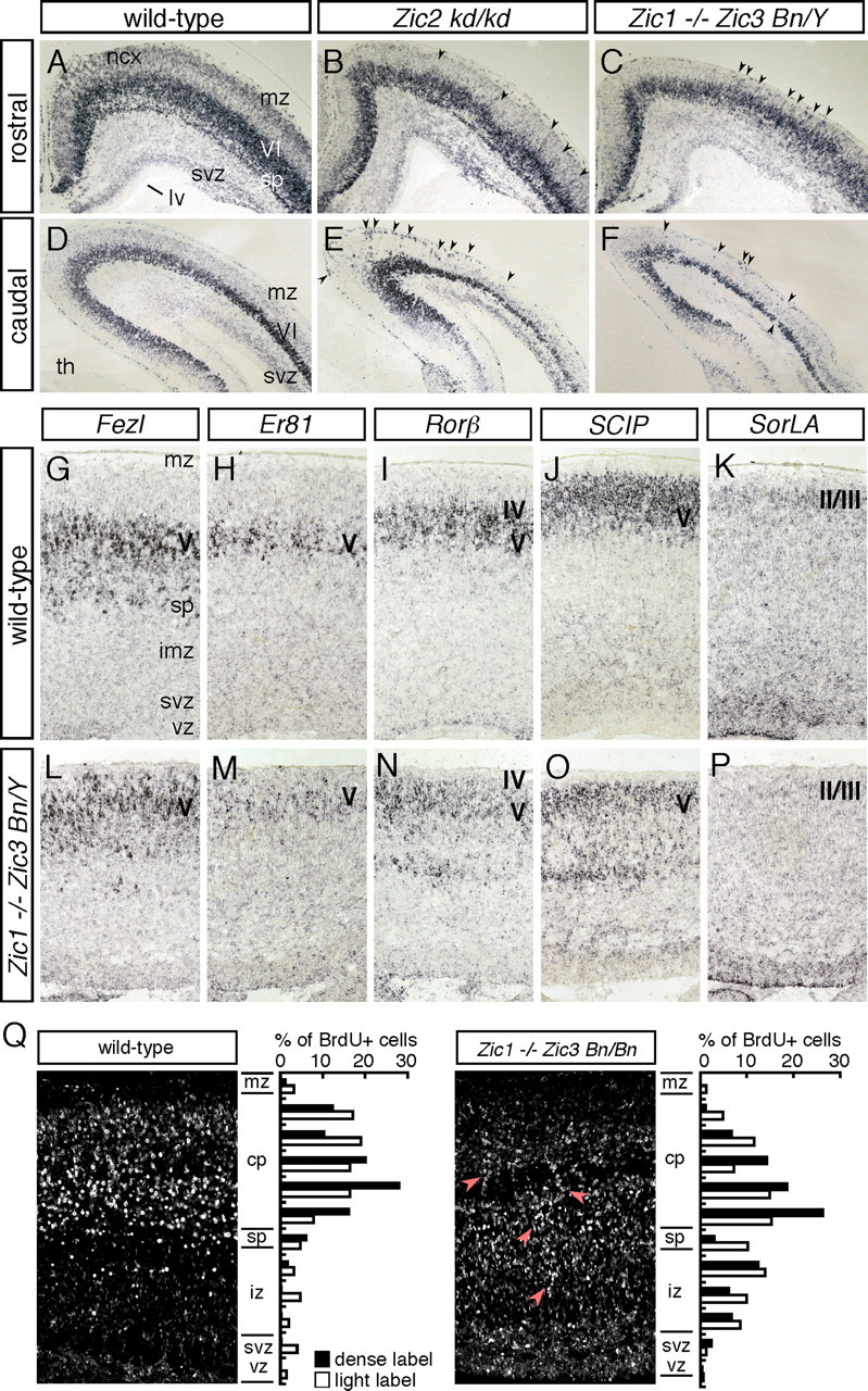
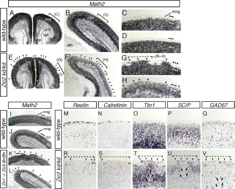
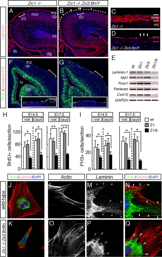
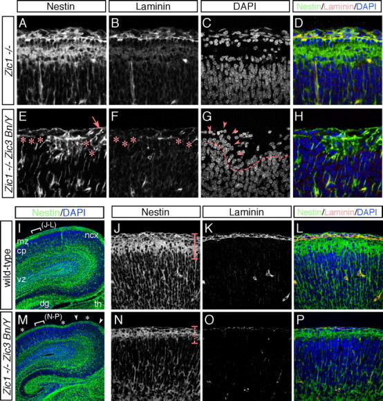
The formation of the highly organized cortical structure depends on the production and correct placement of the appropriate number and types of neurons. The Zic family of zinc-finger transcription factors plays essential roles in regulating the proliferation and differentiation of neuronal progenitors in the medial forebrain and the cerebellum. Examination of the expression of Zic genes demonstrated that Zic1, Zic2, and Zic3 were expressed by the progenitor cells in the septum and cortical hem, the sites of generation of the Cajal-Retzius (CR) cells. Immunohistochemical studies have revealed that Zic proteins were abundantly expressed in the meningeal cells and that the majority of the CR cells distributed in the medial and dorsal cortex also expressed Zic proteins in the mid-late embryonic and postnatal cortical marginal zones. During embryonic cortical development, Zic1/Zic3 double-mutant and hypomorphic Zic2 mutant mice showed a reduction in the number of CR cells in the rostral cortex, whereas the cell number remained unaffected in the caudal cortex. These mutants also showed mislocalization of the CR cells and cortical lamination defects, resembling the changes noted in type II (cobblestone) lissencephaly, throughout the brain. In the Zic1/3 mutant, reduced proliferation of the meningeal cells was observed before the thinner and disrupted organization of the pial basement membrane (BM) with reduced expression of the BM components and the meningeal cell-derived secretory factor. These defects correlated with the changes in the end feet morphology of the radial glial cells. These findings indicate that the Zic genes play critical roles in cortical development through regulating the proliferation of meningeal cells and the pial BM assembly.


Figures

Figure 1.
Figure 1.
During corticogenesis, Zic genes are expressed by the meningeal cells. A–C, Expression of Zic1 (A), Zic2 (B), and Zic3 (C) mRNAs in coronal sections of the head at E14.5. Zic genes are abundantly expressed in the medial neural tissues and marginal brain. Strong expression of Zic1 and Zic2 in the marginal brain is indicated by asterisks. Expression of Zic2 in the dorsal VZ/SVZ is indicated by arrowheads. cpe, Choroid plexus; he, cortical hem; ncx, neocortex; lge, lateral ganglionic eminence; nr, neural retina; poa, preoptic area; oe, olfactory epithelium; th, thalamus. D, E, Distribution of the Zic proteins was examined using a pan-Zic antibody in coronal sections of the head at E14.5. Rostral (D) and caudal (E) sections are shown. Zic protein expression in the marginal brain is indicated by asterisks. cpe, Choroid plexus; hip, hippocampus; lge, lateral ganglionic eminence; lv, lateral ventricle; nr, neural retina; sep, septum; th, thalamus; III, third ventricle. F–J, Expression of Nid-1 (F), Foxc1 (G), Zic1 (H), Zic2 (I), and Zic3 (J) mRNAs in the marginal brain at E16.5. These genes are commonly expressed by the meningeal cells. Zic1 and Zic2 are additionally found to be expressed by the CR cells in the cortical MZ (arrowheads). mng, Meninges; mz, marginal zone. K, Distribution of the Zic proteins (red) and collagen IV (green) in the marginal brain at E16.5. The nuclei of the cells are stained with 4′,6-diamidino-2-phenylindole (DAPI) (blue). Zic-positive CR cells in the cortical MZ are indicated by arrowheads. Inset shows cultured meningeal cells from E16.5 brain. The meningeal fibroblasts are intensely positive for Zic (red) and collagen IV (green).
Figure 2.
Figure 2.
A–D, Distribution of Zic proteins in coronal sections at E12.5. Different rostrocaudal levels of the same brain are indicated (A is the most rostral and D is the most caudal). Zic proteins are abundantly expressed in the septum, choroid plexus, thalamus, dorsal, ventral, and caudal cortical hem. Sections are counterstained with DAPI (blue). cge, Caudal ganglionic eminence; che, caudal cortical hem; cpe, choroid plexus; dhe, dorsal cortical hem; lge, lateral ganglionic eminence; lv, lateral ventricle; ncx, neocortex; sep, septum; tt, tenia tecta; th, thalamus; vhe, ventral cortical hem. E–G, Double-immunofluorescence staining for Zic (red) and p73 (green). High-magnification views of the areas indicated in A, B, and D, respectively. Insets in E and G are higher-magnification views of the areas indicated in E and G, respectively. Typical colabeled cells are indicated by arrowheads. H, Distribution of the Zic proteins in the coronal brain sections at E16.5. The area shown in I–K is indicated by white squares. cp, Cortical plate; dg, dentate gyrus; mz, marginal zone; th, thalamus; vz, ventricular zone. I–K, Distribution of Zic proteins in the dorsal cortical MZ at E16.5 (green). Sections were subjected to costaining for p73 (I′–I″), reelin (J′–J″), and calretinin (K′–K″) (red). Marked overlapping of the Zic-producing nuclei in the cortical MZ with p73-positive (I″), reelin-positive (J″), and calretinin-positive (K″) CR cells. Zic proteins are also produced by the meningeal cells. mng, Meninges; mz, marginal zone.
Figure 3.
Figure 3.
Zic proteins are produced by the majority of the CR cells in the embryonic and postnatal cortical MZs. A–H, Coronal sections of the septum (A, E), dorsal cortex (B, F), hippocampal fissure (C, G), and lateral cortex (D, H) at E16.5 are shown, with double-immunofluorescence staining for Zic (green), p73 (red in A–D), and reelin (red in E–H). dg, Dentate gyrus; hf, hippocampal fissure; ncx, neocortex; pir, piriform cortex; sep, septum. Marked overlapping of the distribution of the Zic+ cells with that of p73+ cells is observed (white arrowheads in A–D). Several p73+ZicCR cells are observed in the lateral/piriform cortex (open arrowheads in D); reelin+Zic+ cells are also observed (white arrowheads in E–H). Several reelin+ Zic cells are observed in the lateral/piriform cortex (open arrowheads in H). The locations of the images in the brain is indicated in the schema in I. I, Schematic representation of the distribution Zic-positive CR cells in coronal mouse brain sections from E16.5 mice at various rostral (a) to caudal (d) levels. p73+ CR cells are shown as black dots in the left half of each schema. On the right, p73+ Zic+ CR cells are represented as blue dots, and p73+ Zic CR cells are represented by red dots. The majority of the p73+ CR cells express Zic proteins, and partial dissociation of Zic and p73 expression is observed in the lateral/piriform cortex at this age. amy, Amygdala; cp, cortical plate; dg, dentate gyrus; hf, hippocampal fissure; hip, hippocampus; lge, lateral ganglionic eminence; mng, meninges; pir, piriform cortex; sep, septum; th, thalamus. J–O, Expression of Zic proteins by CR cells in the cortical MZ at P0 (J–L) and P6 (M–O). Immunofluorescence staining for Zic proteins (green) with p73 (red in J, M) and reelin (red in K, N). The location of the images in the brain is indicated in the schema in L and O. Many reelin-expressing cells expressed Zic, but several cells were also negative for Zic expression (open arrowheads in N); these cells are reelin-expressing interneurons. mng, Meninges; mz, marginal zone of the cerebral cortex. L, O, Schematic representation of the distribution Zic-positive CR cells in P0 (L) and P6 (O) mouse brains at mediocaudal levels. Marked overlapping of expression of the Zic proteins with that of p73 at P0 and P6 (filled arrowheads) is shown. The notations for each cell type are the same as those indicated in I.
Figure 4.
Figure 4.
Reduced number of CR cells in the rostral cortical MZ of Zic1/Zic3 double-mutant mice. A–L, Morphology and marker analysis of the rostral (A, C, E, G), intermediate (B, D, F, H), cortical MZs at E13.0 (A–D) and E15.5 (E–L). In situ hybridization for p73 in the wild-type (A, B, E, F, I, K) and Zic1/3 double-mutant (C, D, G, H, J, L). Coronal sections are shown. I, K and J, L are higher-magnification images of the indicated areas in E and G, respectively. Dashed lines in E and G indicate the lateral ventricles. Reduced numbers of p73+ CR cells contiguous to their sources at E13.0 are indicated by arrowheads in C and D. In the rostral cortex of the Zic1/3 double-mutant mice at E15.5, the MZ has multiple areas lacking in p73+ CR cells (brackets in J and L). CA1, Hippocampal CA1 subfield; hippocampal CA3 subfield; dg, dentate gyrus; dg*, hypoplastic dentate gyrus; dth, dorsal thalamus; dth*, hypoplastic dorsal thalamus; lge, lateral ganglionic eminence; lv, lateral ventricle; mng, meninges; ncx, neocortex; pir, piriform cortex; sep, septum. M–V, In situ hybridization for Reelin (M, N, R, S), Calretinin (O, T), Cxcr4 (P, U), and Cxcl12 (Q, V) in the dorsal and medial regions of the rostral cortex. The regions in M and O–Q, R and T–V, N, and S correspond to the same regions indicated in K, L, I, and J, respectively. Cell-free areas without Reelin+, Calretinin+, or Cxcr4+ CR cells in the cortical MZ of the Zic1/3 mutant mice are indicated by brackets. The reduced expression of Cxcl12 in the meninges is indicated by asterisks in V.
Figure 5.
Figure 5.
A–D, G–J, Abnormal distribution of CR cells in the cortical MZ of the Zic1/Zic3 double-mutant mice. Distribution of Reelin+ (A, G) and p73+ (B–D, H–J) CR cells in the rostromedial (A, G), rostrodorsal (B, H), and caudolateral (C, I) cortex and the caudal hippocampus (D, J) at E15.5. Distribution of CR cells in the coronal brain sections of the wild-type (A–D) and Zic1/3 mutant (G–J) embryos are shown. In the wild-type embryos, Reelin+ and p73+ CR cells are confined to the neocortical MZ (A–C) and hippocampal fissure (D). In the mutants, the CR cells are widely scattered and found in the cortical plate of the neocortex (G–I) and deeper positions in the hippocampal CA3 region (J) (arrowheads). CA1, Hippocampal field CA1; CA3, hippocampal field CA3; cp, cortical plate; dg, dentate gyrus; hf, hippocampal fissure; fm, fimbria; mng, meninges; mz, marginal zone. E, F, K, L, Expression of Cxcl12 in the wild-type mice (E, F) and Zic1/3 mutants (K, L) in the caudal hippocampus (E, K) and caudolateral cortex (F, L) at E15.5. E, F, K, and L are neighboring sections of D, C, J, and I, respectively. Reduced expression of Cxcl12 in the meningeal layer of the Zic1/3 mutants is indicated by asterisks in K and L.
Figure 6.
Figure 6.
Loss and reduced expression of the Zic genes results in cortical lamination defects. A–F, In situ hybridization for Tbr1 in wild-type (A, D), Zic2 kd/kd (B, E), and Zic1/3 double-mutant (Zic1−/− Zic3 Bn/Y) (C, F) mice at E18.5. Coronal sections of the rostral (A–C) and caudal (D–F) brain are shown. In the wild type, Tbr1 is expressed in layer VI, the subplate, and the SVZ at this stage. In the Zic2 kd/kd and Zic1/3 mutants, the Tbr1 expression is disorganized and diffuse (arrowheads), suggesting the existence of cortical lamination defects in these mutants. lge, Lateral ganglionic eminence; lv, lateral ventricle; mz, marginal zones; ncx, neocortex; sp, subplate; th, thalamus; VI, layer VI. G–P, Expression of cortical-layer specific genes in the Zic1/3 mutant neocortex. In situ hybridization for Fezl (G, L), Er81 (H, M), Rorβ (I, N), SCIP (J, O), and SorLA (K, P) in coronal brain sections of E18.5 wild-type (G–K) and Zic1/3 mutant (L–P) mice. Q, Abnormal migration of Zic1/3 (Zic1−/− Zic3 Bn/Bn) mutant neurons. BrdU birthdating analysis reveals perturbed neuronal migration in the Zic1/3 mice. The final destination of the early-born neurons labeled by BrdU at E13.5 is shown in the sagittal sections of the wild-type (left) and Zic1/3 mutant (right) mice at E18.5. BrdU-positive nuclei were detected by immunohistochemistry. The arrows indicate neurons exhibiting aberrant migration. Bar graphs showing the radial distribution of heavily labeled cells (first generation at the time of the BrdU injection) and lightly labeled cells (the majority are second- and perhaps third-generation cells from subsequent progenitor cell divisions) in the neocortex of E18.5 wild-type (left) and Zic1/3 mutant (right) mice. cp, Cortical plate; iz, intermediate zone; mz, marginal zone; sp, subplate; svz, subventricular zone; vz, ventricular zone.
Figure 7.
Figure 7.
Reduced expression of Zic2 and loss of both Zic1 and Zic3 result in cortical lamination defects resembling those in cobblestone (type II) lissencephaly. A–H, In situ hybridization for Math2 in wild-type (A–D) and Zic2 mutant (E–H) brains at E17.5. Coronal sections of the rostral (A, C, E, G) and caudal (B, D, F, H) regions are shown. C, D, G, and H represent higher-magnification views of the indicated areas in A, B, E, and F, respectively. Wild-type brains showed a highly organized multilayered cortex with a hypocellular marginal zone at the surface of the brain (A–D). In contrast, Zic2 mutant brains showed severe disorganization of cortical lamination and neurons invading and obliterating the marginal zone (arrowheads in E, F; asterisk in G, H). cp, Cortical plate; dth, dorsal thalamus; lv, lateral ventricle; mng, meninges; mz, marginal zone of the cerebral cortex; ncx, neocortex; sep, septum. I–L, In situ hybridization for Math2 in wild-type (I, J) and Zic1/3 double-mutant (Zic1−/− Zic3 Bn/Bn) (K, L) mice at E17.5. Dorsal cortex in the rostral (I, K) and caudal (J, L) brain. M–V, In situ hybridization for Reelin (M, R), Calretinin (N, S), Tbr1 (O, T), SCIP (P, U), and GAD67 (Q, V) in wild-type (M–Q) and Zic2 mutant (R–V) mice at E17.5. Images in M–Q and R–V were taken from sequential sections in C and G, respectively. The area indicated by the dashed line in G corresponds to the indicated areas shown in R–V. Altered marginal expressions of the markers are indicated by arrowheads. Disorganized cortical layer structures beneath the marginal defects are indicated by arrows in T–V.
Figure 8.
Figure 8.
The pial BM is defective in the Zic1/3 double-mutant neocortex. A–D, Immunofluorescence detection of laminin (red) in coronal sections of the brain from the wild-type (A, C) and Zic1/3 mutant (B, D) mice at E16.5. The sections are counterstained with DAPI (blue). Control Zic1−/− show a smooth layer of basal lamina-associated laminin at the surface of the brain, as well as a basal lamina surrounding the blood vessels. In contrast, in the Zic1/3 mutant, the laminin layer at the surface of the brain is thinner compared with that in the Zic1−/− sections. C and D are high-magnification views of the indicated areas in A and B, respectively. E, Analysis for transcripts of the meningeal BM components and secreted factor. RT-PCR for Laminin γ1, Nid-1, Foxc1, Perlecan, and Cxcl12; GAPDH served as the internal control. Analysis was performed on cDNAs prepared from the meninges of wild-type (n = 6), Zic1−/− (n = 3), Zic3 Bn/Y (n = 4), and Zic1/3 (Zic1−/− Zic3 Bn/Y) (n = 3) mutants at E15.5. F, G, Decreased numbers of BrdU- and PH3-labeled proliferating meningeal cells in the Zic1/3 double-mutant mice. Proliferating meningeal cells in the S-phase were pulse labeled with BrdU for 1 h at E15.5. Immunohistochemical analysis showed decreased numbers of BrdU-labeled (green) and PH3-labeled (red) cells in the meningeal layer in the Zic1/3 mutants compared with those in the meningeal layer of the wild-type mice. The sections were counterstained with DAPI (blue). Comparable rostral areas in the Zic1/3 mutant and wild-type brains are shown. Insets are high-magnification views of the indicated areas. H, I, Quantification of the number of BrdU-labeled (H) and PH3-labeled (I) cells in the meningeal layers. BrdU- and PH3-labeled cells in the rostral and caudal regions were counted in the wild-type, Zic1−/−, and Zic1/3 mutant mice at E14.5 and E17.5. BrdU- and PH3-labeled meningeal cells were significantly decreased in the Zic1/3 mutants compared with those in the wild-type and Zic1−/− mice. The results are shown as the mean number of multiple comparable sections with SDs. *p < 0.05; **p < 0.01 by t test. J–Q, Primary meningeal fibroblasts from E14.5 wild-type (J, L–N) and Zic1/3 mutants (K, O–Q) were double stained with a laminin (J, K, M, N, P, Q) and F-actin (J, K, L, N, O, Q). N and Q are merged images of L, M and O, P, respectively. Meningeal fibroblasts were cultured for 8 h (J, K) and 3 d (L–Q). Fibroblasts from the Zic1/3 mutants spread less well and showed less proliferative activity (K). Fibrillar and punctate laminin is observed at the periphery in the wild-type meningeal fibroblasts (arrowheads in M, N), whereas it is absent from the meningeal fibroblasts of the Zic1/3 mutants (open arrowheads in P, Q). cp, Cortical plate; dg, dentate gyrus; lge, lateral ganglionic eminence; lv, lateral ventricle; mng, meninges; mz, marginal zone of the cerebral cortex; ncx, neocortex; th, thalamus; vz, ventricular zone; wt, wild type.
Figure 9.
Figure 9.
BM disruption and altered radial glial end feet in the Zic1/3 double-mutant neocortex. A–H, Immunofluorescence staining for nestin (A, E) and laminin (B, F) in the dorsal cortical MZ of the Zic1−/− (A–D) and Zic1/3 mutant (E–H) mice at E17.5. Sections were costained with DAPI (C, G). D and H are merged images of A–C and E–G, respectively. In the control Zic1−/− brain, radial glia showed a parallel arrangement of fibers that terminated in well defined end feet at the pial surface (A). In the Zic1/3 mutants, absence of the nestin-immunoreactive radial glial end feet (asterisks in E) was associated with breakdown of the surface laminin (asterisks in F) and neuronal ectopias (arrowheads in G). In the mutant brains, the radial glial end feet were occasionally found to be detached from the pial surface in the regions of the ectopias. These defects were seen scattered throughout the cortical MZ. I, M, Immunofluorescence staining for nestin in the dorsal cortex of the wild-type (I) and Zic1/3 (M) mice at E17.5. The sections were counterstained with DAPI (blue). The locations of the neuronal ectopias are shown in white arrowheads, and the thinner continuous band of nestin-positive radial glial end feet is indicated by asterisks in M. The locations indicated in J–L and N–P are indicated by white brackets in I and M, respectively. J–L, N–P, Higher magnification of the indicated areas in I and M. Immunofluorescence staining for nestin (J, N) and laminin (K, O) in the wild-type (J–L) and Zic1/3 mutant (N–P) mice. L and P are merged images of J,K and N,O, respectively. The sections were counterstained with DAPI (blue). The thinner continuous band of nestin-positive radial glial end feet in the Zic1/3 mutants is indicated by a dashed line in N.

References

    1. Alcántara S, Ruiz M, D'Arcangelo G, Ezan F, de Lecea L, Curran T, Sotelo C, Soriano E. Regional and cellular patterns of reelin mRNA expression in the forebrain of the developing and adult mouse. J Neurosci. 1998;18:7779–7799. - PMC - PubMed
    1. Alcántara S, Pozas E, Ibañez CF, Soriano E. BDNF-modulated spatial organization of Cajal-Retzius and GABAergic neurons in the marginal zone plays a role in the development of cortical organization. Cereb Cortex. 2005;16:487–499. - PubMed
    1. Aruga J. The role of Zic genes in neural development. Mol Cell Neurosci. 2004;26:205–221. - PubMed
    1. Aruga J, Minowa O, Yaginuma H, Kuno J, Nagai T, Noda T, Mikoshiba K. Mouse Zic1 is involved in cerebellar development. J Neurosci. 1998;18:284–293. - PMC - PubMed
    1. Aruga J, Inoue T, Hoshino J, Mikoshiba K. Zic2 controls cerebellar development in cooperation with Zic1. J Neurosci. 2002a;22:218–225. - PMC - PubMed

Publication types

MeSH terms

LinkOut - more resources