. 2008 May 6;105(18):6548-53.
doi: 10.1073/pnas.0706335105. Epub 2008 Apr 30.

Biogenesis of extracellular microfibrils: Multimerization of the fibrillin-1 C terminus into bead-like structures enables self-assembly

Affiliations

Biogenesis of extracellular microfibrils: Multimerization of the fibrillin-1 C terminus into bead-like structures enables self-assembly

Dirk Hubmacher et al. Proc Natl Acad Sci U S A. .

Abstract

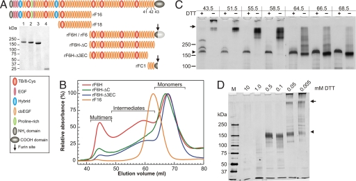
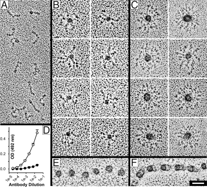
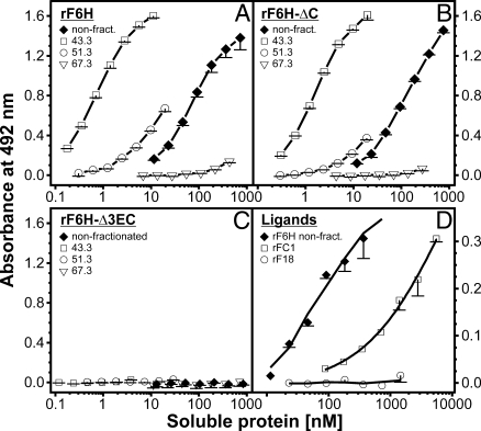
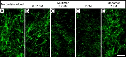
Microfibrils are essential elements in elastic and nonelastic tissues contributing to homeostasis and growth factor regulation. Fibrillins form the core of these multicomponent assemblies. Various human genetic disorders, the fibrillinopathies, arise from mutations in fibrillins and are frequently associated with aberrant microfibril assembly. These disorders include Marfan syndrome, Weill-Marchesani syndrome, Beals syndrome, and others. Although homotypic and heterotypic fibrillin self-interactions are considered to provide critical initial steps, the detailed mechanisms for microfibril assembly are unknown. We show here that the C-terminal recombinant half of fibrillin-1 assembles into disulfide-bonded multimeric globular structures with peripheral arms and a dense core. These globules are similar to the beaded structures observed in microfibrils isolated from tissues. Only these C-terminal fibrillin-1 multimers interacted strongly with the fibrillin-1 N terminus, whereas the monomers showed very little self-interaction activity. The multimers strongly inhibited microfibril formation in cell culture, providing evidence that these recombinant assemblies can also interact with endogenous fibrillin-1. The C-terminal self-interaction site was fine-mapped to the last three calcium-binding EGF domains in fibrillin-1. These results suggest a new mechanism for microfibril formation where fibrillin-1 first oligomerizes via its C terminus before the partially or fully assembled bead-like structures can further interact with other beads via the fibrillin-1 N termini.


Conflict of interest statement

The authors declare no conflict of interest.

Figures

Fig. 1.
Fig. 1.
Properties and multimerization of recombinant fibrillin-1 constructs. (A) Schematic representation of recombinant human fibrillin-1 constructs. The last three cbEGF domains are numbered. (Inset) Coomassie-stained purified and reduced recombinant fibrillin-1 fragments: lane 1, rF6H; lane 2, rF6H-ΔC; lane 3, rF6H-Δ3EC; lane 4, rFC1. Fragments rF6, rF16, and rF18 were described in detail previously (13, 31). rF6H is expressed without C-terminal propeptide (dark gray region of C-terminal domain), and rF6 is expressed with propeptide (additional light gray region of C-terminal domain) that becomes cleaved during or after secretion (arrow). (B) Gel filtration chromatography of recombinant fibrillin-1 fragments (as indicated) demonstrating various degrees of monomeric, intermediate, and multimeric fractions. The absorbance at 280 nm for each fragment was set to 100% for the monomeric peaks. Minor differences between monomeric rF6H and rF6H-ΔC elution volumes are statistically not significant. Note that rF6H-ΔC resulted in variable degrees of multimerization (see Table 1). (C) Individual fractions of the gel filtration chromatography of rF6H shown in B were subjected to SDS gel electrophoresis under reducing (+) or nonreducing (−) conditions and silver-stained. Elution volumes in milliliters on top correlate with elution volumes in B. The two additional bands above the rF6H monomer in the 43.5-ml fraction were determined to be rF6H by reaction with specific antibodies. (D) rF6H multimers were treated with various concentrations of DTT as indicated, alkylated, digested with trypsin, separated by SDS gel electrophoresis, and silver-stained. Note that reduction of the multimers to monomers requires ≈10- to 20-fold less DTT than reduction of intramolecular disulfide bonds as monitored by trypsin degradation of the monomers. In C and D, arrows denote the start of the separating gels and arrowheads indicate the position of monomeric and completely reduced rF6H.
Fig. 2.
Fig. 2.
Structural analysis of rF6H by electron microscopy after rotary shadowing (A–C, E, and F) and by ELISA (D). Fragment rF6H was subjected to gel filtration chromatography (Fig. 1B), and representative fractions of the monomers (A), intermediates (B), and multimers (C) were analyzed. (D) ELISA analysis with an antibody against the C-terminal hexahistidine tag showed significant differences (P < 0.001) in the reactivity of monomers (open symbols) and multimers (filled symbols). A beaded microfibril isolated from skin fibroblasts after collagenase digestion (32) is shown in the open (E) or closed (F) form. Note the similar appearance of the beaded structures in C, E, and F. (Scale bar: 50 nm.)
Fig. 3.
Fig. 3.
N-to-C interaction of fibrillin-1 constructs. In this previously established solid-phase interaction assay (22), the N-terminal half of fibrillin-1 (rF16) was immobilized. Construct rF6H (A) and the deletion constructs rF6H-ΔC (B) and rF6H-Δ3EC (C) were size-fractionated by gel filtration chromatography (Fig. 1B), and individual fractions as indicated in each graph by their respective elution volumes in milliliters were used as soluble ligands. Mixtures of monomers and multimers not subjected to gel filtration were included as controls (filled diamonds). (D) The biotinylated small C-terminal fragment rFC1 bound to rF16 albeit with a significantly lower affinity compared with the rF6H mixture. A biotinylated control fragment rF18 did not interact. Data represent means of triplicates (A–C) or duplicates (D) including standard deviations.
Fig. 4.
Fig. 4.
Inhibition of the fibrillin-1 network produced by fibroblasts. At the time of seeding of human skin fibroblasts into chamber slides, no protein (A), rF6H multimers (B–D), or rF6H monomers (E) were added at the concentrations indicated. The cells were grown for 3 days, and network formation was monitored by indirect immunofluorescence with antibodies against fibrillin-1. Fibronectin and fibulin-2 network formation are shown as controls in Fig. S5C. (Scale bar: 50 μm.)

References

    1. Hubmacher D, Tiedemann K, Reinhardt DP. Fibrillins: From biogenesis of microfibrils to signaling functions. Curr Top Dev Biol. 2006;75:93–123. - PubMed
    1. Robinson P, et al. The molecular genetics of Marfan syndrome and related disorders. J Med Genet. 2006;43:769–787. - PMC - PubMed
    1. Sakai LY, Keene DR, Engvall E. Fibrillin, a new 350-kD glycoprotein, is a component of extracellular microfibrils. J Cell Biol. 1986;103:2499–2509. - PMC - PubMed
    1. Kielty CM. Elastic fibres in health and disease. Expert Rev Mol Med. 2006;8:1–23. - PubMed
    1. Henderson M, Polewski R, Fanning JC, Gibson MA. Microfibril-associated glycoprotein-1 (MAGP-1) is specifically located on the beads of the beaded-filament structure for fibrillin-containing microfibrils as visualized by the rotary shadowing technique. J Histochem Cytochem. 1996;44:1389–1397. - PubMed

Publication types

LinkOut - more resources