

Gastrin-releasing peptide (GRP) in the ovine uterus: regulation by interferon tau and progesterone
- PMID: 18448839
- PMCID: PMC2714990
- DOI: 10.1095/biolreprod.108.068403
Gastrin-releasing peptide (GRP) in the ovine uterus: regulation by interferon tau and progesterone
Abstract
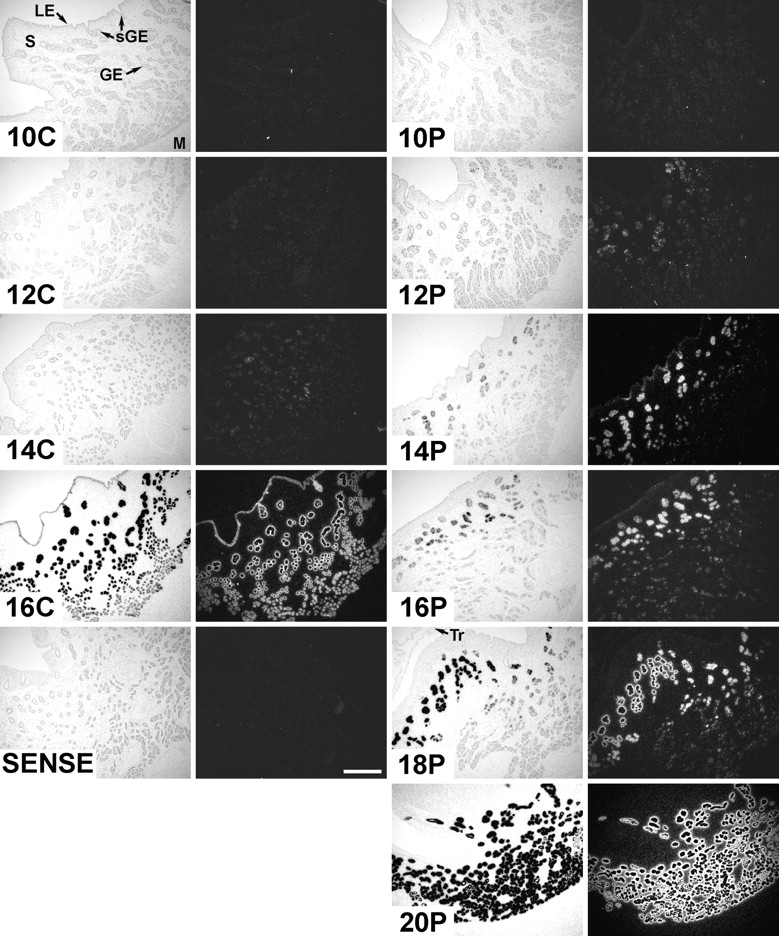
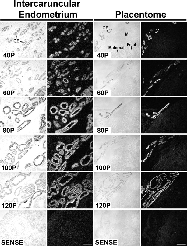
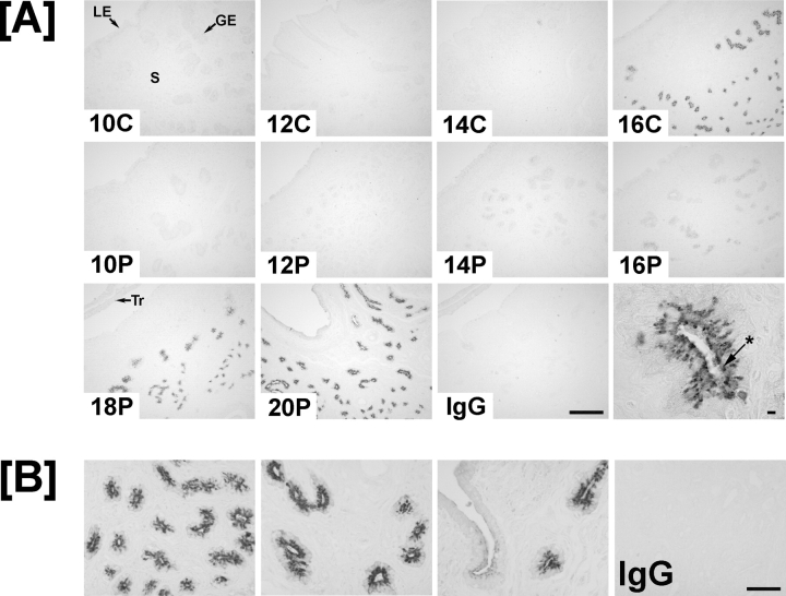
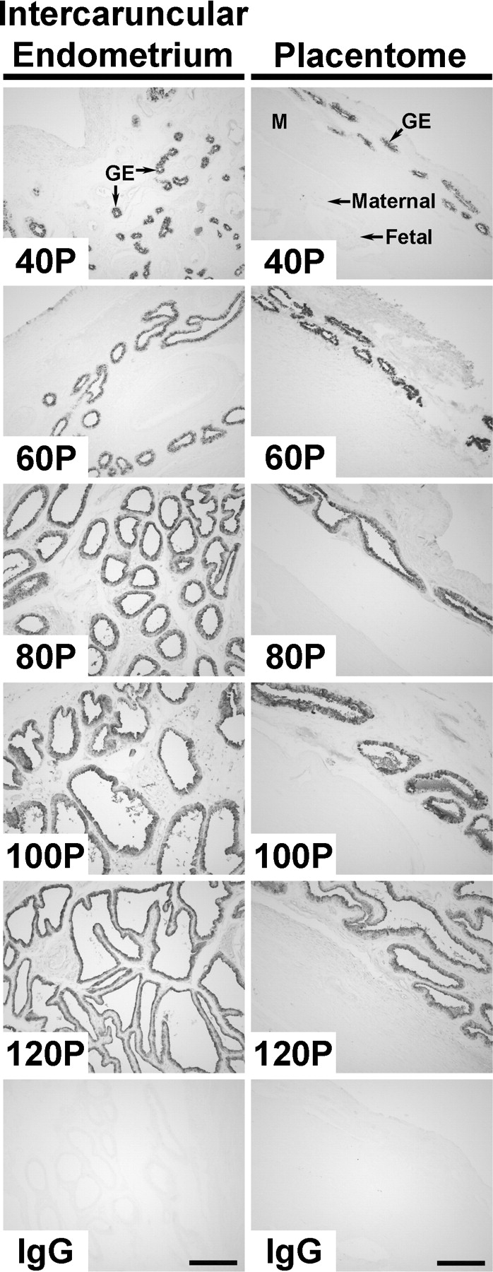
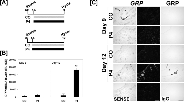
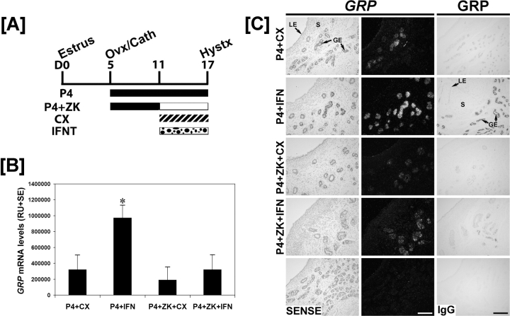
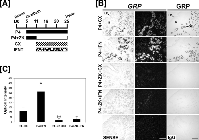
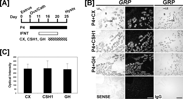
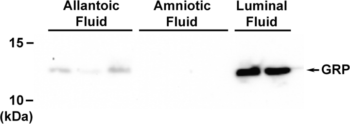
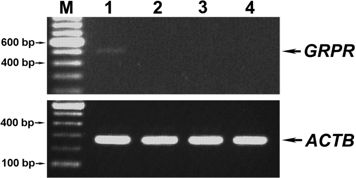
Gastrin-releasing peptide (GRP) is abundantly expressed by endometrial glands of the ovine uterus and processed into different bioactive peptides, including GRP1-27, GRP18-27, and a C-terminus, that affect cell proliferation and migration. However, little information is available concerning the hormonal regulation of endometrial GRP and expression of GRP receptors in the ovine endometrium and conceptus. These studies determined the effects of pregnancy, progesterone (P4), interferon tau (IFNT), placental lactogen (CSH1), and growth hormone (GH) on expression of GRP in the endometrium and GRP receptors (GRPR, NMBR, BRS3) in the endometrium, conceptus, and placenta. In pregnant ewes, GRP mRNA and protein were first detected predominantly in endometrial glands after Day 10 and were abundant from Days 18 through 120 of gestation. Treatment with IFNT and progesterone but not CSH1 or GH stimulated GRP expression in the endometrial glands. Western blot analyses identified proGRP in uterine luminal fluid and allantoic fluid from Day 80 unilateral pregnant ewes but not in uterine luminal fluid of either cyclic or early pregnant ewes. GRPR mRNA was very low in the Day 18 conceptus and undetectable in the endometrium and placenta; NMBR and BRS3 mRNAs were undetectable in ovine uteroplacental tissues. Collectively, the present studies validate GRP as a novel IFNT-stimulated gene in the glands of the ovine uterus, revealed that IFNT induction of GRP is dependent on P4, and found that exposure of the ovine uterus to P4 for 20 days induces GRP expression in endometrial glands.
Figures
Similar articles
-
Progesterone and interferon tau-regulated genes in the ovine uterine endometrium: identification of periostin as a potential mediator of conceptus elongation.Reproduction. 2009 Nov;138(5):813-25. doi: 10.1530/REP-09-0208. Epub 2009 Jul 27. Reproduction. 2009. PMID: 19635739
-
Pregnancy and interferon tau regulate RSAD2 and IFIH1 expression in the ovine uterus.Reproduction. 2007 Jan;133(1):285-95. doi: 10.1530/REP-06-0092. Reproduction. 2007. PMID: 17244754
-
Progesterone and interferon tau regulate hypoxia-inducible factors in the endometrium of the ovine uterus.Endocrinology. 2008 Apr;149(4):1926-34. doi: 10.1210/en.2007-1530. Epub 2008 Jan 3. Endocrinology. 2008. PMID: 18174278 Free PMC article.
-
Biology of progesterone action during pregnancy recognition and maintenance of pregnancy.Front Biosci. 2002 Sep 1;7:d1879-98. doi: 10.2741/spencer. Front Biosci. 2002. PMID: 12161340 Review.
-
Chronicling the discovery of interferon tau.Reproduction. 2017 Nov;154(5):F11-F20. doi: 10.1530/REP-17-0257. Epub 2017 Jul 26. Reproduction. 2017. PMID: 28747540 Free PMC article. Review.
Cited by
-
The proteome of IVF-induced aberrant embryo-maternal crosstalk by implantation stage in ewes.J Anim Sci Biotechnol. 2020 Jan 14;11:7. doi: 10.1186/s40104-019-0405-y. eCollection 2020. J Anim Sci Biotechnol. 2020. PMID: 31956410 Free PMC article.
-
Comparative analysis between endometrial proteomes of pregnant and non-pregnant ewes during the peri-implantation period.J Anim Sci Biotechnol. 2015 Apr 25;6(1):18. doi: 10.1186/s40104-015-0017-0. eCollection 2015. J Anim Sci Biotechnol. 2015. PMID: 26023329 Free PMC article.
-
Alterations in expression of endometrial genes coding for proteins secreted into the uterine lumen during conceptus elongation in cattle.BMC Genomics. 2013 May 10;14:321. doi: 10.1186/1471-2164-14-321. BMC Genomics. 2013. PMID: 23663413 Free PMC article.
-
Altrenogest Supplementation during Early Pregnancy Improves Reproductive Outcome in Pigs.Animals (Basel). 2022 Jul 14;12(14):1801. doi: 10.3390/ani12141801. Animals (Basel). 2022. PMID: 35883348 Free PMC article.
-
Isolation, identification and biological activity of gastrin-releasing peptide 1-46 (oGRP 1-46), the primary GRP gene-derived peptide product of the pregnant ovine endometrium.Peptides. 2010 Feb;31(2):284-90. doi: 10.1016/j.peptides.2009.11.013. Epub 2009 Nov 26. Peptides. 2010. PMID: 19944725 Free PMC article.
References
-
- McDonald TJ, Jornvall H, Nilsson G, Vagne M, Ghatei M, Bloom SR, Mutt V.Characterization of a gastrin releasing peptide from porcine non-antral gastric tissue. Biochem Biophys Res Commun 1979; 90: 227–233. - PubMed
-
- Patel O, Shulkes A, Baldwin GS.Gastrin-releasing peptide and cancer. Biochim Biophys Acta 2006; 1766: 23–41. - PubMed
-
- Fraser M, Carter AM, Challis JR, McDonald TJ.Gastrin releasing peptide immunoreactivity is present in ovine amniotic fluid and fetal and maternal circulations. MRC Group in Fetal and Neonatal Health and Development. Endocrinology 1992; 131: 2033–2035. - PubMed
-
- Fraser M, McDonald TJ, Spindel ER, Fahy M, Hill D, Challis JR.Gastrin-releasing peptide is produced in the pregnant ovine uterus. Endocrinology 1994; 135: 2440–2445. - PubMed
-
- Giraud A, Parker L, Taupin D, Hardy K, Shulkes A.Mammalian bombesin as a hormone in ovine pregnancy: ontogeny, origin, and molecular forms. Am J Physiol 1993; 265: E866–E873. - PubMed
Publication types
MeSH terms
Substances
Grants and funding
LinkOut - more resources
Full Text Sources
