. 2008 Jul 1;112(1):150-8.
doi: 10.1182/blood-2007-12-129395. Epub 2008 Apr 30.

SDF-1/CXCR4 and VLA-4 interaction regulates homing in Waldenstrom macroglobulinemia

Affiliations

SDF-1/CXCR4 and VLA-4 interaction regulates homing in Waldenstrom macroglobulinemia

Hai T Ngo et al. Blood. .

Abstract

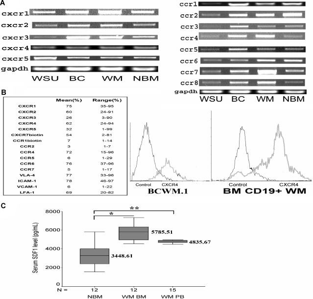
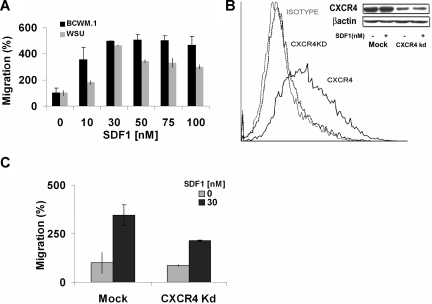
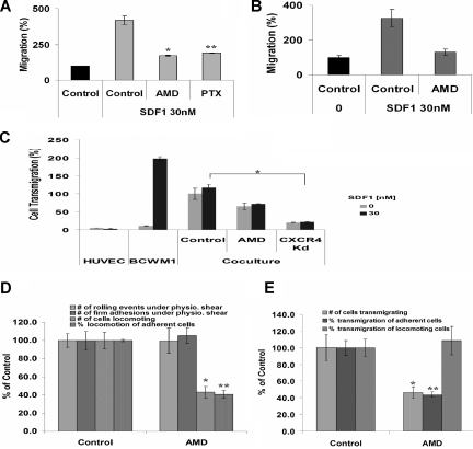
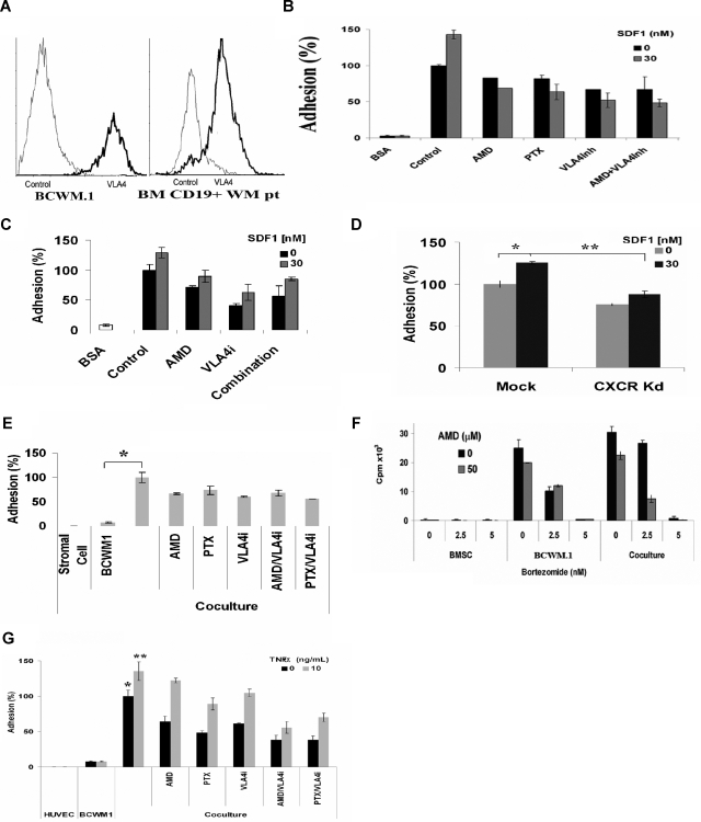
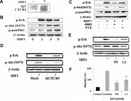
Waldenstrom macroglobulinemia (WM) is characterized by widespread involvement of the bone marrow at the time of diagnosis, implying continuous homing of WM cells into the marrow. The mechanisms by which trafficking of the malignant cells into the bone marrow has not been previously elucidated. In this study, we show that WM cells express high levels of chemokine and adhesion receptors, including CXCR4 and VLA-4. We showed that CXCR4 was essential for the migration and trans-endothelial migration of WM cells under static and dynamic shear flow conditions, with significant inhibition of migration using CXCR4 knockdown or the CXCR4 inhibitor AMD3100. Similarly, CXCR4 or VLA-4 inhibition led to significant inhibition of adhesion to fibronectin, stromal cells, and endothelial cells. Decreased adhesion of WM cells to stromal cells by AMD3100 led to increased sensitivity of these cells to cytotoxicity by bortezomib. To further investigate the mechanisms of CXCR4-dependent adhesion, we showed that CXCR4 and VLA-4 directly interact in response to SDF-1, we further investigated downstream signaling pathways regulating migration and adhesion in WM. Together, these studies demonstrate that the CXCR4/SDF-1 axis interacts with VLA-4 in regulating migration and adhesion of WM cells in the bone marrow microenvironment.


Figures

Figure 1
Figure 1
Chemokine receptors expression in WM. (A) CXCR1, CXCR2, CXCR3, CXCR4, CXCR5, CCR1, CCR2, CCR3, CCR4, CCR5, CCR6, CCR7, and CCR8 expression in WM cell lines, including WM-WSU (WSU), BCWM.1 (BC), a representative patient sample (WM) and a representative normal bone marrow CD19+ cells (NBM). A total of 3 patient samples and 3 normal marrow samples were tested. GAPDH was used as control. (B) Chemokine receptors and adhesion molecule expression and CXCR4 expression in 10 patient samples using flow cytometry. The table shows the mean and range of expression of all the CXCR, CCR receptors, and adhesion molecules. The right panel shows BCWM.1 cell line and CD19+ WM cells from a primary patient using flow cytometry and compared with IgG1 control. (C) Enzyme-linked immunosorbent assay for SDF-1 level (pg/mL) demonstrates that the level of SDF-1 is significantly higher in the bone marrow of WM patients (N = 12, *P = .001.) and peripheral blood of WM patients (N = 15, **P = .004) compared with healthy volunteer control BM supernatants (N = 12).
Figure 2
Figure 2
Migration of BCWM.1 and CXCR4 knockdown cell lines. (A) Transwell migration assay using BCWM.1 and WM-WSU (WSU) WM cell lines. SDF-1 0 to 100 nM was placed in the lower chambers, and migration was determined after 4 hours. Maximum migration occurred at approximately 30 nM with no further increase in migration beyond 30 nM. (B) CXCR4 knockdown BCWM.1 cell line (CXCR4Kd) was generated using lentivirus infection. Flow cytometry for CXCR4 expression was performed using IgG1 isotype control, CXCR4 Kd cell line, and mock BCWM.1 showing no CXCR4 expression in the Kd cell line. Similarly, Western blotting demonstrates negligible CXCR4 expression in the Kd cell line compared with mock BCWM.1 (C) Transwell migration assay using CXCR4Kd cell line compared with Mock-infected BCWM.1 cell line showing significant migration with SDF-1 30 nM, whereas the CXCR4 Kd cell line showed minimal migration in response to SDF-1.
Figure 3
Figure 3
AMD3100 inhibits migration and transendothelial migration under static and shear flow conditions in BCWM.1. (A) Transwell migration assay was performed using no SDF-1 (▤ for control) or with the presence of SDF-1 30 nM (). BCWM.1 pretreated with AMD3100 20 μM for 2 hours inhibited migration by 50% compared with control in the presence of SDF-1 30 nM. Similarly, pretreatment of BCWM.1 with PTX 200 ng/mL for 2 hours inhibited migration in response to SDF-1 similar to AMD3100. Data show an average of 3 independent experiments (*P < .05, **P < .05). (B) Migration of CD19+ primary WM cells from 3 patients demonstrated significant migration in response to SDF-1 30 nM compared with control (no SDF-1). Pretreatment of the cells with AMD3100 20 μM for 2 hours showed significant reduction of migration compared with untreated cells (C) Transendothelial migration of BCWM.1. HUVECs were grown on the inner wells of the Boyden chamber migration wells until confluent. BCWM.1 untreated or pretreated with AMD3100 20 μM for 2 hours or CXCR4 knockdown BCWM.1 cell line were placed in the upper chambers with or without SDF-1 30 nM in the lower chambers. The number of cells that migrated to the lower chambers was counted after 4 hours. As shown, AMD3100 and CXCR4Kd showed decreased migration compared with control. The presence of SDF-1 did not induce significant increase in migration in the presence of endothelial cells (that secrete SDF-1), indicating that endothelial cells induce transmigration through the SDF-1 axis (*P = .04). (D) Shear flow chamber assay for rolling, firm adhesion to endothelial cells, and locomotion. Pretreatment of BCWM.1 cells with AMD3100 (20 μM for 2 hours) had no effect on rolling and firm adhesion (which are regulated by selectins) but had significant inhibition on the number of locomoting cells and the percentage of locomotion of adherent cells. Bars represent percent of untreated controls. Data show an average of 3 independent experiments (*P = .001, **P = .001). (E) Shear flow chamber assay for transendothelial migration showing the number of cells transmigrating as well as the percentage of cells transmigrating from the adherent and locomoting cell populations. Pretreatment of BCWM.1 cells with AMD3100 (20 μM for 2 hours) induced significant inhibition on transendothelial migration, specifically on the adherent cell population. Data show an average of 3 independent experiments (*P = .01, **P = .004).
Figure 4
Figure 4
CXCR4 and VLA-4 regulate adhesion to fibronectin, stromal cells, and endothelial cells in WM and sensitize WM cells to therapeutic agents. (A) Flow cytometry for VLA-4 expression showing that BCWM.1 and CD19+ primary WM cells (N = 10) have high surface expression of VLA-4. (B) Adhesion assay with fibronectin (VLA-4 ligand). BCWM.1 showed significant increase in adhesion to fibronectin compared with BSA used as control. SDF-1 30 nM induced further increase in adhesion. Pretreatment of BCWM.1 for 2 hours with AMD3100 20 μM, PTX 200 ng/mL, and anti–VLA-4 antibody 10 ng/mL significantly inhibit adhesion, even in the presence of SDF-1 30 nM. (C) Adhesion of primary CD19+ cells (N = 3) alone or with SDF-1, showing increased adhesion in response to SDF-1 30 nM. Pretreatment of the cells with AMD3100 20 μM, anti–VLA-4 antibody 10 ng/mL, or the combination showed inhibition of adhesion, even in the presence of SDF-1. (D) CXCR4 knockdown cell line showed lower adhesion compared with mock-infected BCWM.1, even in the presence of 30 nM SDF-1. Data show an average of 3 independent experiments (*P = .04, **P = .01). (E) Inhibition of adhesion to stromal cells. Coculture of stromal cells with BCWM.1 resulted in increased adhesion (*P = .008). Pretreatment of BCWM.1 with AMD3100, anti–VLA-4 antibody, PTX, or the combination of AMD3100/anti–VLA-4 antibody, or the combination of PTX/anti–VLA-4 antibody resulted in significant inhibition of adhesion, even in the presence of SDF-1. The combination of AMD3100 and anti–VLA-4 antibody did not induce further decrease in adhesion, indicating that the 2 receptors use the same pathway to regulate adhesion. (F) Coculture of stromal cells with BCWM.1. Bortezomib 2.5 and 5 nM inhibited proliferation in BCWM.1, but less when the cells were cocultured with stromal cells, indicating that stromal cells confer resistance to WM cells. However, when AMD3100 20 μM was added 2 hours before bortezomib, it increased the sensitivity of cells to bortezomib in the coculture experiments at bortezomib 2.5 nM. (G) Adhesion assay to endothelial cells. HUVECs were cultured for 24 hours. BCWM.1 were labeled with calcein AM and cocultured with HUVEC for 4 hours, with or without SDF-1. BCWM.1 showed increased adhesion to endothelial cells, and AMD3100, anti–VLA-4 inhibitor, PTX, and the combination of AMD3100/anti–VLA-4 antibody or anti–VLA-4 antibody/PTX showed decreased adhesion, even in the presence of SDF-1 (*P = .009, **P = .009).
Figure 5
Figure 5
CXCR4 and VLA-4 cointeract and regulate downstream P13K and ERK/MAPK pathways. (A) Coimmunoprecipitation using anti–VLA-4 antibody and blotting for CXCR4 antibody, showing that SDF-1 30 nm for 3 minutes induces coimmunoprecipitation of CXCR4 with VLA-4 indicating direct interaction of these 2 receptors. (B) Immunoblotting of BCWM.1 stimulated with SDF-1 30 nM for 1, 2, 3, and 5 minutes. SDF-1 induced activation of pERK and pAkt within 1 and 3 minutes, whereas PKC activation occurred at 3 to 5 minutes. Anti–β-actin was used as a loading control. (C) Immunoblotting of BCWM.1 with SDF-1 30 nM for 3 minutes and in the presence of AMD3100 20 μM (pretreated for 2 hours followed by SDF-1 activation at the last 3 minutes). AMD3100 inhibited pERK, pAkt, and pPKC activation, even in the presence of SDF-1. (D) Immunoblotting for pERK and pAkt using the mock-infected or CXCR4-knockdown BCWM.1 cell line showing that SDF-1 30 nM for 3 minutes does not significantly activate pERK and pAkt in the CXCR4-knockdown cell line. (E) Immunoblotting with BCWM.1 showing activation of pERK and pAkt by SDF-1 30 nM for 3 minutes, whereas the MEK inhibitor PD098059 20 μM (for 20 minutes) inhibited pERK activation but not pAkt, and the PI3K inhibitor LY294002 (for 15 minutes) 25 μM inhibited pAkt and pERK activation, in the presence of SDF-1 30 nM for 3 minutes. (F) Migration assay of BCWM.1 in response to 30 nM SDF-1 using PD098059 20 μM for 20-minute pretreatment, or LY294002 25 μM for 15-minute pretreatment, showing that PD098059 and LY294002 inhibit migration of BCWM.1 in response to SDF-1. Data show an average of 3 independent experiments (*P = .01, **P = .05).

References

    1. Ghobrial IM, Gertz MA, Fonseca R. Waldenstrom macroglobulinaemia. Lancet Oncol. 2003;4:679–685. - PubMed
    1. Vijay A, Gertz MA. Waldenstrom macroglobulinemia. Blood. 2007;109:5096–5103. - PubMed
    1. Ghobrial IM, Fonseca R, Gertz MA, et al. Prognostic model for disease-specific and overall mortality in newly diagnosed symptomatic patients with Waldenstrom macroglobulinaemia. Br J Haematol. 2006;133:158–164. - PubMed
    1. Baggiolini M. Chemokines and leukocyte traffic. Nature. 1998;392:565–568. - PubMed
    1. Murphy PM. Chemokines and the molecular basis of cancer metastasis. N Engl J Med. 2001;345:833–835. - PubMed

Publication types

MeSH terms