

Using an onboard kilovoltage imager to measure setup deviation in intensity-modulated radiation therapy for head-and-neck patients
- PMID: 18449150
- PMCID: PMC5722619
- DOI: 10.1120/jacmp.v8i4.2439
Using an onboard kilovoltage imager to measure setup deviation in intensity-modulated radiation therapy for head-and-neck patients
Abstract
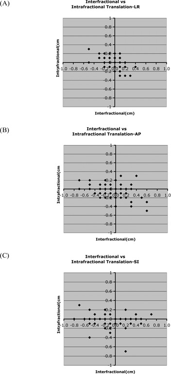
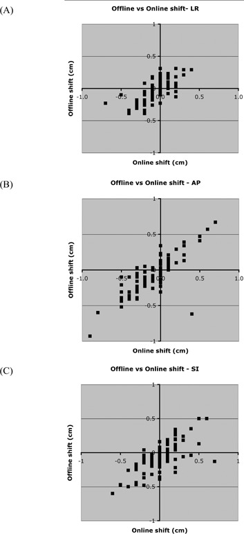
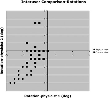
The purpose of the present study was to use a kilovoltage imaging device to measure interfractional and intrafractional setup deviations in patients with head-and-neck or brain cancers receiving intensity-modulated radiotherapy (IMRT) treatment. Before and after IMRT treatment, approximately 3 times weekly, 7 patients were imaged using the Varian On-Board Imager (OBI: Varian Medical Systems, Palo Alto, CA), a kilovoltage imaging device permanently mounted on the gantry of a Varian 21EX LINAC (Varian Medical Systems). Because of commissioning of the remote couch correction of the OBI during the study, online setup corrections were performed on 2 patients. For the other 5 patients, weekly corrections were made based on a sliding average of the measured data. From these data, we determined the interfractional setup deviation (defined as the shift from the original setup position suggested by the daily image), the residual error associated with the weekly correction protocol, and the intrafractional setup deviation, defined as the difference between the post-treatment and pretreatment images. We also used our own image registration software to determine interfractional and intrafractional rotational deviations from the images based on the template-matching method. In addition, we evaluated the influence of inter-observer variation on our results, and whether the use of various registration techniques introduced differences. Finally, translational data were compared with rotational data to search for correlations. Translational setup errors from all data were 0.0 +/- 0.2 cm, -0.1 +/- 0.3 cm, and -0.2 +/- 0.3 cm in the right-left (RL), anterior-posterior (AP), and superior-inferior (SI) directions respectively. Residual error for the 5 patients with a weekly correction protocol was -0.1 +/- 0.2 cm (RL), 0.0 +/- 0.3 cm (AP), and 0.0 +/- 0.2 cm (SI). Intrafractional translation errors were small, amounting to 0.0 +/- 0.1 cm, -0.1 +/- 0.2 cm, and 0.0 +/- 0.1 cm in the RL, AP, and SI directions respectively. In the sagittal and coronal views respectively, interfractional rotational errors were -1.1 +/- 1.7 degrees and -0.5 +/- 0.9 degrees, and intrafractional rotational errors were 0.3 +/- 0.6 degrees and 0.2 +/- 0.5 degrees. No significant correlation was seen between translational and rotational data. The OBI image data were used to study setup error in the head-and-neck patients. Nonzero systematic errors were seen in the interfractional translational and rotational data, but not in the intrafractional data, indicating that the mask is better at maintaining head position than at reproducing it.
Figures


References
-
- Manning MA, Wu Q, Cardinale RM, et al. The effect of setup uncertainty on normal tissue sparing with IMRT for head‐and‐neck cancer. Int J Radiat Oncol Biol Phys. 2001;51(5):1400–1409. - PubMed
-
- Samuelsson A, Mercke C, Johansson KA. Systematic setup errors for IMRT in the head and neck region: effect on dose distribution. Radiother Oncol. 2003;66(3):303–311. - PubMed
-
- Astreinidou E, Bel A, Raaijmakers CP, Terhaard CH, Lagendijk JJ. Adequate margins for random setup uncertainties in head‐and‐neck IMRT. Int J Radiat Oncol Biol Phys. 2005;61(3):938–944. - PubMed
-
- Hess CF, Kortmann RD, Jany R, Hamberger A, Bamberg M. Accuracy of field alignment in radiotherapy of head and neck cancer utilizing individualized face mask immobilization: a retrospective analysis of clinical practice. Radiother Oncol. 1995;34(1):69–72. - PubMed
-
- Pisani L, Lockman D, Jaffray D, Yan D, Martinez A, Wong J. Setup error in radiotherapy: on‐line correction using electronic kilovoltage and megavoltage radiographs. Int J Radiat Oncol Biol Phys. 2000;47(3):825–839. - PubMed
Publication types
MeSH terms
LinkOut - more resources
Full Text Sources
Medical
