. 2008 Sep;15(17):1210-22.
doi: 10.1038/gt.2008.73. Epub 2008 May 1.

Efficient entry inhibition of human and nonhuman primate immunodeficiency virus by cell surface-expressed gp41-derived peptides

Affiliations

Efficient entry inhibition of human and nonhuman primate immunodeficiency virus by cell surface-expressed gp41-derived peptides

R C Zahn et al. Gene Ther. 2008 Sep.

Abstract

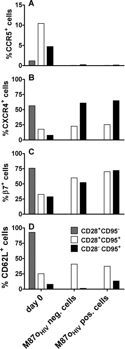
Membrane-anchored C-peptides (for example, maC46) derived from human immunodeficiency virus type 1 (HIV-1) envelope glycoprotein gp41 effectively inhibit HIV-1 entry in cell lines and primary human CD4+ cells in vitro. Here we evaluated this gene therapy approach in animal models of AIDS. We adapted the HIV gp41-derived maC46 vector construct for use in rhesus monkeys. Simian immunodeficiency virus (SIV and SHIV) sequence-adapted maC46 peptides, and the original HIV-1-derived maC46 expressed on the surface of established cell lines blocked entry of HIV-1, SIVmac251 and SHIV89.6P. Furthermore, primary rhesus monkey CD4+ T cells expressing HIV sequence-based maC46 peptides were also protected from SIV entry. Depletion of CD8+ T cells from PBMCs enhanced the yield of maC46-transduced CD4+ T cells. Supplementation with interleukin-2 (IL-2) increased transduction efficiency, whereas IL-7 and/or IL-15 provided no additional benefit. Phenotypic analysis showed that maC46-transduced and expanded cells were predominantly central memory CD4+ T cells that expressed low levels of CCR5 and slightly elevated levels of CD62L, beta7-integrin and CXCR4. These findings show that maC46-based cell surface-expressed peptides can efficiently inhibit primate immunodeficiency virus infection, and therefore serve as the basis for evaluation of this gene therapy approach in an animal model for AIDS.


Figures

Figure 1
Figure 1
Sequence variability of HIV, SHIV and SIV envelope protein in the C46 region of the HR2 of gp41 Env. HIV sequences were derived from 176 HIV-1 clade B strains (A) and compared to the HXB2 envelope region (aa 117-163 of gp41). SHIV sequences were derived from 13 rhesus monkeys infected with SHIV (B) and compared to the SHIV89.6P gp41 region (aa 117-163). SIV sequences from 27 rhesus monkeys infected with SIVmac239 or SIVmac251 (C) were compared to gp41 of SIVmac239 (aa 112-157 of gp41). The bars represent the percent amino acid change at a specific position and are color coded for each amino acid.
Figure 2
Figure 2
Inhibition of replication of HIV, SIV and SHIV in C46-expressing cell lines. PM-1 cells were transduced with retroviral vectors encoding for the antiviral maC46 sequence derived from HIV, SIV, or SHIV (A, D, and G: M87oHIV; B, E, and H: M87oSHIV; C, F, and I: M87oSIV). PM-1 cells transduced with the inactivate form of the C46 peptide were used as control cells. PM-1 cells were infected with HIV-1 NL4-3 (A-C), SIVmac251 (D-F), or SHIV89.6P (G-I) and the amount of p24 or p27 antigen in culture supernatants was determined by ELISA. The data shown are representative graphs from three experiments that were each performed in duplicate.
Figure 3
Figure 3
Efficacy of entry inhibition of HIV, SIV and SHIV afforded by low and high maC46-expressing variants of retroviral vectors. PM-1 cell lines expressing high levels of maC46 were generated by fluorescence activated cell sorting. PM-1 cell lines with a low expression of maC46 were generated by selection with G418. Control PM-1 cells were transduced with a retroviral vector coding for IRES-Neo and selected with G418. (A) Expression level of maC46 was detected by 2F5 mAb staining. (B-D) Relative level of transduction compared to control PM-1 cells. Lentiviruses coding for eGFP were either pseudotyped with HIV-1 JRFL, SIVmac251 or SHIV89.6P envelope protein. Transduction efficacy was determined by measuring eGFP expression by flow cytometry.
Figure 4
Figure 4
maC46 prevents infection of rhesus monkey CD4+ T cells with SIVmac251. Peripheral blood T lymphocytes from a naïve rhesus monkey were either transduced with the inactive C46 control vector or with M87oHIV and infected with SIVmac251. The expression of the maC46 peptide was determined by 2F5 mAb staining. The percentage of SIVmac251 infected cells was determined by anti-SIV p27 mAb staining. (A) Background p27 staining in non-transduced and C46 control vector-transduced, uninfected CD4+ T cells. (B) Detection of p27 antigen in non-transduced and C46 control vector transduced SIVmac251-infected CD4+ T cells. (C) Detection of p27 antigen in non-transduced and M87oHIV vector transduced, SIVmac251-infected CD4+ T cells. The percentage of p27 positive cells is indicated in each graph (top: transduced cells; bottom: non-transduced cells).
Figure 5
Figure 5
Expansion, M87oHIV transduction and phenotype of maC46-transduced CD4+ T cells. CD8+ lymphocyte-depleted and total lymphocytes obtained from peripheral blood were transduced with the M87oHIV retroviral vector and cultured with anti-CD3 and anti-CD28 coated beads for 7 days. (A) Rate of expansion of CD4+ T cells 7 days after initiation of in vitro culture. Cell counts were determined by hemacytometer. The black bar represents the bulk culture, the white bar the CD8+ cell-depleted culture. (B) Transduction efficacy of CD4+ T cells with the retroviral vector M87oHIV. The cells were transduced twice with a multiplicity of infection (MOI) of 1.3-2 or more than a MOI of 3.3. M87oHIV expression was determined by staining with a C46 specific mAb. The black bar represents the bulk culture, the white bar the CD8+ cell-depleted culture. (C) and (D) Phenotype of expanded CD4+ T cells. On day 7 after culture, cells were stained with anti-CD28 and anti-CD95 mAbs to determine T cell subsets and with 2F5 to determine C46 expression. The black bars represent M87oHIV-expressing cells, the white bars M87oHIV negative cells. The bars in all graphs represent median values, error bars the interquartile range (n = 4-10).
Figure 6
Figure 6
Phenotype of M87oHIV-transduced CD4+ T cells. Peripheral blood from 4 or 5 naïve rhesus monkeys was used to determine the expression of CCR5 (A), CXCR4 (B), β7-integrin (C) and CD62L (D) on CD4+ T cell subsets prior to transduction and on day 7 after transduction and in vitro culture. The CD28 and CD95 paradigm was used to determine the lymphocyte maturation-associated T cell subsets. PBMC separated by Ficoll gradient were used to determine cell surface expression of β7-integrin, CXCR4 and CD62L on day 0. The cell surface expression of CCR5 on day 0 was determined by whole blood staining. The bars in all graphs represent median values.
Figure 7
Figure 7
Effect of different cytokines on efficacy of expansion and level of transduction of CD4+ T cells. PBMC were depleted of CD8+ cells and expanded for 7 days with anti-CD3 and anti-CD28-coated paramagnetic beads. The different cytokines or combinations of cytokines were added on day 0. Cell culture supernatants were partially replaced with fresh media containing the different cytokines on days 4, 5 and 6. On days 4 and 5 cells were transduced with M87oHIV retroviral vectors. (A) Fold expansion of CD4+ T cells expanded in different cytokines or cytokine combinations relative to IL-2 treated cells that were set to an expansion factor of 1. (B) M87oHIV transduction efficacy of CD4+ T cells expanded in different cytokines or cytokine combinations. The bars represent median values from 5 animals. (C-F) Side and Forward Scatter of one representative dot plot of CD4+ T cells expanded in media without cytokines (C), supplemented with IL-2 (D), IL-7 (E), and IL-7 + IL-15 (F).

Similar articles

Cited by

References

    1. Puls RL, Emery S. Therapeutic vaccination against HIV: current progress and future possibilities. Clin Sci. 2006;110:59–71. - PubMed
    1. von Laer D, Hasselmann S, Hasselmann K. Gene therapy for HIV infection: what does it need to make it work? J Gene Med. 2006;8:658–667. - PubMed
    1. Strayer DS, Akkina R, Bunnell BA, Dropulic B, Planelles V, Pomerantz RJ, et al. Current status of gene therapy strategies to treat HIV/AIDS. Mol Ther. 2005;11:823–842. - PubMed
    1. von Laer D, Hasselmann S, Hasselmann K. Impact of gene-modified T cells on HIV infection dynamics. J Theor Biol. 2006;238:60–77. - PubMed
    1. Gallo SA, Finnegan CM, Viard M, Raviv Y, Dimitrov A, Rawat SS, et al. The HIV Env-mediated fusion reaction. Biochim Biophys Acta. 2003;1614:36–50. - PubMed

Publication types

MeSH terms

Substances