. 2008 Oct;457(1):25-36.
doi: 10.1007/s00424-008-0501-x. Epub 2008 May 1.

Differential contribution of cardiac sarcomeric proteins in the myofibrillar force response to stretch

Affiliations

Differential contribution of cardiac sarcomeric proteins in the myofibrillar force response to stretch

Younss Ait Mou et al. Pflugers Arch. 2008 Oct.

Abstract

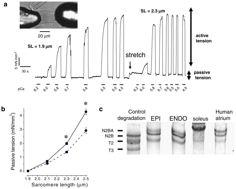
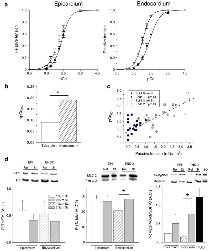
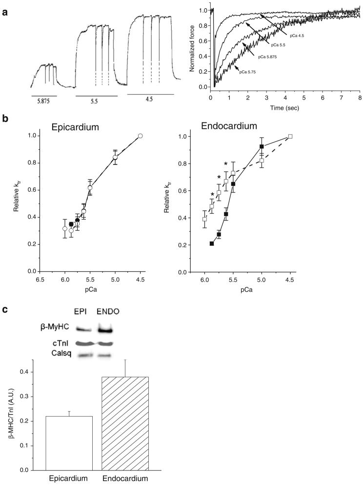
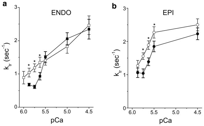
The present study examined the contribution of myofilament contractile proteins to regional function in guinea pig myocardium. We investigated the effect of stretch on myofilament contractile proteins, Ca(2+) sensitivity, and cross-bridge cycling kinetics (K (tr)) of force in single skinned cardiomyocytes isolated from the sub-endocardial (ENDO) or sub-epicardial (EPI) layer. As observed in other species, ENDO cells were stiffer, and Ca(2+) sensitivity of force at long sarcomere length was higher compared with EPI cells. Maximal K (tr) was unchanged by stretch, but was higher in EPI cells possibly due to a higher alpha-MHC content. Submaximal Ca(2+)-activated K (tr) increased only in ENDO cells with stretch. Stretch of skinned ENDO muscle strips induced increased phosphorylation in both myosin-binding protein C and myosin light chain 2. We concluded that transmural MHC isoform expression and differential regulatory protein phosphorylation by stretch contributes to regional differences in stretch modulation of activation in guinea pig left ventricle.


Figures

Fig. 1
Fig. 1
Passive and active properties of permeabilized guinea pig ventricular myocyte isolated in sub-epicardium (EPI) and sub-endocardium (ENDO). a Original recording of force during myocyte activation. b Passive properties of the cells were determined by sequentially stretching the cell to various SL in relaxing solution. EPI cells (filled circles) are significantly more compliant than ENDO cells (filled squares; n=14 cells per region). c Titin isoform content was investigated on a gradient SDS gel (2.7–7%); rat cardiac tissue incubated with trypsin was used as a control of titin degradation, rat soleus for the N2A isoform and human atrium for the N2BA and N2B titin isoforms; guinea pig cardiac tissue expressed both N2B and N2BA isoforms (n=5 animals, in duplicate). *P<0.05
Fig. 2
Fig. 2
Relationship between the relative tension and calcium in ENDO and EPI cells at two sarcomere lengths. a Changes of calcium sensitivity of the contractile machinery are indicated by the leftward shift of the tension–pCa curve when cells were stretched from 1.9 μm SL (closed symbols) to 2.3 μm SL (open symbols; n=14 cells per region). b Stretch-induced calcium sensitization (ΔpCa50) was more prominent in ENDO cells. c Relationship between calcium sensitivity and passive tension for all data from ENDO and EPI at two sarcomere lengths. Passive tension was measured in relaxing solution prior each activation procedure from slack length to 1.9 or 2.3 μm SL (y=5.47+ 0.011x, r=−0.82, P<0.001). d Skinned muscle strips dissected from the sub-epicardial layer (full bar) or the sub-endocardial layer (hatched bar) were quick-frozen either at slack length (open bar) or after stretch to 20–30% of the slack length (grey bar). Left: immunoblots with anti-cardiac TnI and anti-cTnI phosphorylated by PKA showed that TnI phosphorylation was not affected by stretch (n= 6 animals/group). Middle: for MLC-2 phosphorylation quantification, each isoform was separated and expressed as a percentage of the total amount (phosphorylated + non-phosphorylated forms). Right: Immunoblots with anti-cardiac MyBP-C and anti-phospho Ser282 cMyBP-C showed that cMyBP-C phosphorylation level was increased by stretch (n=6 animals/group). Isoprenaline stimulation (Iso) of intact myocytes was used as control for full phosphorylation of MyBP-C. *P<0.05
Fig. 3
Fig. 3
Release/restretch experiments on a single skinned cardiac myocyte. a Original tracing of the tension showing the application of three successive large (20%) release/restretch maneuvers of cell length elicited at three pCas (5.875, 5.5, and 4.5). Tension reached zero level after this procedure; however, the frequency response of the chart recorder was not sufficient to allow this to be recorded before the cell was rapidly restretched; for clarity the fast initial tension releases have been retouched (dotted lines). Right: averaged records of tension for determination of Ktr at four calcium concentrations. For each Ca2+ concentration, force before applying the slack release–restretch maneuver was used for normalization to emphasize the changes in the rate of tension redevelopment with Ca2+. b Relationship between relative Ktr and pCa in EPI (n=8 cells, circle) and ENDO (n=8 cells, square) cells at SL=1.9 μm (closed symbols, line) and SL=2.3 μm (open symbols, dashed line). c Left: β-Myosin heavy chain expression in cardiomyocytes isolated from sub-epicardium (open bar) or sub-endocardium (grey bar) as determined by Western Blot was expressed relative to the total TnI content in each group (n=5 hearts per group analyzed in triplicate). Calsequestrin (Cals) was used as an additional internal control of loading. *P<0.05
Fig. 4
Fig. 4
Passive tension dependency of the rate constant of tension redevelopment in ENDO (a) and EPI (b) myocytes. Ktr was measured in myocytes at short sarcomere length (closed symbols) and at a long sarcomere length developing a passive tension of about 2.4 mN/mm2 (open symbols) corresponding to a stretch of 2.3 μm SL for ENDO cells (n=14) and about 2.45 μm SL for EPI cells (n=10). *P<0.05

Similar articles

Cited by

References

    1. Allen DG, Kentish JC. Calcium concentration in the myoplasm of skinned ferret ventricular muscle following changes in muscle length. J Physiol. 1988;407:489–503. - PMC - PubMed
    1. Balnave CD, Allen DG. The effect of muscle length on intracellular calcium and force in single fibres from mouse skeletal muscle. J Physiol. 1996;492:705–713. - PMC - PubMed
    1. Borbely A, van der Velden J, Papp Z, Bronzwaer JG, Edes I, Stienen GJ, Paulus WJ. Cardiomyocyte stiffness in diastolic heart failure. Circulation. 2005;111:774–781. - PubMed
    1. Brenner B, Eisenberg E. Rate of force generation in muscle: correlation with actomyosin ATPase activity in solution. Proc Natl Acad Sci USA. 1986;83:3542–3546. - PMC - PubMed
    1. Bugaisky LB, Anderson PG, Hall RS, Bishop SP. Differences in myosin isoform expression in the subepicardial and subendocardial myocardium during cardiac hypertrophy in the rat. Circ Res. 1990;66:1127–1132. - PubMed

Publication types

MeSH terms